
Myocardial infarction pathology laq
gross pathology myocardial infarction heart cut section
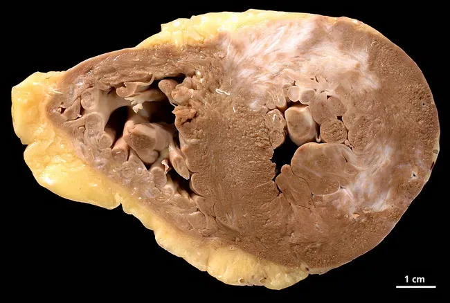
Gross pathology image of a human heart cross-section (short-axis/transverse) from a 73-year-old female with a history of arterial hypertension, chronic smoking, and three-vessel coronary artery disease. The specimen reveals chronic myocardial infarction scar throughout the myocardium with areas of subacute necrosis on both ventricles. The ventricles show concentric hypertrophy characterized by thickened ventricular walls and reduced cavity size, consistent with long-standing pressure overload. On cut surface, extensive fibrous replacement tissue is seen in the LV and RV walls, with pale, firm areas indicating scar tissue; endocardial surfaces may show thinning in infarcted zones. The presence of bilateral ventricular involvement suggests diffuse ischemic injury, possibly past infarcts with replacement fibrosis. The gross morphology corresponds to ischemic cardiomyopathy spectrum; pathophysiology includes atherosclerotic CAD, hypertension-induced hypertrophy, and chronic subendocardial ischemia. This combination results in remodeling and scar formation, predisposing to arrhythmias and heart failure. The image is from pathorama.ch, illustrating classic post-infarction changes: myocardial scar, subacute necrosis, and hypertrophic remodeling. Clinically, such pathology would correlate with prior chest pain episodes, elevated troponin in the past, and radiographic evidence of cardiomegaly; autopsy or pathology reporting would emphasize chronic infarct burden, LV dysfunction, and risk stratification for sudden cardiac death. Suitable for education on myocardial infarction progression and necrosis patterns.
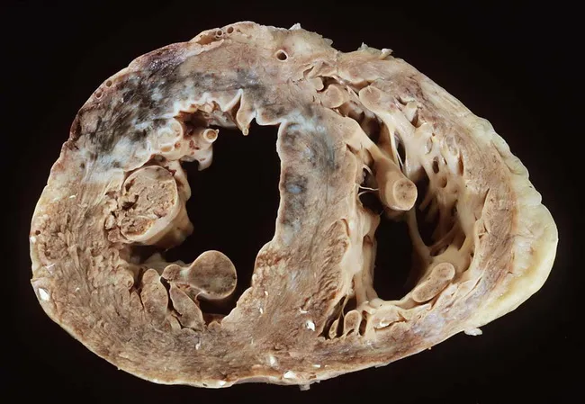
Gross pathology image of an extracted human heart cut in transverse section to expose the ventricles. Modality: macroscopic inspection during autopsy or surgical specimen; technique: serial cross-sectional dissection of the ventricles revealing infarcted and fibrotic zones. Anteriorly in the anteroseptal left ventricular wall there is a partially healed infarction with thinning, scar formation and mottled pallor consistent with prior ischemic injury. In the anterolateral/lateral left ventricular wall, a subacute infarct is evident, showing fibrous connective tissue with adjacent edema and discoloration. The right ventricle demonstrates marked fibrosis and myocardial remodeling with patchy, firm trabeculations. Overall, the myocardium exhibits biventricular hypertrophy with thickened walls and increased myocardial mass; left ventricular wall thickening may appear concentric but can reflect remodeling from chronic hypertension and ischemic injury. The combination of age (82-year-old female) and cardiovascular risk factors-smoking, essential hypertension, dyslipidemia—supports a long-standing coronary atherosclerotic disease. Notable clinical context includes postmenopausal elevation of coronary risk. The image highlights the structural consequences of myocardial infarction, scar replacement, and concentric/eccentric hypertrophy in the setting of ischemic cardiomyopathy. This resource is useful for education on infarct evolution, ventricular remodeling, and CAD risk assessment in aging female patients. Correlation with clinical history strengthens diagnostic interpretation and teaching value.
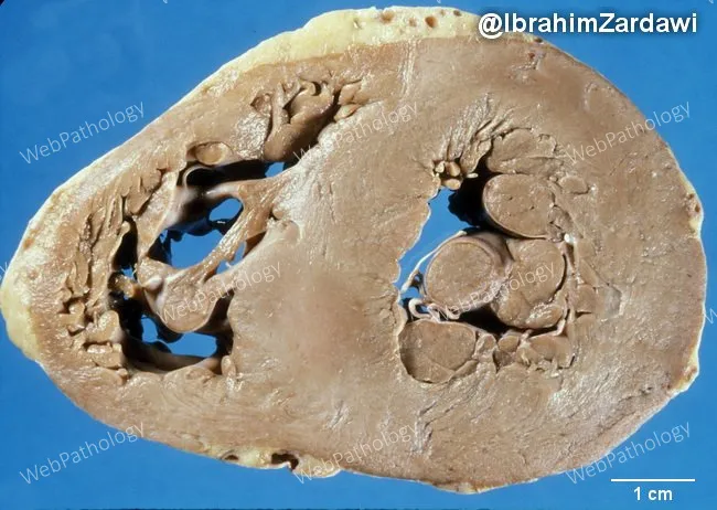
Gross pathology photograph of a human heart in transverse mid-ventricular section, illustrating macroscopic features of cardiac amyloidosis. The myocardium appears pale, waxy, and firm, consistent with amyloid deposition replacing normal myocardial tissue. Concentric left ventricular hypertrophy is evident, with thickened LV wall while chamber cavity is reduced in caliber relative to normal. The right ventricle is less involved in this cut, and the endocardial surfaces show smooth contour without focal infarction. These findings reflect an infiltrative cardiomyopathy process where extracellular amyloid proteins infiltrate the interstitium, causing stiff, noncompliant myocardium and diastolic dysfunction. The gross resemblance to waxy infiltration, in combination with preserved coronary architecture, supports a diagnosis of cardiac amyloidosis rather than primary hypertrophic hypertrophy or hypertensive remodeling alone. This image serves educational and diagnostic contexts for surgical pathology, cardiology, and radiology reviews, and is useful for teaching about late-stage restrictive cardiomyopathy. In clinical practice, these macroscopic cues prompt confirmatory microscopic evaluation, immunohistochemical typing, and systemic evaluation for underlying plasma cell dyscrasia or transthyretin amyloidosis. Potential uses include medical education, pathology correlation, radiology-pathology conferences, and research on infiltrative heart disease and cardiomyopathy phenotypes. Correlative clinical data and ancillary testing guide prognosis and treatment choices in suspected infiltrative cardiomyopathy patients today.
microscopic histology myocardial infarction coagulative necrosis neutrophils
"myocardial infarction" pathogenesis
contraction band necrosis reperfusion injury myocardium histology
Myocardial Infarction – Pathology (Long Answer Question)
Definition
Etiology / Risk Factors
- Underlying cause in >90% of cases: coronary atherosclerosis
- Non-atherosclerotic causes (~10%): coronary vasospasm, embolism (mural thrombus in AF, valve vegetations), vasculitis, amyloid deposition, sickle cell stasis, cocaine-induced spasm
- Risk rises with age; men > women (gap narrows after menopause); ~10% of MIs occur before age 40 and 45% before age 65 (Robbins & Kumar Basic Pathology, p. 318)
Pathogenesis
- An atheromatous plaque undergoes sudden change - erosion or rupture (endothelial injury, intraplaque hemorrhage, mechanical stress), exposing subendothelial collagen and necrotic plaque contents to blood.
- Platelets adhere, aggregate, and are activated - releasing thromboxane A2, ADP, serotonin - causing further aggregation and vasospasm.
- Tissue factor activates the coagulation cascade, adding to the growing thrombus.
- Within minutes, the enlarging thrombus can completely occlude the lumen.
- Loss of aerobic metabolism within seconds → ATP depletion → loss of contractility within 2 minutes
- Ischemia >20-40 minutes → irreversible injury and coagulative necrosis
- Necrosis begins in the subendocardium (last area perfused, highest intramural pressure) and spreads as a "wavefront" toward the epicardium over 6-12 hours if untreated (transmural infarct)
| Feature | Time of onset |
|---|---|
| ATP depletion begins | Seconds |
| Loss of contractility | <2 minutes |
| ATP at 50% of normal | 10 minutes |
| ATP at 10% of normal | 40 minutes |
| Irreversible injury | 20-40 minutes |
| Microvascular injury | >1 hour |
Coronary artery distribution and infarct location
| Vessel occluded | Frequency | Region infarcted |
|---|---|---|
| Left anterior descending (LAD) | 40-50% | Anterior LV wall, anterior septum, apex |
| Right coronary artery (RCA) | 30-40% | Inferior/posterior LV, posterior septum, ± RV free wall |
| Left circumflex (LCX) | 15-20% | Lateral LV wall (except apex) |
Types / Patterns of Infarction
- Transmural infarct: full thickness of the wall, corresponds to a single epicardial artery territory, associated with occlusive thrombus over atheroma
- Subendocardial (non-transmural) infarct: limited to inner 1/3-1/2 of wall; caused by global hypoperfusion (shock, severe stenosis with increased demand) superimposed on chronic stenosis; may be circumferential rather than in one vascular territory
- Microinfarcts: due to small vessel disease (vasculitis, embolization of small particles)
- Clinically classified by biomarker/ECG criteria: STEMI vs NSTEMI, and by Universal Classification (Type 1 spontaneous plaque rupture, Type 2 supply-demand mismatch, Type 3 sudden death, Type 4/5 procedure-related)
Morphology
Gross changes (correlate with time since onset)
| Time | Gross appearance |
|---|---|
| <12 hours | Usually none visible; TTC (triphenyl tetrazolium chloride) staining shows a pale unstained zone against brick-red viable myocardium |
| 12-24 hr | Reddish-blue discoloration (congestion, trapped blood) |
| 1-3 days | Mottled with yellow-tan center |
| 3-7 days | Hyperemic (red) border of granulation tissue; soft yellow-tan center |
| 7-10 days | Maximally yellow-tan and soft, with depressed red-tan margins (most vulnerable to rupture) |
| 10-14 days | Red-gray depressed borders |
| 2-8 weeks | Progressive gray-white fibrous scarring from the border inward |
| >2 months | Scarring complete - dense white/gray fibrous scar |
Microscopic changes
| Time | Light microscopy |
|---|---|
| 0.5-4 hr | Usually none; occasional wavy fibers at border |
| 4-12 hr | Early coagulative necrosis, edema, hemorrhage |
| 12-24 hr | Ongoing coagulative necrosis; pyknotic nuclei, myocyte hypereosinophilia, marginal contraction band necrosis, early neutrophilic infiltrate |
| 1-3 days | Coagulative necrosis with loss of nuclei/striations; brisk neutrophilic infiltrate |
| 3-7 days | Disintegration of dead fibers; dying neutrophils; early macrophage phagocytosis at border |
| 7-10 days | Well-developed phagocytosis of dead cells; granulation tissue at margins |
| 10-14 days | Well-established granulation tissue - new vessels + collagen |
| 2-8 wk | Increased collagen deposition, decreased cellularity → scar |
| >2 months | Dense collagenous scar |
Reperfusion and its modification of infarcts
- Reperfusion injury: paradoxical injury from oxidative stress, calcium overload, and inflammation, producing contraction band necrosis (hypercontracted sarcomeres)
- Myocardial "stunning": transient reversible contractile dysfunction after reperfusion
- Hemorrhagic infarction: blood extravasation into the necrotic zone through damaged microvasculature
Complications of Myocardial Infarction
| Complication | Typical timing | Notes |
|---|---|---|
| Arrhythmia | Within hours (most common cause of death) | VF is the leading cause of pre-hospital death |
| Cardiogenic shock / CHF | Early, if >40% of LV involved | Pump failure |
| Fibrinous pericarditis | 2-4 days | Due to transmural necrosis irritating epicardium |
| Free wall (myocardial) rupture | 3-7 days (softest, weakest phase) | Causes hemopericardium/tamponade, often fatal |
| Papillary muscle rupture | 3-7 days | Acute mitral regurgitation |
| Interventricular septal rupture | 3-7 days | Left-to-right shunt, new murmur |
| Mural thrombus | Variable, especially with LV aneurysm or akinetic segments | Risk of systemic embolization/stroke |
| Ventricular aneurysm | Weeks later (healed thin scar) | Risk of mural thrombus, arrhythmia, rarely rupture (low, since it's fibrous) |
| Dressler syndrome | Weeks to months | Autoimmune pericarditis |
| Chronic ischemic heart disease / progressive CHF | Months to years | From loss of contractile mass and remodeling |
Diagnosis (pathology-clinical correlation)
- Cardiac troponin I/T: rises 2-4 hours, peaks 24-48 hr, remains elevated 7-10 days - most sensitive/specific biomarker
- CK-MB: rises faster but less specific, useful for detecting reinfarction (returns to baseline sooner)
- ECG: ST elevation (transmural/STEMI) vs ST depression/T inversion (subendocardial/NSTEMI); Q waves develop with transmural necrosis
- Gross/histologic diagnosis at autopsy uses TTC staining (for infarcts >2-3 hr old) and the histologic timeline above