
Biliary tract and system, imaging of biliary tract and gallstone disease
Reading File
This is a broad visual/anatomical/clinical topic. I'll run parallel searches across the library.
Finding Sources
Finding Sources
Finding Sources
Excellent. The key sections are in Sleisenger & Fordtran and Yamada's. Let me read the most relevant blocks in parallel.
Reading File
Reading File
Finding Sources
The block18 from Sleisenger contains the full imaging chapter. Let me read more of it, and also get the gallbladder/gallstone disease section from Schwartz's, and the biliary disease chapter from Sleisenger block 17.
Reading File
Reading File
Reading File
Excellent content. Now let me get the detailed gallstone formation/disease content from Schwartz's and the gallbladder anatomy from it, plus more of the imaging chapter from Sleisenger block 18, and fetch the key images.
Reading File
Reading File
Loading Image
Now let me get the gallstone formation content from Schwartz's, the gallbladder diagnostic studies section, and search for more images and additional key content.
Reading File
Finding Sources
Finding Sources
Excellent. I now have comprehensive content. Let me fetch the cholescintigraphy and more detailed gallstone content.
Reading File
Reading File
I now have comprehensive content from multiple authoritative sources. Let me fetch the cholescintigraphy image and the Schwartz's gallstone formation section.
Loading Image
Reading File
I now have all the information needed for a comprehensive response. Let me compile it.
Biliary Tract and System: Imaging and Gallstone Disease
Anatomy of the Biliary Tract
The gallbladder is a pear-shaped sac measuring 7-10 cm long with a capacity of 30-50 mL (can distend to 300 mL when obstructed). It sits in a fossa on the inferior surface of the liver and is divided into four regions:
| Region | Features |
|---|---|
| Fundus | Rounded blind end, extends 1-2 cm beyond liver margin, contains most smooth muscle |
| Body | Main storage area, rich in elastic tissue allowing distension |
| Infundibulum (Hartmann's pouch) | Mucosal outpouching at junction of body and neck - site where gallstones commonly lodge |
| Neck | Lies in deepest gallbladder fossa, connects to cystic duct |
Cantle's line (a vertical plane from gallbladder fossa anteriorly to the IVC posteriorly) divides the liver into right and left lobes.
The cystic artery supplying the gallbladder is a branch of the right hepatic artery in >90% of cases, nearly always found within the hepatocystic triangle (Calot's triangle).
The bile duct system includes:
- Right and left hepatic ducts joining to form the common hepatic duct (CHD)
- CHD + cystic duct = Common bile duct (CBD)
- CBD opens into the duodenum at the ampulla of Vater, regulated by the sphincter of Oddi
Sphincter of Oddi
The sphincter of Oddi spans 4-6 mm, has a basal resting pressure ~13 mmHg above duodenal pressure, and shows phasic contractions at ~4/min with amplitude 12-140 mmHg. It regulates bile/pancreatic juice flow, prevents duodenal regurgitation into the biliary tree, and diverts bile into the gallbladder. Relaxation occurs in response to CCK, glucagon, and secretin.
Imaging of the Biliary Tract
Imaging is of utmost importance in planning management for biliary disorders, regardless of whether an endoscopic or percutaneous approach will be taken.
1. Transabdominal Ultrasound (US)
The first-line imaging modality. It is universally available, involves no radiation, and requires no contrast agents.
Capabilities:
- Detects intrahepatic ductal dilatation
- Images the gallbladder - highly sensitive for gallstones
- Detects extrahepatic biliary obstruction with accuracy 78-98%
- Elicits the sonographic Murphy's sign: increased pain when the transducer is pressed over the gallbladder - highly suggestive of acute cholecystitis
- Additional findings in cholecystitis: gallbladder wall thickening (>3 mm), pericholecystic fluid, gallbladder enlargement >5 cm
Limitations:
- For choledocholithiasis (CBD stones): specificity is high but sensitivity does not exceed 68% and is often <50%, especially for small stones with non-dilated ducts
- Accuracy for level of obstruction: 27-95%; for cause of obstruction: 23-88%
- Limited ability to distinguish malignant from benign causes
- Reduced resolution in obese patients
Advances include 3D/4D imaging, contrast-enhanced US, and elastography
2. MRCP (Magnetic Resonance Cholangiopancreatography)
An MRI-based, noninvasive study exploiting the high T2-signal of bile (bright signal), with solid material such as stones appearing as dark filling defects.
Performance for choledocholithiasis:
- Sensitivity: 81-100%
- Specificity: 96-100%
- High overall diagnostic accuracy
Also highly accurate for benign and malignant strictures, and thorough evaluation of intrahepatic bile ducts. False-positives may arise from pneumobilia. IV contrast (gadodiamide/gadopentetate dimeglumine) can be added; in post-liver transplant patients, mangafodipir trisodium (excreted in bile) enhances detection of leaks and strictures.
Fig. 70.1 - Choledocholithiasis on MRCP and ERCP:
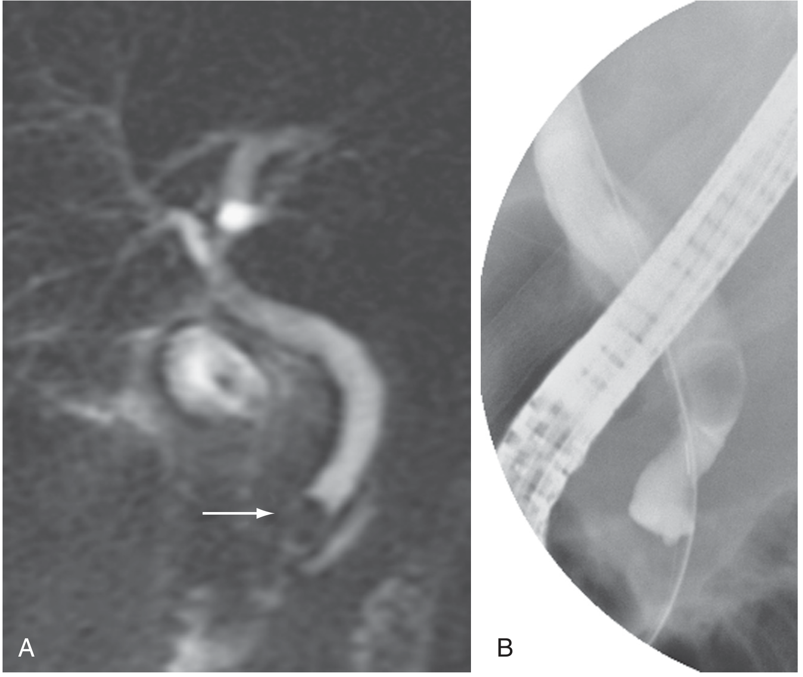
A: MRCP shows a filling defect in the distal bile duct (arrow). B: Corresponding ERCP confirming the same filling defect. (Sleisenger & Fordtran, Fig. 70.1)
3. Multidetector CT Cholangiography
CT can identify biliary obstruction and is often used for broader abdominal assessment, but it is less specific than MRCP for choledocholithiasis. Plain radiographs may demonstrate biliary air (pneumobilia) in emphysematous cholecystitis or biliary-enteric fistula, though these are more reliably seen on other modalities.
4. Cholescintigraphy (HIDA/DISIDA Scan)
A radionuclide imaging test most useful for suspected acute cholecystitis. IV-administered 99mTc-labeled HIDA (hydroxyl iminodiacetic acid) or DISIDA (diisopropyl iminodiacetic acid) is taken up by hepatocytes and excreted into bile.
Normal scan: Radioactivity in gallbladder, bile duct, and small intestine within 30-60 minutes.
Abnormal ("positive") scan: Non-visualization of the gallbladder with preserved excretion into bile duct or small intestine - indicates cystic duct obstruction (hallmark of acute cholecystitis).
- Accuracy for acute cholecystitis: 92% (superior to US)
- False positives in fasting or critically ill patients (decreased gallbladder motility)
- DISIDA can image patients with bilirubin as high as 20 mg/dL
- Morphine augmentation: If gallbladder not visualized at 60 min, IV morphine raises sphincter of Oddi pressure, forcing preferential flow into gallbladder if cystic duct is patent - scan repeated at 30 minutes
Cholescintigraphy serial scan (normal study):
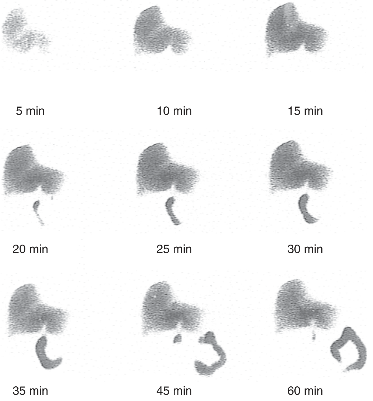
Serial cholescintigraphy: radiotracer fills the liver (5-15 min), progressing to visualize the gallbladder and bile duct (20-35 min), with filling of the duodenum by 45-60 min. (Sleisenger & Fordtran, Fig. 65.10)
5. Endoscopic Retrograde Cholangiopancreatography (ERCP)
ERCP is the gold standard for biliary stone removal and provides both diagnostic and therapeutic capability. It is one of the most technically demanding endoscopic procedures. Indications include choledocholithiasis, biliary strictures (benign/malignant), PSC, bile leaks, and sphincter of Oddi dysfunction.
Adverse events (5 main types):
- Sedation-related
- Post-ERCP pancreatitis (highest risk: young women with suspected SOD; reduced by prophylactic pancreatic duct stenting and rectal NSAIDs)
- Bleeding (risk increased with coagulopathy, anticoagulation within 72h)
- Perforation (<1%, may require surgery)
- Infection/cholangitis (primarily when drainage is inadequate)
6. Endoscopic Ultrasound (EUS)
EUS uses a specialized 30° endoscope with a radial or linear ultrasound transducer. Key advantages:
- Less invasive than ERCP (no sphincter cannulation needed)
- Useful for visualizing the retroduodenal bile duct (difficult on transabdominal US)
- Identifies choledocholithiasis
- Evaluates tumors near/behind the duodenum and resectability
- Linear EUS scope allows EUS-guided FNA, therapeutic injections, and drainage (EUS-guided biliary drainage, EUS-BD)
In patients with surgically altered anatomy (Roux-en-Y), EUS-guided biliary drainage achieves technical success ~98% vs. ~65% with enteroscopy-assisted ERCP.
7. Percutaneous Transhepatic Cholangiography (PTC)
An invasive procedure where intrahepatic bile ducts are accessed percutaneously under fluoroscopic guidance. Antegrade cholangiography and interventions (tissue sampling, drain/stent placement, balloon dilation) can be performed. Reserved for patients in whom ERCP is not feasible:
- Roux-en-Y or gastric bypass anatomy
- Extrahepatic stricture not traversable endoscopically
Risks: bleeding, cholangitis, bile leak, catheter-related complications.
8. Oral Cholecystography (OCG)
Now has limited application - largely replaced by US. Useful only for:
- Evaluating patients being considered for medical dissolution therapy or lithotripsy
- Evaluating unsuspected gallbladder disease (adenomyomatosis, cholesterolosis) when US is non-diagnostic
Gallstone Disease (Cholelithiasis)
Epidemiology
Gallstones affect 10-15% of adults at autopsy. Risk is related to diet, age, sex, BMI, and ethnicity (highest in Native American and Latin American populations).
Risk factors:
- Pregnancy
- Non-HDL hyperlipidemia
- Crohn's disease
- Hemolytic disorders (hereditary spherocytosis, sickle cell disease, thalassemia) - pigment stones
- Ileal resection (terminal ileum resorbs bile salts)
- Rapid weight loss/bariatric surgery
- Gastric/duodenal surgery (alters neuro-hormonal biliary regulation)
Types of Gallstones
| Type | Composition | Key Features |
|---|---|---|
| Cholesterol (most common) | >50% cholesterol monohydrate | Require supersaturated bile; radiolucent on X-ray |
| Pigment (black) | Bilirubin polymers + calcium | Hemolytic disease, cirrhosis; radiopaque |
| Pigment (brown) | Calcium bilirubinate | Infection, bile stasis; form in bile ducts |
Pathogenesis of Cholesterol Gallstones
Three factors are required:
- Bile supersaturation with cholesterol (lithogenic bile)
- Crystal nucleation - cholesterol monosaturation with mucin glycoproteins acting as nucleating agents
- Gallbladder stasis - impaired emptying allows crystal growth and aggregation
Clinical Presentations and Diagnosis
Biliary colic: Steady, severe pain in the epigastric/right subcostal area (not truly "colicky"), often radiating to the right shoulder/interscapular region. Duration typically 30 minutes to several hours. Note that the left subcostal area is not an unusual site of pain.
Diagnosis:
- Abdominal ultrasound is the standard first-line test - highly sensitive for gallstones (>95% sensitivity), demonstrating echogenic foci with posterior acoustic shadowing
- Stones found incidentally on CT or plain films in asymptomatic patients should be left in place
- Sludge with typical biliary pain: cholecystectomy is warranted
- Stones not visible but typical symptoms present: consider EUS or MRCP
Complications
| Complication | Key Features |
|---|---|
| Acute cholecystitis | Cystic duct obstruction → inflammation; US shows wall thickening, pericholecystic fluid, sonographic Murphy's sign; HIDA most accurate (92%) |
| Choledocholithiasis | CBD stones; elevated bilirubin + ALP; MRCP or EUS for diagnosis; ERCP for treatment |
| Cholangitis | Charcot's triad (fever, jaundice, RUQ pain); Reynold's pentad adds shock + altered consciousness; urgent biliary decompression |
| Gallstone pancreatitis | US first; ERCP if concurrent cholangitis or persistent obstruction |
| Gallstone ileus | Rare; fistula between gallbladder and bowel; rigler's triad on X-ray (pneumobilia, SBO, ectopic stone) |
| Hydrops of gallbladder | Impacted cystic duct stone without infection; gallbladder fills with clear white mucin |
| Mirizzi syndrome | Extrinsic CBD compression by stone in cystic duct or Hartmann's pouch |
Non-Surgical Management
Oral dissolution therapy (ursodeoxycholic acid/UDCA):
- Reduces biliary cholesterol secretion, desaturates bile
- Effective only for radiolucent (cholesterol) stones in a functioning gallbladder with a patent cystic duct
- Small stones respond more quickly (better surface-to-volume ratio)
- High recurrence rate on stopping therapy
- Patient selection (Box 66.1):
- Symptomatic (biliary pain) without complications
- Normal gallbladder function (confirmed by OCG, cholescintigraphy, or functional US)
- Radiolucent stones, <1.5 cm, <3 in number, no calcification on CT
- Not indicated for asymptomatic stones or complications (cholecystitis, pancreatitis, cholangitis)
Surgical treatment:
- Laparoscopic cholecystectomy is the gold standard
- ~90% of patients with typical symptoms are rendered symptom-free
- Atypical symptoms (dyspepsia, flatulence, bloating) have lower success rates
Summary of Imaging Modality Choice
| Clinical Question | Best Modality |
|---|---|
| Gallstones in gallbladder | Transabdominal US (first-line) |
| Suspected acute cholecystitis | US (initial); HIDA scan (most accurate, 92%) |
| CBD stones / choledocholithiasis | MRCP (81-100% sensitivity); EUS if MRCP equivocal |
| CBD access + stone removal | ERCP (diagnostic + therapeutic) |
| Biliary stricture characterization | MRCP + EUS with FNA |
| Malignant obstruction, altered anatomy | PTC (when ERCP not feasible) |
| Pre-dissolution therapy evaluation | OCG or functional US |
Sources:
- Sleisenger and Fordtran's Gastrointestinal and Liver Disease, Chapter 70 (Imaging/Endoscopic Treatment of Biliary Tract), Chapter 65-66 (Gallstone Disease and Dissolution)
- Schwartz's Principles of Surgery, 11th ed., Chapter 32 (Gallbladder and Extrahepatic Biliary System)
- Yamada's Textbook of Gastroenterology, 7th ed.
This is a shared conversation. Sign in to Orris to start your own chat.