
Down syndrome
"Down syndrome"[MeSH Terms]
Down syndrome trisomy 21 karyotype facial features
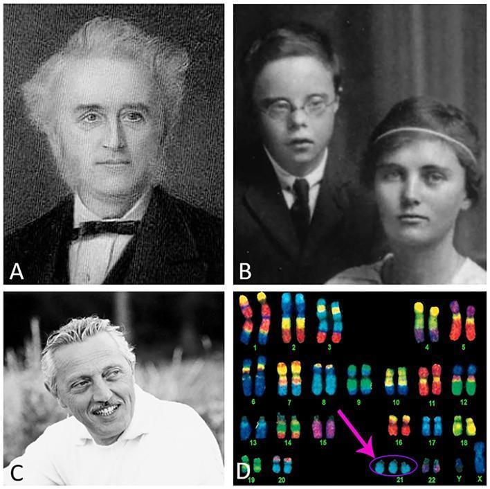
A composite figure illustrating the historical and genetic context of Down syndrome (Trisomy 21). Panel A shows a portrait of John Langdon Down, who first described the clinical features. Panel B is a black-and-white clinical photograph of two individuals; the male exhibits characteristic dysmorphic facial features of Down syndrome, including upslanting palpebral fissures, epicanthic folds, and a flattened nasal bridge. Panel C depicts Dr. Jérôme Lejeune, the geneticist who identified the chromosomal basis of the condition. Panel D shows a spectral karyotype (SKY) or multicolor fluorescence in situ hybridization (m-FISH) arrangement. All autosomal pairs are numbered 1 through 22, along with X and Y sex chromosomes. A pink arrow highlights the genetic hallmark of the disorder: three copies of chromosome 21 (Trisomy 21) instead of the typical homologous pair. This educational graphic links clinical phenotype with its underlying cytogenetic pathology, suitable for medical genetics and pediatrics curricula.
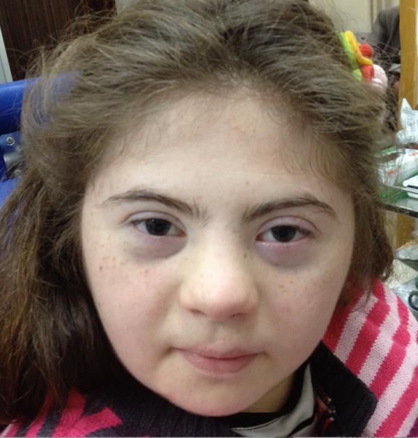
Clinical photograph of a 15-year-old female patient exhibiting phenotypic facial features characteristic of Down syndrome (Trisomy 21). The image demonstrates a flattened facial profile with a notably low-set and depressed nasal bridge. Ocular findings include upslanting palpebral fissures and prominent bilateral epicanthic folds (medial epicanthal skin folds) covering the inner canthi. Additional craniofacial features include a relatively small oral cavity and mild brachycephaly. The patient has light skin with fine freckling across the midface. This image serves as a teaching tool for medical genetics and clinical pediatrics, illustrating the classic dysmorphic facial features associated with Trisomy 21 in an adolescent. The clinical context suggests an association with Morning Glory Syndrome (MGS), though the internal ocular pathologies are not visible in this external facial view.
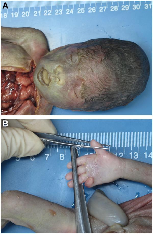
This composite clinical photograph displays the postnatal morphological features of a fetus with trisomy 21 (Down syndrome). Panel A provides a facial profile view illustrating several classic dysmorphic features, including a flat facial profile, telecanthus/hypertelorism (widely spaced eyes), a markedly depressed nasal bridge, and macroglossia resulting in a protruding tongue. Panel B shows the right hand of the fetus being examined with surgical forceps, highlighting a single transverse palmar crease (simian crease), a common soft marker associated with various chromosomal abnormalities. The images are set against a blue background with a metric ruler for scale, typical of a clinical pathology or autopsy setting. These findings serve as physical manifestations of the underlying genetic duplication of 21q22.12-q22.3, which includes critical regions associated with the Down syndrome phenotype.
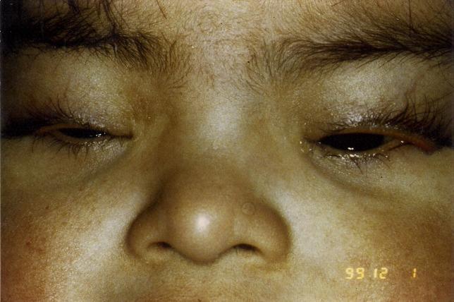
Clinical photograph of a pediatric patient exhibiting characteristic dysmorphic facial features and ophthalmologic abnormalities associated with Down syndrome (Trisomy 21). The image primarily demonstrates bilateral congenital ectropion of the upper eyelids, characterized by an outward eversion of the lid margins and exposure of the palpebral conjunctiva. Other visible craniofacial findings include bilateral epicanthal folds (skin folds covering the medial canthus), a flattened nasal bridge, and midface hypoplasia. There is also evident periorbital edema and an upward slanting of the palpebral fissures. This visual case illustrates the rare manifestation of congenital upper lid ectropion in the context of anterior lamellar shortening and general horizontal lid laxity. The photograph is essential for teaching neonatal and pediatric ophthalmology diagnostic signs and the phenotypic spectrum of Trisomy 21.
Down Syndrome (Trisomy 21)
Overview
- Robbins & Cotran Pathologic Basis of Disease, p. 163
- Thompson & Thompson Genetics and Genomics in Medicine, 9th ed., p. 103
Cytogenetics: Three Mechanisms
| Type | Frequency | Karyotype | Maternal Age Effect |
|---|---|---|---|
| Trisomy 21 (free) | ~95% | 47,XX or XY,+21 | Yes - strong |
| Robertsonian translocation | ~4% | 46 chromosomes (21q fused to chr 14 or 22) | No |
| Mosaic | ~1-2% | Mix of 46 and 47 chromosome cells | No |
- Thompson & Thompson Genetics, p. 103-104
- Robbins, p. 162-163
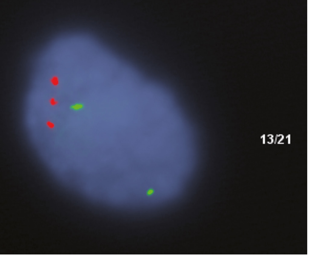
FISH Confirmation of Trisomy 21
Clinical Features
Facial & Physical
- Flat facial profile, oblique (upslanting) palpebral fissures, epicanthic folds
- Brachycephaly with flat occiput; short neck with loose nape skin
- Short, broad hands; single transverse palmar crease (simian crease); fifth-finger clinodactyly
- Short stature; hypotonia (often the first sign in the newborn)
Cognitive
- Intellectual disability ranging from mild to moderate
- Delay typically becomes obvious by end of the first year
- Wide variability - many individuals attend school and develop partial self-reliance
- Mosaics can have near-normal intelligence
Cardiac (present in ~40-50%)
- Atrioventricular septal defects (43%)
- Ventricular septal defects (32%)
- Atrial septal defects (19%)
- Tetralogy of Fallot (6%)
Gastrointestinal
- Duodenal atresia and tracheoesophageal fistula occur at much higher rates than in the general population
- Esophageal atresia and small bowel atresias also reported
Hematologic / Oncologic
- 20-fold increased risk of precursor B-cell ALL
- 500-fold increased risk of acute myeloid leukemia (particularly acute megakaryoblastic leukemia)
- Transient myeloproliferative disorder (TMD): Up to 10% of neonates with Down syndrome develop peripheral blood leukocytosis with blasts; TMD usually resolves spontaneously but progresses to acute megakaryoblastic leukemia in 23-30% of cases. Driven by acquired GATA1 mutations during fetal life.
Neurological / Alzheimer Disease
- Virtually all patients with trisomy 21 older than age 40 develop neuropathologic changes of Alzheimer disease (cortical atrophy, neurofibrillary tangles, amyloid plaques)
- This is explained by triplication of the APP gene on chromosome 21 - overproduction of amyloid precursor protein leads to early amyloid-beta accumulation
Immune
-
Abnormal T-cell function predisposes to serious pulmonary infections
-
Increased risk of thyroid autoimmunity
-
Robbins, p. 163-164
-
Goldman-Cecil Medicine
Molecular Pathogenesis
- Gene overexpression - most protein-coding genes on chr 21 are overexpressed ~1.5x, including APP (Alzheimer risk) and genes regulating hematopoiesis (GATA1 pathway)
- Mitochondrial dysfunction - ~10% of overexpressed genes regulate mitochondrial function; mitochondria show abnormal cristae, reactive oxygen species generation, and increased apoptosis
- Noncoding RNAs - chromosome 21 has the highest density of long noncoding RNAs (lncRNAs) of any chromosome; their roles are still being characterized
Prenatal Diagnosis
- Cell-free fetal DNA (cfDNA) / NIPT: ~5-10% of cell-free DNA in maternal blood is fetal; next-generation sequencing of chromosome 21 gene dosage is a powerful non-invasive screening tool
- Positive screens should be confirmed by conventional karyotyping from chorionic villus sampling (CVS) or amniocentesis
- Karyotyping is also essential to distinguish free trisomy from translocation type (different recurrence risk and genetic counseling implications)
Prognosis & Longevity
Recent Evidence (2024-2026)
- Alzheimer biomarkers: Blood-based biomarkers for Alzheimer disease in Down syndrome were reviewed in a 2025 meta-analysis (PMID 40219863), identifying promising plasma phospho-tau and amyloid markers for earlier detection
- Respiratory support: A 2025 systematic review (PMID 39909501) examined respiratory complications and support strategies, reinforcing that lung disease remains a major morbidity