
from Guyton and Hall textbook of medical physiology, 4th South Asia edition write a 10 mark long answer question of the following questions. Cardiac cycle define pressure and volume changes in left ventricle diagram third and fourth heart sound
Reading File
Finding Sources
Finding Sources
Finding Sources
Reading File
Reading File
Reading File
Reading File
Reading File
Finding Sources
Reading File
Loading Image
I now have all the content needed. Let me compile the comprehensive 10-mark long answer question from Guyton and Hall.
10-Mark Long Answer Question
Q. Define the cardiac cycle. Describe the pressure and volume changes in the left ventricle during the cardiac cycle with a labelled diagram. Add a note on the third and fourth heart sounds.
(Guyton and Hall Textbook of Medical Physiology, 4th South Asia Edition)
DEFINITION
The cardiac cycle refers to all the cardiac events that occur from the beginning of one heartbeat to the beginning of the next heartbeat. Each cycle is initiated by the spontaneous generation of an action potential in the sinoatrial (SA) node. At a normal heart rate of 72 beats/min, one cardiac cycle lasts approximately 0.833 seconds.
The cardiac cycle consists of two major phases:
- Systole - the period of ventricular contraction and ejection
- Diastole - the period of ventricular relaxation and filling
DIAGRAM
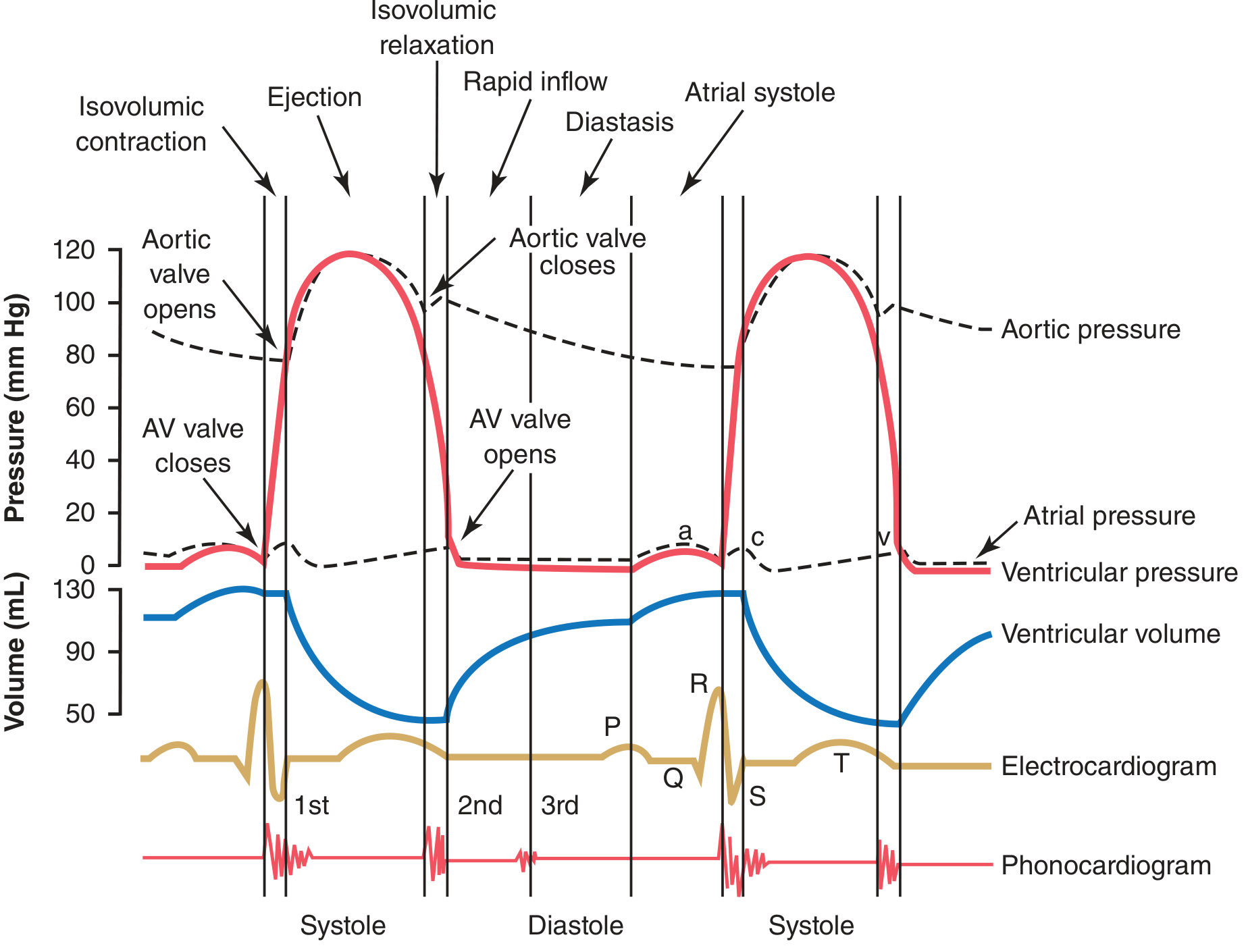
Fig. 9.8 - Events of the cardiac cycle for left ventricular function, showing changes in left atrial pressure, left ventricular pressure, aortic pressure, ventricular volume, the electrocardiogram, and the phonocardiogram. (Guyton & Hall)
PHASES OF THE CARDIAC CYCLE WITH PRESSURE AND VOLUME CHANGES
A. DIASTOLE (Ventricular Filling)
1. Period of Rapid Filling (Early Diastole)
When ventricular systole ends, ventricular pressure falls rapidly to its low diastolic value (close to 0 mm Hg in the left ventricle). The pressure in the left atrium, which has been accumulating blood during systole, is now slightly higher. This pressure difference forces open the AV (mitral) valves, and blood rushes rapidly into the ventricles.
- Ventricular volume: Rises rapidly from approximately 50 mL (end-systolic volume) toward 130 mL (end-diastolic volume)
- Left ventricular pressure: Remains low, near 0-5 mm Hg
- This phase occupies approximately the first third of diastole and accounts for the majority of ventricular filling (70-80%)
2. Diastasis (Middle Third of Diastole)
During the middle third of diastole, only a small amount of blood flows into the ventricles. Blood continues to flow from the veins through the atria directly into the ventricles.
- Ventricular volume: Rises slowly
- Ventricular pressure: Remains low
3. Atrial Systole (Last Third of Diastole)
The atria contract, giving an additional thrust to blood inflow - this acts as a "booster pump" and contributes an additional 20-30% of ventricular filling.
- Ventricular volume: Reaches end-diastolic volume (EDV) = approximately 130 mL
- Atrial pressure: Shows the a wave (4-6 mm Hg rise in right atrium; 7-8 mm Hg in left atrium)
- AV valves remain open throughout diastole
B. SYSTOLE (Ventricular Contraction and Ejection)
4. Period of Isovolumic (Isometric) Contraction
Ventricular contraction begins immediately after the QRS complex on the ECG. Ventricular pressure rises rapidly, forcing the AV valves to close (producing the 1st heart sound). However, the ventricular pressure has not yet exceeded aortic pressure (~80 mm Hg), so the aortic valve remains closed.
- Duration: approximately 0.02-0.03 seconds
- Ventricular pressure: Rises steeply from near 0 mm Hg toward 80 mm Hg
- Ventricular volume: No change (remains at ~130 mL) - this is the hallmark of this phase
- Both AV and semilunar valves are closed
- The atrial pressure curve shows the c wave, caused mainly by bulging of the AV valves backward toward the atria
5. Period of Ejection (Ventricular Ejection)
When left ventricular pressure rises slightly above 80 mm Hg, it exceeds aortic diastolic pressure, forcing the aortic valve open. Blood is ejected into the aorta.
- Rapid ejection phase (first third of ejection): ~70% of the ejected blood leaves in this phase; LV pressure peaks at ~120 mm Hg; aortic pressure also peaks at ~120 mm Hg
- Reduced ejection phase (remaining two-thirds): Ejection rate slows; LV pressure begins to fall
- Ventricular volume: Falls from ~130 mL to ~50 mL; approximately 60% of EDV is ejected (this is the ejection fraction, normally ~60%)
- Stroke volume = EDV - ESV = 130 - 50 = ~80 mL
- The atrial pressure curve shows the v wave (blood accumulating in atria while AV valves are closed)
C. TRANSITION TO DIASTOLE
6. Period of Isovolumic (Isometric) Relaxation
After ejection, the ventricles begin to relax (marked by the T wave on ECG). When ventricular pressure falls below aortic pressure, the aortic valve snaps shut (producing the 2nd heart sound). There is a brief period of elastic recoil in the aorta - this produces a small notch in the aortic pressure tracing called the dicrotic notch.
- The ventricular pressure continues to fall but has not yet dropped below atrial pressure, so AV valves remain closed
- Ventricular volume: Unchanged at ~50 mL (end-systolic volume)
- Both AV and semilunar valves are again closed
- Duration: Very brief, ~0.03-0.06 seconds
Then ventricular pressure falls below atrial pressure, AV valves open, and the next cycle of rapid filling begins.
PRESSURE AND VOLUME SUMMARY TABLE
| Phase | LV Pressure | LV Volume | Valves Open |
|---|---|---|---|
| Rapid filling | Low (~0-5 mmHg) | Rapidly rising | Mitral |
| Diastasis | Low | Slowly rising | Mitral |
| Atrial systole | Low | Rises to 130 mL | Mitral |
| Isovolumic contraction | Rising (0 → 80 mmHg) | Unchanged (~130 mL) | None |
| Ejection | Peaks at ~120 mmHg | Falls to ~50 mL | Aortic |
| Isovolumic relaxation | Falling rapidly | Unchanged (~50 mL) | None |
ATRIAL PRESSURE WAVES (a, c, v)
- a wave: Due to atrial contraction (late diastole)
- c wave: Due to bulging of AV valves toward atria at onset of ventricular contraction
- v wave: Due to slow accumulation of blood in atria while AV valves are closed during ventricular systole
THIRD AND FOURTH HEART SOUNDS
Third Heart Sound (S3)
- Timing: Occurs at the beginning of the middle third of diastole (early-to-mid diastole), following S1 and S2
- Mechanism: Oscillation of blood back and forth between the walls of the ventricles, initiated by the inrush of blood from the atria during rapid ventricular filling. This is analogous to water rushing into a paper bag and reverberating back and forth between its walls.
- The reason S3 does not occur in early diastole is that the ventricles are not filled sufficiently at that point to create even the small elastic tension necessary for reverberation.
- Frequency: Very low - usually below the audible range for the human ear. It can often be recorded by phonocardiography but is not heard with a stethoscope in most adults.
- Clinical significance:
- Normal in children, adolescents, and young adults (physiological S3)
- In older adults, an S3 generally indicates systolic heart failure - it is heard as an S3 gallop
- An S3 gallop in a patient with heart failure indicates volume overload and reduced ejection fraction
Fourth Heart Sound (S4)
- Timing: Occurs just before S1, at the end of diastole - coincides with atrial contraction
- Mechanism: Caused by the inrush of blood into the ventricles when the atria contract, initiating vibrations similar to those of the third heart sound. It is essentially the vibration caused by forceful atrial systole against a stiff, non-compliant ventricle.
- Frequency: Very low - usually 20 cycles/sec or less; rarely heard with a stethoscope because of its weakness. It can be recorded on phonocardiogram.
- Clinical significance:
- S4 (S4 gallop) is common in patients who derive benefit from atrial contraction for ventricular filling as a result of decreased ventricular wall compliance and increased resistance to ventricular filling
- Commonly heard in older patients with left ventricular hypertrophy (e.g., due to hypertension, aortic stenosis)
- Also seen in hypertrophic cardiomyopathy, acute myocardial infarction, and severe anaemia
- An S4 is always pathological in adults - it indicates reduced ventricular compliance
Comparison of S3 and S4
| Feature | S3 | S4 |
|---|---|---|
| Timing | Early-mid diastole (after S2) | Late diastole (before S1) |
| Cause | Rapid ventricular filling reverberation | Forceful atrial contraction against stiff ventricle |
| Frequency | Very low (<40 cycles/sec) | Very low (≤20 cycles/sec) |
| Heard with stethoscope | Rarely | Rarely |
| Normal in | Children/young adults | Not normal in adults |
| Pathological significance | Systolic heart failure (volume overload) | Diastolic dysfunction, LV hypertrophy |
| Gallop rhythm produced | S3 gallop (ventricular gallop) | S4 gallop (atrial gallop) |
KEY POINTS TO REMEMBER
- The cardiac cycle at 72 beats/min lasts ~0.833 seconds; systole occupies ~40% and diastole ~60%
- Left ventricular pressure ranges from near 0 mmHg (diastole) to ~120 mmHg (peak systole)
- Aortic pressure ranges from ~80 mmHg (diastolic) to ~120 mmHg (systolic)
- EDV ~130 mL; ESV ~50 mL; Stroke Volume ~80 mL; Ejection Fraction ~60%
- Two phases have no change in ventricular volume - isovolumic contraction and isovolumic relaxation
- S3 = volume overload/systolic failure; S4 = pressure overload/diastolic dysfunction
Source: Guyton and Hall Textbook of Medical Physiology, 4th South Asia Edition, Chapter 9 (pp. 126-130) and Chapter 23 (pp. 289-290)
This is a shared conversation. Sign in to Orris to start your own chat.