
Say about this report analysis
Reading File
Running Command
cd /home/daytona/workspace/attachments/81ccc45f-53e9-424b-a1a4-714682c7b199/ && python3 -c " import pdfplumber with pdfplumber.open('11407260013_SmartReport (2).pdf') as pdf: print(f'Total pages: {len(pdf.pages)}') for i, page in enumerate(pdf.pages): text = page.extract_text() print(f'--- PAGE {i+1} ---') print(text) print() "
Here is a thorough analysis of the Smart Health Report for Mrs. Phoolpati Sharma (70 years, Female), tested on 14-July-2026.
Patient Overview
| Detail | Value |
|---|---|
| Name | Mrs. Phoolpati Sharma |
| Accession No. | 11407260013 |
| Age / Gender | 70 years, Female |
| Date of Test | 14-07-2026 |
Summary of Findings
The report covers 6 health panels: Glucose, Liver Function, Kidney Function, Hematology, Cholesterol, Thyroid, and Urinalysis.
🔴 Abnormal / Very Abnormal Findings
1. Elevated Blood Sugar (Glucose Panel - Abnormal)
| Test | Result | Normal Range | Status |
|---|---|---|---|
| FBS (Fasting Blood Sugar) | 141 mg/dL | 70-100 | 🔴 Very High |
| PPBS (Post-Prandial) | 127 mg/dL | <140 (Normal) | 🟡 Borderline |
- FBS of 141 mg/dL is in the diabetic range (>126 mg/dL). This is a primary concern.
- The report recommends HbA1c testing to confirm long-term sugar control, along with anti-diabetic medication if advised by a doctor.
2. Low Hemoglobin / Anemia (Hematology - Very Abnormal)
| Test | Result | Normal Range | Status |
|---|---|---|---|
| Haemoglobin (Hb) | 9.9 gms/dl | 12.0-16.0 | 🔴 Low |
| RBC Count | 4.07 milli./cu.mm | 4.5-5.9 | 🔴 Low |
| HCT (Packed Cell Volume) | 30.2% | 33.0-51.0 | 🔴 Low |
| MCV | 74.3 fL | 80.0-100.0 | 🔴 Low (Microcytic) |
| MCH | 24.5 pg | 26.0-34.0 | 🔴 Low |
| RDW-CV | 18.2% | 11.0-16.0 | 🔴 High |
- The pattern of low Hb + low MCV + low MCH + high RDW is a classic picture of Iron Deficiency Anemia (or mixed nutritional anemia).
- Additional tests recommended: Serum Iron, Ferritin, Vitamin B12.
3. High White Blood Cell Count (Leukocytosis)
| Test | Result | Normal Range | Status |
|---|---|---|---|
| Total WBC Count | 13,360 /cumm | 4,500-11,000 | 🔴 High |
| Absolute Lymphocyte Count | 4,943 /cumm | 1,080-4,840 | 🔴 Marginally High |
- Elevated WBCs suggest the immune system is fighting an infection or inflammation. The report recommends a Peripheral Smear to evaluate further.
- Neutrophils (55%) and other differential counts are within normal limits.
4. High Platelet Count (Thrombocytosis)
| Test | Result | Normal Range | Status |
|---|---|---|---|
| Platelet Count | 4.86 Lakh/cumm | 1.5-4.5 Lakh | 🔴 High |
- Elevated platelets can be a reactive response to anemia (iron deficiency), infection, inflammation, or other causes. A Peripheral Smear is recommended.
5. Low HDL Cholesterol ("Good" Cholesterol)
| Test | Result | Normal Range | Status |
|---|---|---|---|
| HDL Cholesterol | 35 mg/dL | >40 mg/dL | 🔴 Low |
| Cholesterol/HDL Ratio | 4.46 | <3.5 Optimal | 🟠 Low Risk Zone |
- Low HDL, especially in a diabetic elderly woman, increases cardiovascular risk.
- Triglycerides (148 mg/dL) are borderline (<150 normal) - just within range.
🟢 Normal / Reassuring Findings
| Panel | Key Tests | Status |
|---|---|---|
| Kidney Function | Creatinine 0.71 mg/dL, eGFR 86.3, Calcium 8.96 mg/dL | ✅ Normal |
| Liver Function | SGPT 22 U/L (range 0-34) | ✅ Normal |
| Thyroid | TSH 1.75 µIU/mL (range 0.55-4.78) | ✅ Normal |
| Cholesterol Total | 156 mg/dL (<200) | ✅ Normal |
| LDL Cholesterol | 91 mg/dL (<100) | ✅ Normal |
| Triglycerides | 148 mg/dL (<150) | ✅ Just Normal |
| Urine | No glucose, no protein, no blood, no bacteria | ✅ Essentially Normal |
Key Clinical Concern Clusters
Cluster 1 - Likely Diabetes:
FBS 141 mg/dL is diagnostic of diabetes (>126 mg/dL fasting). PPBS of 127 mg/dL adds further concern. HbA1c is urgently needed to gauge 3-month average sugar control.
Cluster 2 - Microcytic Anemia (likely Iron Deficiency):
Low Hb (9.9), low MCV (74.3), low MCH (24.5), and high RDW (18.2) all point toward iron deficiency or combined iron + B12/folate deficiency. Serum Iron, Ferritin, and Vit B12 testing is needed to confirm the type.
Cluster 3 - Active Infection or Inflammation:
Elevated WBCs (13,360) alongside high platelets (4.86 lakh) and slightly turbid urine could suggest a low-grade infection (though urine bacteria are absent). A Peripheral Smear will help rule out more serious causes.
Cluster 4 - Cardiovascular Risk:
Despite normal total cholesterol and LDL, the low HDL (35 mg/dL) combined with diabetes significantly elevates the risk of heart disease and stroke in a 70-year-old woman.
Recommended Next Steps (from the report)
Immediate - Consult a General Physician for:
- High blood sugar (possible diabetes)
- Low hemoglobin (anemia)
- High WBCs (possible infection)
- High platelet count
Within 3 Months - Repeat Tests:
- HbA1c
- FBS & PPBS
- Complete Blood Count (CBC)
- Peripheral Smear
Additional Tests (if not done):
- HbA1c, Urine Albumin-Creatinine Ratio (for diabetic kidney monitoring)
- Serum Iron, Ferritin (for anemia workup)
- Vitamin B12
- Peripheral Smear
- Ophthalmologist referral (for diabetic eye screening)
Diet Guidance Summary
The personalized diet plan focuses on:
- Low sugar diet - avoid refined carbs, white rice, sugar, potatoes, bananas
- Iron-rich foods - cooked greens (broccoli, cabbage), bajra, rajma, tomato paste
- Vitamin B12-rich foods - eggs, lean chicken, fish (salmon, tuna)
- Folate-rich foods - fenugreek leaves, beetroot, citrus fruits
- HDL-boosting foods - avocado, olive oil, almonds, walnuts, fatty fish
- Drink 8-10 glasses of water daily
- Avoid fried foods, packaged foods, alcohol, and sweets
Bottom Line
Mrs. Phoolpati Sharma, at 70 years, presents with four primary issues that need urgent attention: probable diabetes (FBS 141 mg/dL), moderate anemia (Hb 9.9), leukocytosis suggesting active infection/inflammation, and thrombocytosis. Her kidney, liver, and thyroid are functioning normally, which is positive. The priority is to confirm diabetes with HbA1c, investigate the anemia type with iron studies and B12 levels, and get a Peripheral Smear to evaluate the high WBC and platelet count. She should see a general physician promptly.
Disclaimer: This is an analysis of the lab report only. It does not replace a doctor's clinical evaluation. Please consult a qualified physician before making any health decisions.
Peripheral smear ?
A peripheral smear (also called a peripheral blood smear or blood film) is a test where a drop of blood is spread on a glass slide, stained, and examined under a microscope. It gives a detailed visual picture of all blood cells - their shape, size, and appearance.
Searching sources...
Reading File
Finding Sources
Searching the Web
peripheral blood smear test what it shows purpose indications
Reading File
Reading File
Loading Image
Loading Image
What is a Peripheral Smear?
A peripheral blood smear (also called a blood film) is a simple but powerful microscopic test. A small drop of blood is spread as a thin film on a glass slide, stained with a special dye (Romanowsky/Leishman stain), and examined under a microscope by a pathologist or hematologist.
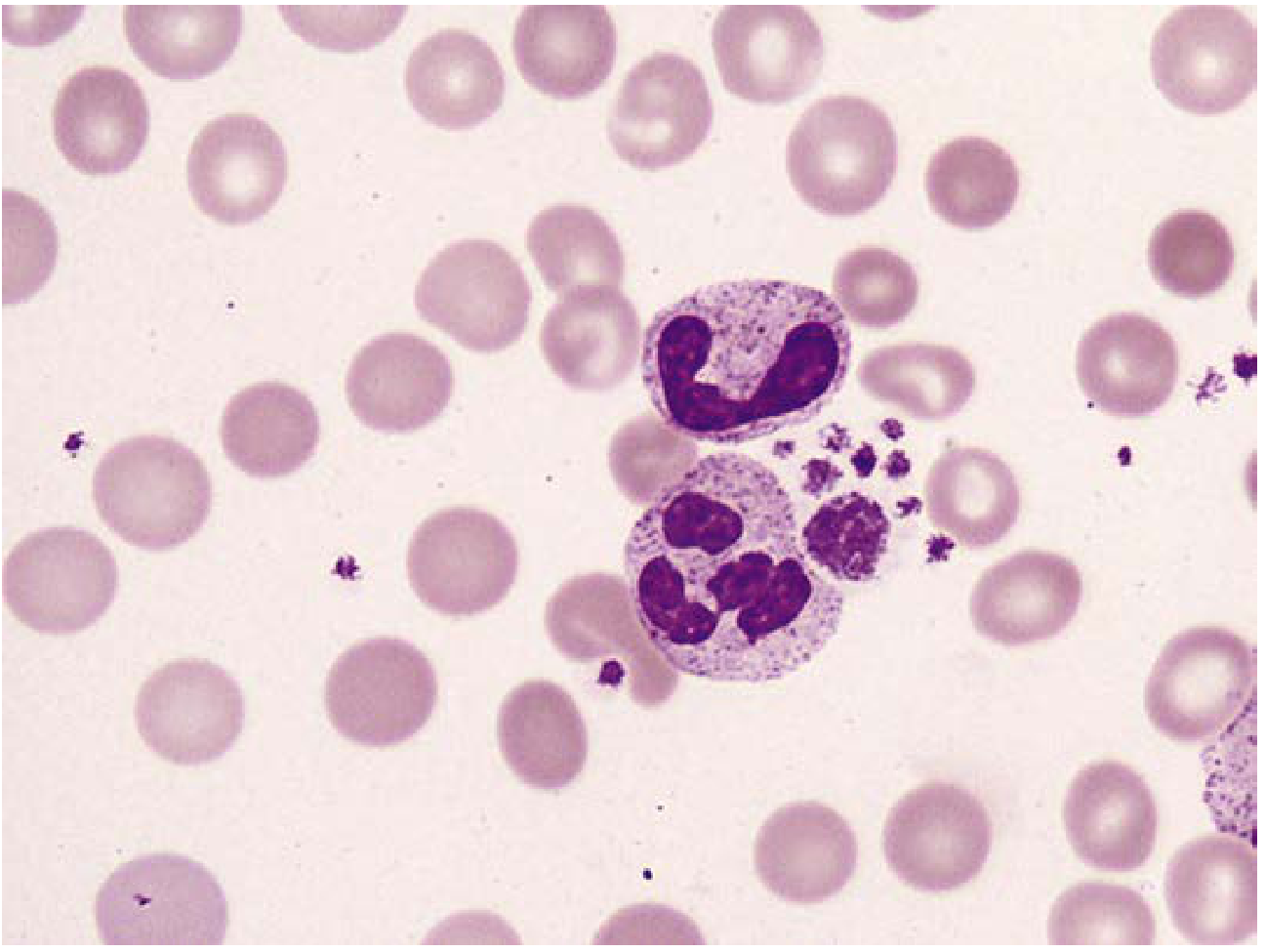
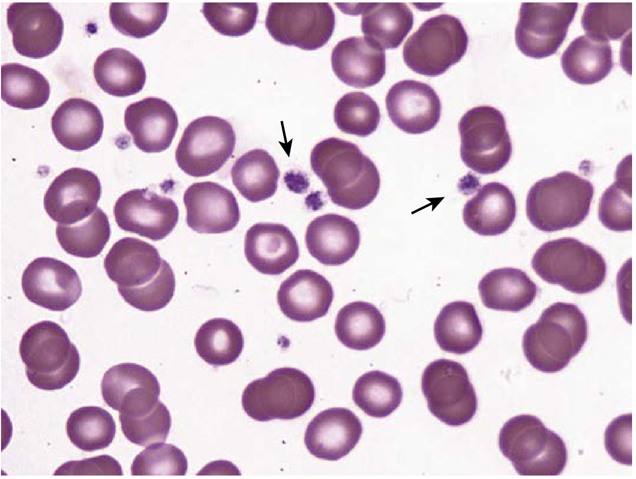
Here is what a normal peripheral smear looks like under the microscope:
White blood cells (neutrophils) among red blood cells:
Platelets (arrows) among red blood cells:
What Does It Examine?
The peripheral smear gives a visual assessment of all three types of blood cells:
1. Red Blood Cells (RBCs)
The doctor looks at:
- Size - are they too small (microcytic) or too large (macrocytic)?
- Shape - are they normal discs, or abnormal (sickle cells, target cells, fragmented cells)?
- Color - pale (hypochromic = iron deficiency) or normal?
- Variation - anisocytosis (varied sizes), poikilocytosis (varied shapes)
2. White Blood Cells (WBCs)
- What types are present and in what proportion (neutrophils, lymphocytes, monocytes, eosinophils)?
- Are there any immature or abnormal forms (blast cells = leukemia)?
- Are neutrophils hypersegmented (seen in B12/folate deficiency)?
- Any toxic granulation or Dohle bodies (severe infection)?
3. Platelets
- Are they too many, too few, or abnormal in size?
- Giant platelets suggest certain blood disorders
- Platelet clumping can be spotted
Why is it Recommended for Mrs. Phoolpati Sharma?
Her report showed three abnormalities that a peripheral smear will help explain:
| Abnormality | What the Smear Will Look For |
|---|---|
| Low Hb (9.9), low MCV (74.3), high RDW (18.2) | Small, pale (hypochromic microcytic) RBCs - confirming iron deficiency anemia. Or mixed anemia (small + large cells together). |
| High WBC (13,360) | Whether the elevation is due to infection (reactive - mostly neutrophils with toxic changes), or a more serious cause like leukemia (abnormal blast cells). |
| High Platelets (4.86 lakh) | Whether platelets look reactive and normal in shape, or if there are giant/abnormal platelets suggesting a bone marrow disorder. |
How is it Done?
- A small blood sample is drawn (same finger prick or vein draw)
- A drop is placed on a glass slide and spread thin
- It is air-dried and stained
- A trained pathologist examines it under a microscope
- Results typically come back within same day to 24 hours
What Can It Diagnose?
- Iron deficiency anemia - small, pale RBCs
- B12 / Folate deficiency - large RBCs (megaloblasts), hypersegmented neutrophils
- Infections - reactive neutrophilia, toxic changes
- Malaria - parasites visible inside RBCs
- Leukemia - blast cells (immature WBCs)
- Thalassemia - target cells, microcytes
- Hemolytic anemia - fragmented RBCs (schistocytes)
- Clotting disorders - abnormal platelets
Bottom Line for This Patient
Given Mrs. Sharma's profile - anemia + high WBCs + high platelets + suspected diabetes - the peripheral smear will help the doctor understand:
- What type of anemia she has (iron deficiency vs. B12/folate vs. mixed)
- Whether the high WBCs are a simple infection response or something that needs deeper investigation
- Whether the high platelets are reactive (secondary to anemia/infection) or a primary blood disorder
It is a low-cost, rapid, and highly informative test - essentially a "window" into what is happening inside the blood cells. The doctor will likely order it alongside Serum Iron, Ferritin, and B12 levels for a complete picture.
- Henry's Clinical Diagnosis and Management by Laboratory Methods, pp. 641-645
This is a shared conversation. Sign in to Orris to start your own chat.