
What is dd of pain start from testis and goes to back and abdomen become tense and get relief self after few min
differential diagnosis testicular pain radiating back abdomen intermittent self resolving colic
Differential Diagnosis: Testicular Pain Radiating to Back/Abdomen, Abdomen Tense, Self-Resolving in Minutes
1. Ureteric Calculus (Ureteric Colic) - MOST LIKELY
- Pain starts suddenly, griping in character, extremely severe
- Patient cannot find a comfortable position (writhes/tosses in bed)
- Abdomen becomes tense/rigid (peritonism from referred pain)
- Pain ceases as suddenly as it appeared - self-resolving in minutes
- Testis may become retracted upward (cremaster spasm)
- Associated nausea, vomiting, profuse sweating
- Haematuria (microscopic or gross) may be present
"At a lower level, pain commences rather anteriorly just above the iliac crest and is referred along the two branches of the genitofemoral nerve to the testis in the male... The testis becomes retracted by the spasm of the cremaster."
- S. Das Manual on Clinical Surgery, p.587
"If the stone is in the lower ureter, pain may radiate to the ipsilateral testicle in men..."
- National Kidney Foundation Primer on Kidney Diseases, p.470
2. Intermittent Testicular Torsion - IMPORTANT (Do Not Miss)
- Episodes of acute scrotal/testicular pain that self-resolve in minutes to hours - this is the defining feature
- 30-50% of patients with acute testicular torsion have prior self-limiting episodes
- Nausea/vomiting may or may not be present
- Between attacks: physical exam and Doppler ultrasound may be normal
- This is a pre-torsion warning - risk of complete (irreversible) torsion and testicular loss if not surgically fixed
"Episodes of self-limited acute scrotal pain precede acute testicular torsion in 30% to 50% of patients... typically begin and resolve acutely with durations of minutes to hours."
- Campbell-Walsh-Wein Urology
"Occasionally the testis untwists spontaneously without surgical treatment and 'intermittent' testicular torsion should be considered as a cause of testicular pain in adolescents."
- Bailey & Love's Surgery, 28th Ed.
3. Other Differentials to Consider
| Condition | Distinguishing Features |
|---|---|
| Acute appendicitis (right-sided) | Right iliac fossa tenderness, fever, raised WBC; pain does not fully resolve spontaneously |
| Inguinal hernia (obstructed/irreducible) | Groin lump palpable; does not fully self-resolve |
| Epididymo-orchitis | Gradual onset, fever, dysuria/discharge, tenderness persistent on palpation |
| Torsion of testicular appendage | Tender "blue dot" at upper pole of testis; pain partially resolves but persists |
| Abdominal aortic aneurysm (AAA) | Elderly patient; back pain + pulsatile abdominal mass; does NOT fully self-resolve - a dangerous mimic of renal colic |
| Genitofemoral / ilioinguinal neuralgia | Burning/shooting nerve-type pain; no systemic features |
| Muscular/skeletal pain | Related to movement; positional |
| Herpes zoster | Later develops dermatomal vesicular rash |
Key Differentiating Points at a Glance
| Feature | Ureteric Colic | Intermittent Torsion |
|---|---|---|
| Pain character | Griping, colicky | Acute, severe |
| Haematuria | Yes (microscopic/gross) | No |
| Cremaster retraction | Yes (stone-level) | Possible |
| Self-resolution | Yes (minutes) | Yes (minutes-hours) |
| Age group | Any | Adolescents/young adults |
| Recurrence pattern | Yes, recurs until stone passes | Yes, before complete torsion |
| Urgency | Semi-urgent | Urgent - risk of organ loss |
| Investigation | Urine dipstick + non-contrast CT | Doppler USS of scrotum |
Bottom Line
Find recent imaging guidelines for kidney stones vs testicular emergencies
"urolithiasis" AND "imaging" AND "guideline"
"testicular torsion" AND "ultrasound" AND "diagnosis"
PMID: 40268592
PMID: 39488348
PMID: 40368720
PMID: 35238603
"renal colic" AND "computed tomography" AND "ultrasound" AND "emergency"
"ACR appropriateness criteria" AND "urolithiasis" AND "imaging"
EAU AUA 2024 2025 guidelines imaging kidney stones first line ultrasound CT
PMID: 38040458
PMID: 33451883
Recent Imaging Guidelines: Kidney Stones vs. Testicular Emergencies
KIDNEY STONES (Urolithiasis) - Imaging Guidelines
1. ACR Appropriateness Criteria - Acute Onset Flank Pain / Suspicion of Urolithiasis (2023 Update)
| Clinical Scenario | Recommended Imaging | Rating |
|---|---|---|
| Acute flank pain, first presentation | Non-contrast CT (NCCT) | Usually Appropriate |
| Known stone disease, recurrent symptoms | NCCT | Usually Appropriate |
| Signs of obstruction only | Ultrasound | May be appropriate |
| Pregnancy (1st trimester) | Ultrasound first, then MRI without contrast | Usually Appropriate |
| Inconclusive NCCT | CT urogram or MRI with contrast | May be appropriate |
| Plain radiograph (KUB) | Limited role; less sensitive than NCCT | May be appropriate in follow-up |
2. EAU Guidelines on Urolithiasis (2025) - Most Current
- Step 1 - Ultrasound first: Recommended as the first-line modality (especially in children, pregnant patients, and for detecting hydronephrosis/obstruction)
- Step 2 - Low-dose NCCT: Gold standard to confirm stone diagnosis after initial ultrasound, providing precise stone size, density (HU), and skin-to-stone distance for treatment planning
- Immediate imaging (NCCT) is mandatory with: fever + obstruction (infected stone = surgical emergency), solitary kidney, or uncertain diagnosis
- Low-dose CT protocols (reducing radiation by 50-75%) are preferred over standard-dose CT
- Contrast CT/CT urogram only if stone removal is planned and collecting system anatomy needs delineation
3. AUA Imaging Guidance for Ureteral Calculous Disease
- Ultrasound is the preferred initial modality in children (radiation concerns) and all pregnant patients
- Low-dose CT is used if ultrasound is non-diagnostic in children
- MRI without contrast is second-line in first-trimester pregnancy (fetus most susceptible to radiation)
NCCT Performance for Urolithiasis (from Campbell-Walsh-Wein Urology)
| Metric | Value |
|---|---|
| Sensitivity | 96-100% |
| Specificity | 92-100% |
| Low-dose CT sensitivity | ~97% |
| Low-dose CT specificity | ~96% |
| Radiation reduction with low-dose protocol | 50-75% per scan |
TESTICULAR EMERGENCIES - Imaging Guidelines
1. ACR Appropriateness Criteria - Acute Scrotal Pain Without Trauma (2024 Update)
- Colour Doppler Ultrasound (CDUS) is always the first and usually only imaging needed
- No role for CT or MRI in primary assessment of acute scrotum
- Differential includes torsion, epididymo-orchitis, appendage torsion, hernia
Critical caveat: If clinical suspicion for testicular torsion is high, do NOT delay surgery for ultrasound. Explore immediately. Ultrasound is for equivocal or low-suspicion cases only.
2. Ultrasound Diagnostic Accuracy for Testicular Torsion - 2026 Meta-Analysis
| Ultrasound Modality | Sensitivity (95% CI) | Specificity (95% CI) |
|---|---|---|
| Colour Doppler Sonography (CDS) alone | 95.3% (91.4-97.5) | 98.3% (96.2-99.3) |
| CDS + B-mode combined | Up to 100% sensitivity | High specificity |
3. TWIST Score - Risk Stratification Before Imaging (2022 Meta-Analysis)
| TWIST Parameter | Points |
|---|---|
| Testicular swelling | 2 |
| Hard testis | 2 |
| High-riding testis | 1 |
| Absent cremasteric reflex | 1 |
| Nausea/vomiting | 1 |
| Total | 7 |
| Score | Risk | Action |
|---|---|---|
| 0-2 | Low (65% of cases) | No torsion - manage conservatively, sensitivity 98.4% |
| 3-4 | Intermediate (20%) | Ultrasound required |
| 5-7 | High (15%) | Immediate surgical exploration - specificity 97.5% |
4. Ultrasound Findings in Testicular Torsion (Campbell-Walsh-Wein Urology)
| Finding | Significance |
|---|---|
| Absent/reduced Doppler colour flow or waveforms | Diagnostic (86% sensitive, 100% specific) |
| Parenchymal heterogeneity vs. contralateral testis | Suggests necrosis - high risk of orchiectomy |
| Homogeneous echogenicity | Lower risk of orchiectomy |
| Whirlpool sign (twisted cord) | Highly specific for torsion |
| Normal flow | Does NOT rule out torsion (false negatives exist) |
Head-to-Head Comparison
| Feature | Kidney Stone | Testicular Torsion |
|---|---|---|
| First-line imaging | Low-dose NCCT (adults) / USS (children, pregnant) | Colour Doppler Ultrasound |
| Gold standard | Non-contrast CT | Colour Doppler USS + B-mode combined |
| Role of plain X-ray | Limited (misses radiolucent stones) | No role |
| Role of MRI | Pregnant patients (2nd-line after USS) | No routine role |
| Can imaging be skipped? | Yes - if recurrent known stone with typical symptoms | YES - if TWIST score 5-7, go straight to theatre |
| Time pressure | Hours to days | Minutes to hours - "time is testis" |
| Key guideline body | EAU 2025 / ACR 2023 | ACR 2024 / EAU (via AUA) |
Key Takeaway for the Clinical Scenario (Testicular Pain + Back/Abdominal Radiation + Self-Resolving)
- Perform urine dipstick - microscopic haematuria strongly supports ureteric colic
- Get a Colour Doppler Ultrasound of the scrotum - to rule out intermittent torsion (even if currently pain-free, blood flow may be abnormal)
- Get a low-dose non-contrast CT abdomen/pelvis - to identify ureteric stone, its exact position and size
- Calculate TWIST score - if high (5-7), do not wait for imaging; refer urgently for orchidopexy
- EAU Urolithiasis Guidelines 2025 (PMID: 40268592)
- ACR Appropriateness Criteria - Urolithiasis 2023 (PMID: 38040458)
- ACR Appropriateness Criteria - Acute Scrotal Pain 2024 (PMID: 39488348)
- Ultrasound for Testicular Torsion - Meta-analysis 2026 (PMID: 40368720)
- TWIST Score Meta-analysis, J Urol 2022 (PMID: 35238603)
- B-mode + CDS Systematic Review 2021 (PMID: 33451883)
- Campbell-Walsh-Wein Urology (textbook), Urolithiasis and Testicular Torsion chapters
A 65 year lady hai pain in knee pain occure during walking no morning stiffness
differential diagnosis knee pain elderly female mechanical no morning stiffness osteoarthritis
Differential Diagnosis: 65-Year-Old Female, Knee Pain on Walking, No Morning Stiffness
Key Clinical Features to Analyze
| Feature | Clinical Meaning |
|---|---|
| Age 65, female | Strong risk profile for OA (post-menopausal, cartilage loss accelerated) |
| Pain during walking | Mechanical pattern - pain with use, relieved by rest |
| No morning stiffness | Against inflammatory arthritis (RA has >1 hour morning stiffness) |
| Self-limiting / episodic? | Suggests mechanical rather than persistent inflammatory |
#1 Most Likely: Osteoarthritis (OA) of the Knee
- Pain worse with activity (walking, stairs, standing) - mechanical pain
- Pain relieved by rest
- Morning stiffness is absent or very brief (<30 minutes) - key differentiator from RA
- Crepitus on movement (grating/grinding sensation)
- Bony enlargement around the joint (osteophytes)
- Gradual onset over months to years
- May have joint swelling (cool, non-inflammatory effusion)
- Worse at end of day
- Age >50
- Female sex (post-menopausal estrogen loss accelerates cartilage breakdown)
- Possibly obesity (major risk factor; weight loss slows progression)
- Medial tibiofemoral (most common) - causes varus deformity (bow legs)
- Lateral tibiofemoral - valgus deformity
- Patellofemoral - pain worse on stairs and sitting for long periods
"Obesity is a known risk factor for osteoarthritis of the knee, and weight loss has been shown to slow the progression of the disease."
- Campbell's Operative Orthopaedics, 15th Ed 2026
Differential Diagnosis - Full List
Inflammatory Causes (LESS likely - no morning stiffness)
| Condition | Why Consider | Why Less Likely Here |
|---|---|---|
| Rheumatoid Arthritis | Knee commonly involved | Morning stiffness >1 hour is hallmark; typically bilateral; systemic features (fatigue, hand joints) |
| Pseudogout (CPPD) | Most common in elderly; knee is favourite joint | Acute attacks, not purely mechanical; may show chondrocalcinosis on X-ray |
| Gout | Can affect knee | Less common in women post-menopause without diuretics; typically acute, very painful attacks |
| Reactive arthritis | Post-infective | Requires trigger (GI/urogenital infection); usually younger |
| Psoriatic arthritis | Affects large joints | Skin/nail changes; usually other joints involved |
Mechanical/Structural Causes
| Condition | Features | Notes |
|---|---|---|
| Meniscal tear | Pain on walking, joint line tenderness, may lock/give way | More common after twisting injury; McMurray test +ve |
| Patellofemoral syndrome | Anterior knee pain, worse on stairs and prolonged sitting | Younger patients usually; can occur in elderly |
| Pes anserine bursitis | Medial knee pain, worse on stairs; common in obese elderly women | Often coexists with OA; point tenderness 2-3 cm below medial joint line |
| Anserine bursitis + OA | Very common combination in this demographic | Tender spot on inner knee separate from joint line |
| Loose body in joint | Intermittent locking, giving way | Cartilage fragments; X-ray may show |
| Ligament laxity/injury | Instability, pain on weight-bearing | Usually post-traumatic |
| Baker's cyst | Posterior knee swelling and pain | Secondary to OA or meniscal tear; may rupture |
Referred Pain / Systemic
| Condition | Features |
|---|---|
| Hip OA (referred pain) | Hip pathology commonly refers pain to knee; always examine the hip in knee pain |
| Lumbar spine disease (L3/L4 radiculopathy) | Anterior/medial knee pain from femoral nerve; check back |
| Paget's disease of bone | Bone pain, deformity; ALP elevated |
| Bone metastases | Night pain, constant, not purely mechanical; weight loss |
| Osteoporotic fracture | Sudden onset; may be spontaneous in elderly osteoporotic women |
Most Likely Diagnosis vs. Key Differentiators
65F + mechanical knee pain + NO morning stiffness
         ↓
Primary diagnosis: Osteoarthritis (OA)
         ↓
Always also check:
• Hip for referred pain (hip OA refers to knee)
• Medial knee tenderness 3 cm below joint line → Pes anserine bursitis
• Posterior knee bulge → Baker's cyst
• Any locking/giving way → Meniscal tear
Investigations
| Investigation | Purpose |
|---|---|
| Standing weight-bearing X-ray knee (AP + lateral) | Gold standard for OA - shows joint space narrowing, osteophytes, subchondral sclerosis |
| X-ray hip (both hips) | Rule out hip OA as source of referred knee pain |
| CBC, ESR, CRP | If inflammatory cause suspected; usually normal in OA |
| Rheumatoid factor + anti-CCP | Only if RA features present |
| Serum uric acid | If gout suspected |
| Calcium, Phosphate, ALP | If CPPD or Paget's considered |
| MRI knee | If meniscal tear, ligament injury, or avascular necrosis suspected |
| Synovial fluid analysis | If acute effusion - to distinguish OA (non-inflammatory), gout, pseudogout, septic arthritis |
Radiological Features of Knee OA (X-ray)
- L - Loss of joint space (medial compartment most common)
- O - Osteophytes (bony spurs at joint margins)
- S - Subchondral sclerosis (increased bone density below cartilage)
- S - Subchondral cysts (geodes)
Bottom Line
- Pes anserine bursitis (very common in obese elderly women with OA - coexists frequently)
- Hip OA with referred knee pain (always examine the hip before treating the knee)
Tell me everything about osteoarthritis features investigation and management
"osteoarthritis" AND "management guidelines"
"osteoarthritis" AND "OARSI" OR "ACR" AND "treatment recommendations"
"knee osteoarthritis" AND "treatment" AND "exercise"
PMID: 38212040
Osteoarthritis - Complete Overview
Definition
"Although the term osteoarthritis implies an inflammatory disease, it is primarily a degenerative disorder of articular cartilage, with inflammation acting as a secondary contributor."
- Robbins & Kumar Basic Pathology
Pathogenesis
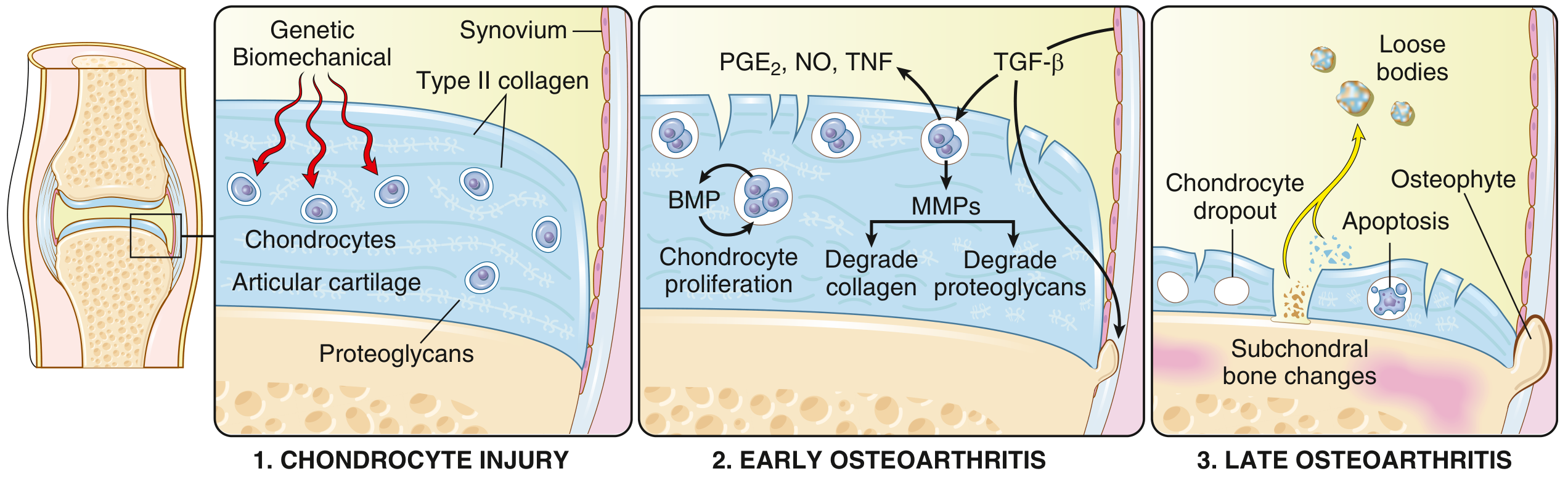
- Chondrocyte apoptosis and dropout
- Full-thickness cartilage loss
- Loose bodies (joint mice) from dislodged cartilage/bone fragments
- Subchondral bone changes (sclerosis, cysts)
- Osteophyte formation at joint margins
- Bone eburnation - exposed subchondral bone becomes polished like ivory
Types
| Type | Description |
|---|---|
| Primary (Idiopathic) OA | No identifiable cause; associated with aging, genetics, female sex, obesity |
| Secondary OA | Due to an identifiable underlying cause (see below) |
- Trauma (acute or chronic repetitive)
- Metabolic: Hemochromatosis, Wilson disease, Ochronosis
- Endocrine: Acromegaly, Hypothyroidism, Hyperparathyroidism, Diabetes
- Crystal deposition: Gout, CPPD (pseudogout)
- Inflammatory arthritis (burnt-out RA)
- Avascular necrosis
- Congenital joint deformity (e.g., DDH, slipped upper femoral epiphysis)
- Ligamentous laxity (Ehlers-Danlos syndrome)
Epidemiology & Risk Factors
- Affects >50 million people worldwide; women disproportionately affected
- Prevalence increases exponentially after age 50; ~40% of people over 70 are affected
- Knee OA is the most common form in clinical practice
- Older age
- Female sex (especially post-menopausal)
- Family history / genetic predisposition (GDF5, RUNX2, SMAD3 loci)
- Black race, lower socioeconomic status
- Obesity - strongest modifiable risk factor for knee OA (causative via BMI shown in Mendelian randomization)
- Joint injury (previous fracture, meniscal tear, ligament injury)
- Repetitive occupational loading
- Metabolic syndrome, higher leptin levels
Joints Commonly Involved
Knees (most common weight-bearing joint affected)
Hips
Lower lumbar and cervical vertebrae
Proximal interphalangeal joints (PIP) and
Distal interphalangeal joints (DIP) of fingers
First carpometacarpal (CMC) joint of thumb
First metatarsophalangeal joint (big toe - hallux rigidus)
Clinical Features
Symptoms
| Symptom | Details |
|---|---|
| Joint pain | Worse with use/activity (mechanical), relieved by rest - cardinal feature |
| Morning stiffness | Present but brief (<30 minutes) - key differentiator from RA (>1 hour) |
| Start-up pain / gelling | Pain/stiffness after sitting still, eases after a few steps |
| Crepitus | Grating/grinding sensation on joint movement |
| Joint swelling | Cool, non-inflammatory effusion (bony swelling from osteophytes) |
| Restricted range of movement | Progressive limitation of joint motion |
| Instability/giving way | In knee OA due to muscle weakness or ligament laxity |
| Night pain | Occurs in advanced disease only |
Signs
- Bony enlargement of joint margins (osteophytes palpable)
- Heberden nodes - osteophytes at DIP joints of fingers (more common in women)
- Bouchard nodes - osteophytes at PIP joints of fingers
- Varus deformity (bow legs) - medial compartment knee OA (commonest)
- Valgus deformity (knock knees) - lateral compartment knee OA
- Crepitus on passive joint movement
- Tenderness along joint line
- Antalgic gait in hip or knee OA
- No significant warmth or systemic features (unlike RA)
- No joint fusion (ankylosis) - unlike RA
OA vs RA - Comparison
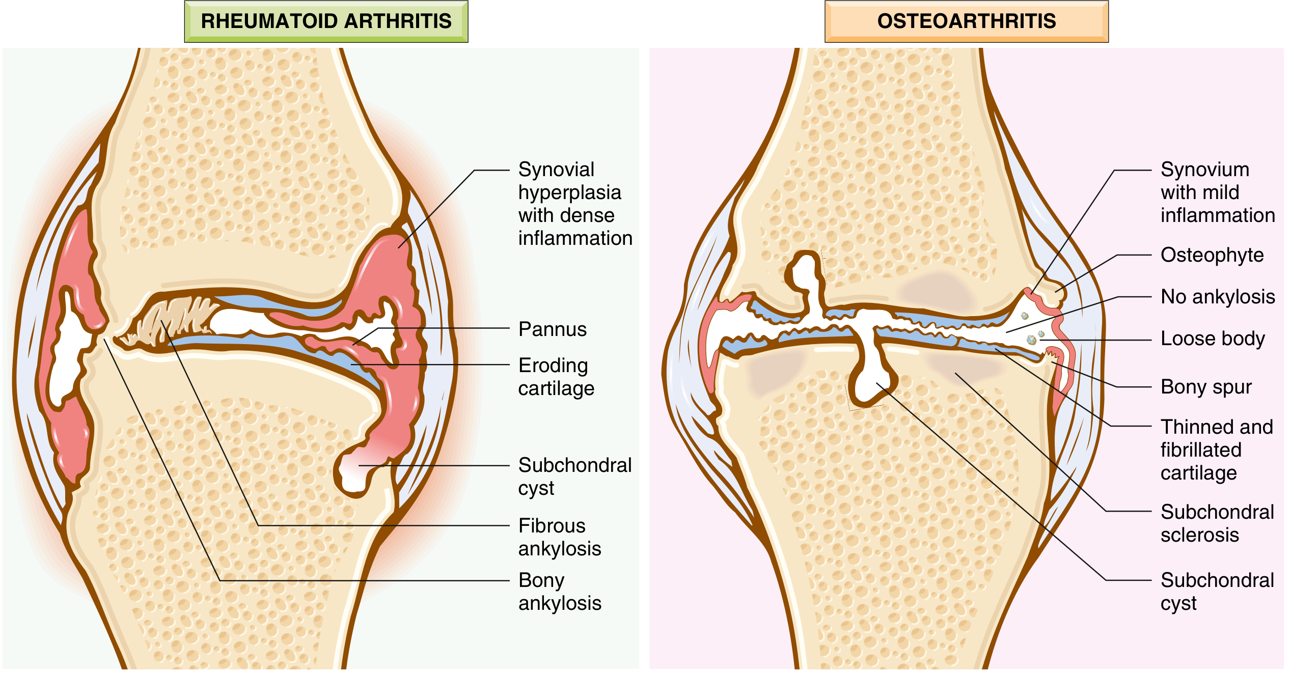
| Feature | Osteoarthritis | Rheumatoid Arthritis |
|---|---|---|
| Mechanism | Mechanical cartilage degeneration | Autoimmune (T cells, ACPA, RF) |
| Joints involved | Weight-bearing (knee, hip), DIP | Small joints of hands first; symmetric; PIP, MCP |
| Morning stiffness | <30 minutes | >1 hour |
| Swelling | Hard (bony osteophytes) | Soft (synovial proliferation) |
| Systemic features | None | Yes (fatigue, fever, extra-articular) |
| Serum antibodies | None | RF, anti-CCP (ACPA) |
| Inflammation | Secondary, mild | Primary, severe |
| Ankylosis | Does NOT occur | Yes (fibrous then bony) |
| Pannus | No | Yes |
Histopathology / Morphology
- Early: Cartilage fibrillation (vertical clefts), chondrocyte proliferation into clusters
- Advanced: Full-thickness cartilage loss, chondrocyte dropout
- Bone eburnation: Exposed subchondral bone polished like ivory by opposing surface
- Subchondral cysts: Synovial fluid forced into bone via micro-fractures (ball-valve mechanism)
- Osteophytes: Fibrocartilage-capped bone outgrowths at joint margins; gradually ossify
- Loose bodies (joint mice): Dislodged fragments of cartilage/bone floating in joint
- Synovium: Only mildly congested/fibrotic with scattered chronic inflammatory cells - contrast with RA's dense synovitis
Investigations
Bloods (Usually Normal in OA)
| Test | Result in OA | Purpose |
|---|---|---|
| ESR / CRP | Normal | To exclude inflammatory arthritis |
| Rheumatoid factor | Negative | Exclude RA |
| Anti-CCP | Negative | Exclude RA |
| Serum uric acid | Normal | Exclude gout |
| FBC | Normal | Baseline; rule out anaemia of chronic disease |
| ALP | Normal (elevated in Paget's) | |
| Blood glucose | Screen for metabolic association |
Imaging
- L - Loss of joint space (narrowing, typically medial compartment in knee)
- O - Osteophytes (bony spurs at joint margins)
- S - Subchondral sclerosis (increased bone density beneath cartilage)
- S - Subchondral cysts (geodes - round radiolucent areas)
- Varus/valgus malalignment
- Loose bodies
- Note: Radiographic severity correlates poorly with pain and disability
- Not routine; used if meniscal tear, ligament injury, or AVN suspected
- Shows bone marrow lesions (BMLs) - correlate with pain
- Reveals synovitis, cartilage integrity, and soft tissue abnormalities
- Detects effusion, synovitis, Baker's cyst
- Guides intra-articular injections
| Feature | OA | Inflammatory/Septic |
|---|---|---|
| Appearance | Clear/yellow (non-inflammatory) | Turbid/opaque |
| WBC | <2000/mm³ | >2000 (inflammatory), >50,000 (septic) |
| Crystals | None (unless coexistent CPPD) | Urate (gout), CPP (pseudogout) |
Management
Step 1 - Non-Pharmacological (First-Line, Always)
- Individualised multicomponent management plan - tailored to patient's needs, comorbidities, preferences
- Education and self-management - patient empowerment about disease, activity, weight
- Exercise therapy (STRONGEST recommendation)
- Land-based exercise (strengthening + aerobic) - strong evidence
- Low-impact aerobics: swimming, cycling, walking
- Quadriceps strengthening especially important in knee OA
- Aquatic/hydrotherapy for those unable to tolerate land-based exercise
- Tailored dosage and progressive intensity
- Weight loss - if BMI >25; moderate-strong evidence (reduces load on joint ~4x body weight at knee)
- Walking aids - cane (in contralateral hand), walking frame
- Footwear and insoles - appropriate footwear; lateral wedge insoles for medial compartment OA
- Work advice - activity modification, pacing
- Behaviour change techniques - improve adherence to lifestyle interventions
| Evidence Level | Intervention |
|---|---|
| Strong | Physical activity, low-impact aerobic strengthening exercise, NSAIDs |
| Moderate | Weight loss (BMI >25) |
| Inconclusive | Electrotherapy, manual therapy, bracing, intra-articular corticosteroids, PRP, acetaminophen, opioids |
| Moderate-Strong Against | Acupuncture, insoles, glucosamine/chondroitin, hyaluronic acid, needle lavage |
Step 2 - Pharmacological
| Drug | Use | Notes |
|---|---|---|
| Paracetamol (Acetaminophen) | Mild pain, step 1 | Ceiling dose 4g/day; less effective than previously thought for OA |
| Topical NSAIDs (diclofenac gel, ibuprofen gel) | Knee/hand OA | First-line for peripheral joints; fewer GI side effects |
| Oral NSAIDs (ibuprofen, naproxen, celecoxib) | Moderate-severe pain | Most effective analgesic; use with PPI; caution in elderly (renal, GI, CV risk) |
| Topical capsaicin | Adjunct in knee OA | Depletes substance P |
| Duloxetine (SNRI) | Central sensitization, chronic pain | OARSI/ACR recommend for OA with central pain features |
| Tramadol | Short-term; moderate-severe pain | Avoid long-term; addiction risk |
| Strong opioids | Last resort before surgery | Short-term only; significant risk in elderly |
| Injection | Evidence | Duration |
|---|---|---|
| Corticosteroid (triamcinolone, methylprednisolone) | Moderate; good for acute flares with effusion | 4-12 weeks; max 3-4/year (cartilage damage risk) |
| Hyaluronic acid (viscosupplementation) | Moderate-against; AAOS recommends against | Variable; 6-12 months claimed |
| PRP (Platelet-rich plasma) | Inconclusive | Experimental; not standard of care |
Step 3 - Surgical
| Procedure | When Used | Notes |
|---|---|---|
| Arthroscopic lavage/debridement | Limited role; may help mechanical symptoms (locking) | Not effective for pure OA; no better than placebo in trials |
| Osteotomy (tibial or femoral) | Younger patients (<55-60), unicompartmental OA with malalignment | Realigns weight-bearing axis to offload diseased compartment |
| Unicompartmental knee arthroplasty (UKA) | Single compartment disease; well-preserved ROM | Less invasive; faster recovery; can convert to TKR later |
| Total Knee Replacement (TKR) | Severe tricompartmental OA | Most common elective orthopaedic procedure; 90-95% 10-year survival |
| Total Hip Replacement (THR) | Severe hip OA | Excellent outcomes; >95% 10-year survival |
- Severe pain at rest and night pain
- Significant functional limitation (unable to walk >100m)
- Radiological grade III-IV OA (Kellgren-Lawrence)
- Failed 6-12 months of conservative treatment
Kellgren-Lawrence Grading (X-ray Classification)
| Grade | Features |
|---|---|
| 0 | Normal |
| I | Doubtful narrowing; possible osteophytes |
| II | Definite osteophytes; possible narrowing |
| III | Moderate narrowing; multiple osteophytes; some sclerosis |
| IV | Severe narrowing; large osteophytes; marked sclerosis; possible deformity |
Prognosis
- OA is a progressive, irreversible condition - no disease-modifying treatments exist yet
- Rate of progression is variable and unpredictable
- Radiographic severity correlates poorly with symptoms
- Up to 20% of patients continue to have significant pain after total knee replacement
- Weight loss and exercise can slow progression and significantly improve quality of life
- Robbins & Kumar Basic Pathology - Pathogenesis and Morphology
- Firestein & Kelley's Textbook of Rheumatology - Pain mechanisms and risk factors
- Grainger & Allison's Diagnostic Radiology - Classification and imaging
- Campbell's Operative Orthopaedics, 15th Ed 2026 - Surgical management and AAOS evidence
- EULAR 2023 Non-pharmacological recommendations for hip/knee OA (PMID: 38212040)