
Physeal bar with its management
I'll start by reading the medical library skill, then search for physeal bar content in parallel.
Searching sources...
Reading File
Now I'll search the library database and look for images in parallel:
Excellent. Now I'll read the main physeal bar sections from Campbell's and fetch the physeal bar MRI image in parallel:
Reading File
Reading File
Loading Image
Good. Now let me read the main "traumatic physeal arrest" section and the growth arrest section:
Reading File
Finding Sources
Now let me read the key sections - traumatic physeal arrest, growth arrest, and Miller's partial growth arrest:
Reading File
Reading File
Reading File
Now let me read the asymmetric physeal arrest section and the Langenskiold classification from Campbell's:
Reading File
Finding Sources
Let me search for Langenskiold classification and also read the technique 38.35 (Langenskiold bony bridge resection) completion section:
Finding Sources
Reading File
I now have comprehensive information to write a detailed answer. Let me also fetch the clinical case image showing physeal bar resection:
Loading Image
I now have all the information needed. Here is the comprehensive answer:
Physeal Bar (Bony Bridge): Definition, Pathology, and Management
Definition
A physeal bar (also called a bony bridge or physeal bridge) is an osseous tether that crosses the growth plate (physis), connecting metaphyseal and epiphyseal bone. It develops after injury to physeal cartilage, most commonly following a Salter-Harris fracture, and arrests growth across the affected portion of the physis while the remaining normal physis continues to grow.
Pathophysiology
- Normal physeal cartilage, when damaged, is replaced by bone rather than regenerating
- The bony bridge acts as a tether, preventing that segment of the physis from contributing to longitudinal growth
- The uninjured portion of the physis continues to grow, creating a differential that produces angular deformity or limb-length discrepancy (LLD), or both
- Spontaneous resolution is exceedingly rare
Causes
| Category | Examples |
|---|---|
| Trauma (most common) | Salter-Harris III, IV fractures; crush injuries (Salter-Harris V) |
| Infection | Acute hematogenous osteomyelitis involving the physis |
| Tumors / irradiation | Growth plate involvement |
| Iatrogenic | Improper pin placement, previous surgery crossing the physis |
| Dysplasia | Blount disease (tibia vara), Madelung deformity |
Classification by Location
1. Peripheral Bar
- Located at the edge of the physis
- More accessible surgically
- Produces predominantly angular deformity
- Better prognosis after resection (especially if small and in young patients)
2. Central Bar
- Located in the central area of the physis
- Surrounded on all sides by normal physeal cartilage
- Produces predominantly limb-length discrepancy / shortening
- More challenging to access; approached via a metaphyseal cortical window or osteotomy
- Arthroscopically assisted resection has been described
3. Combined (Mixed) Bar
- Most cases have components of both shortening and angular deformity
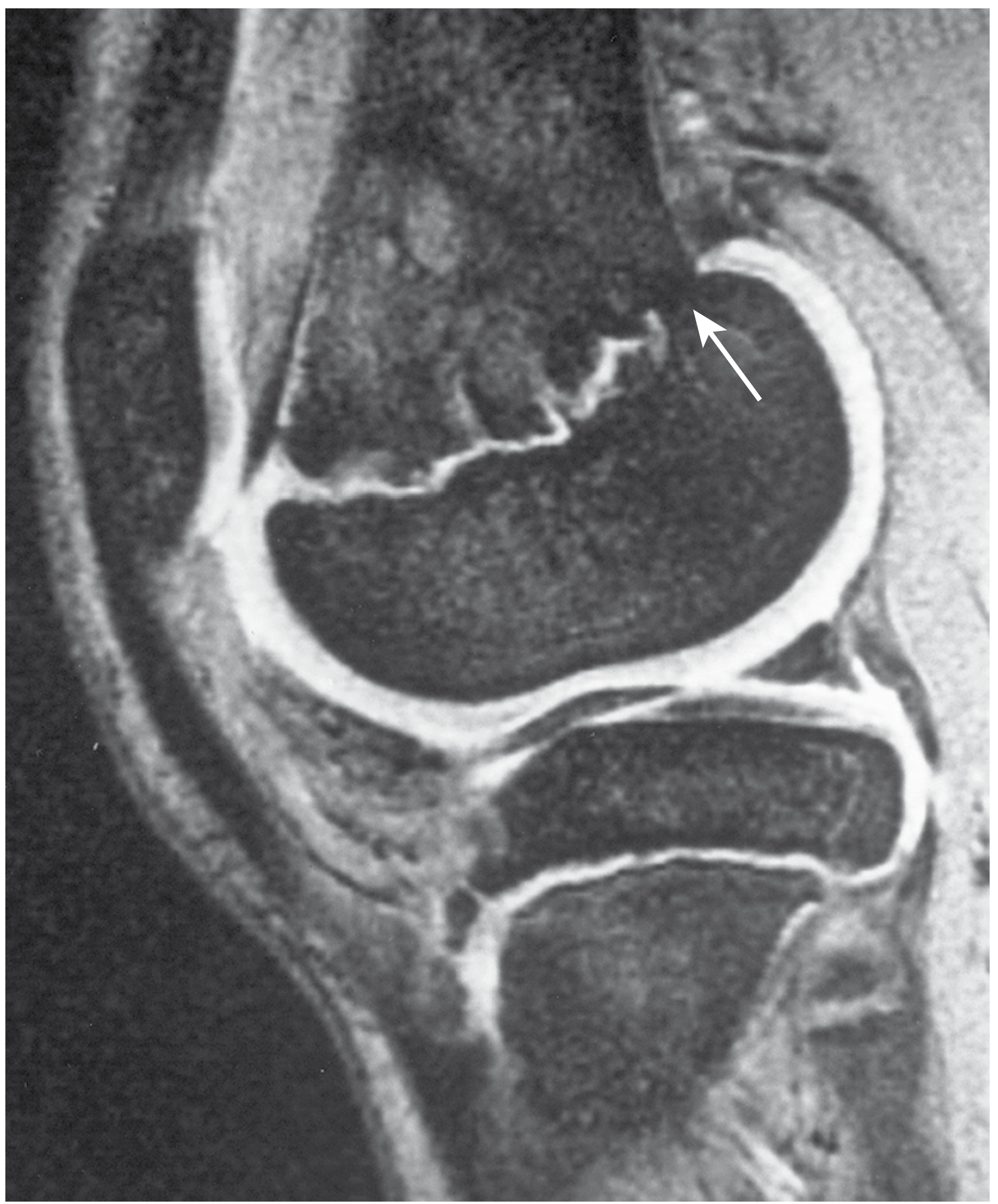
MRI of Physeal Bar at the Knee
Below is a gradient-echo sagittal MRI of the knee in a 12-year-old boy demonstrating interruption of the physis by a physeal bar (arrow):
Diagnosis / Imaging
| Modality | Role |
|---|---|
| Plain radiograph | Initial assessment; reveals angular deformity or growth arrest line |
| CT (3D reconstruction) | Best for mapping bar size, location, and percentage of physis involved; essential for preoperative planning |
| MRI | Superior soft tissue contrast; gradient-echo sequences show cartilage; useful for small or central bars |
| Tomography | Historical but still mentioned for assessment in two planes |
Preoperative mapping using 3D CT or MRI is mandatory before any surgical resection to define:
- Location (peripheral vs. central)
- Percentage of physis involved
- Remaining normal physeal area
Decision-Making Algorithm
The key determinants of management are:
- Percentage of physis involved by the bar
- Age of the child / growth remaining
- Location of the bar (peripheral vs. central)
- Expected deformity at skeletal maturity
Bar identified on CT/MRI
|
├── Near skeletal maturity / minimal growth remaining
| └── Observation / shoe lift / epiphysiodesis
|
├── Bar < 50% physis + young child + significant morbidity expected
| └── BAR RESECTION (with fat/Silastic interposition)
| ± concurrent osteotomy for established angular deformity
|
└── Bar > 50% physis OR large / central / inaccessible
└── Completion epiphysiodesis (ipsilateral)
+ Contralateral epiphysiodesis (for LLD)
OR Limb lengthening (if LLD >5 cm expected)
Indications for Bar Resection
From Campbell's Operative Orthopaedics 15th Ed (2026):
- Child young enough to have significant growth remaining (>2 cm)
- Bar involves <50% of the physis (ideally <30%)
- Bar is peripheral and accessible
- Progressive angular deformity or LLD is expected
- Results are unpredictable; unsuccessful outcomes occur in 10% to 40% of patients
- Young patients with small peripheral bars have the highest success rates
Surgical Techniques
Technique 1: Peripheral Bar Resection - Langenskiold Technique (38.35)
Indication: Peripheral bar, >50% normal physis remaining
Steps:
- Preoperative exact localization and sizing by MRI, CT, and tomography in two planes
- Expose periphery of physis near the bony bridge with a bloodless field (tourniquet); use microscope or binocular loupe
- Remove overlying periosteum and resect the bony bridge until normal periphery of the physis is reached on both sides, with physeal cartilage visible all around the cavity - no bridge remnant left, no unnecessary normal cartilage removal
- Release tourniquet; obtain hemostasis
- Fill cavity with autogenous fat from subcutaneous tissue (gluteal fold or groin), dividing into pieces if irregular
- Suture ligament/muscle/subcutaneous tissue over defect to retain the fat graft
- Close in layers; plaster splint; limit weight-bearing until healed
Technique 2: Bony Bridge Resection + Angulation Osteotomy - Ingram Technique (38.36)
Indication: When concurrent angular deformity correction is needed; bar need not be strictly peripheral
Key steps:
- Perform an osteotomy at the level of the bony bridge, adjacent and parallel to the physis (opening-wedge, same side as the bar)
- Place a guide pin parallel to the physis under fluoroscopy, adjacent to the bar
- Open the osteotomy widely with a laminar spreader
- Using a dental burr and operating microscope/loupe, resect the white sclerotic bony bridge until normal physeal cartilage appears on all sides (dental mirror aids visualization)
- Fill defect with free fat graft or silicone
- Secure the osteotomy after inserting a wedge of autogenous bone to correct deformity; smooth pins hold osteotomy
Technique 3: Peripheral Bar Resection - Birch Technique (38.37)
Steps:
- Carefully expose the peripheral junction of the bar and healthy perichondral ring at one or both edges - use fluoroscopy throughout to stay at the physeal level and not drift into metaphysis/epiphysis
- Continue resection until physis is visible from each edge of healthy perichondrium throughout the depth; alternatively, use periosteal stripping + fluoroscopic guidance to develop a cavity directed toward the physis
- Fill defect with autogenous fat from local subcutaneous tissue or a separate groin/buttock incision
Technique 4: Central Physeal Bar Resection (38 approach)
- Approached from the metaphyseal marrow cavity through a metaphyseal cortical window or an osteotomy
- Arthroscopically assisted central bar resection: scope identifies normal cartilage after dental burr resection in the defect
- More technically demanding; results are less predictable than for peripheral bars
Interposition Materials (After Resection)
The physeal defect does not regenerate. Interposition material prevents re-bridging by bone:
| Material | Notes |
|---|---|
| Autogenous fat | Most commonly used; secured with surrounding tissue suture |
| Physeal graft (iliac crest) | Shown to be superior to Silastic and fat in some studies |
| Silastic (silicone) | Used in Ingram technique; alternative to fat |
The remaining normal physeal cartilage surrounding the resection cavity can produce bone in a more linear and orderly fashion after removal of the bar.
Management of Residual / Predicted Deformity
Bar resection usually does not correct established angular deformity by itself, but it reduces the rate of recurrence and the number of osteotomies needed over the child's growth period.
| Condition | Treatment |
|---|---|
| Angular deformity (concurrent) | Osteotomy at time of bar resection (Ingram) or staged |
| LLD at maturity <2 cm | Observation or shoe lift |
| LLD 2-5 cm | Contralateral epiphysiodesis |
| LLD >5 cm | Limb lengthening (intramedullary nail preferred for accuracy and lower complication rate vs. external fixator) |
| Bar >50% physis | Completion of ipsilateral epiphysiodesis + contralateral epiphysiodesis or limb lengthening |
| Recurrence of deformity after osteotomy | CT to assess for residual/recurrent physeal bar |
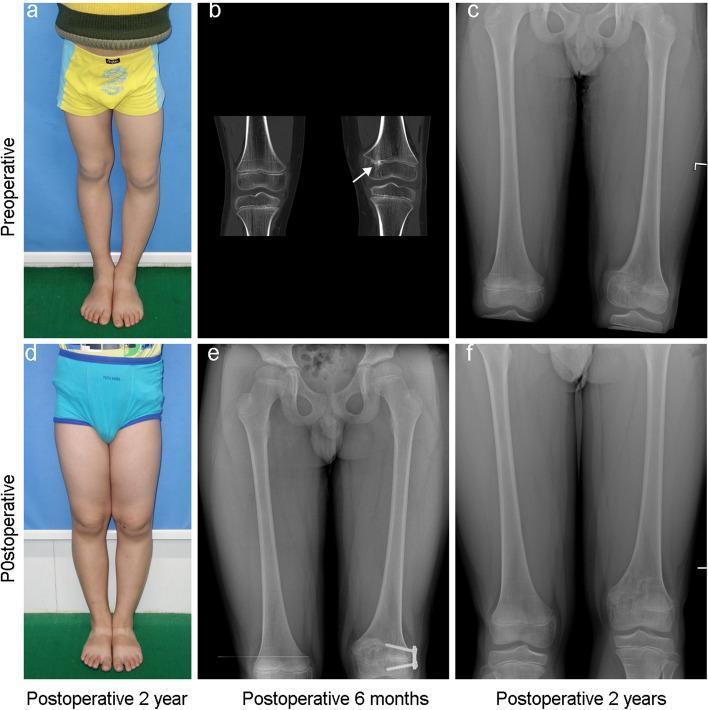
Clinical Case Example: Physeal Bar Resection
The images below show a 9-year-old with a physeal bar causing valgus deformity at the knee, managed with bar resection and guided growth:
Prognosis and Key Points Summary (Miller's Review)
From Miller's Review of Orthopaedics 9th Edition:
- Physeal bar resection with fat/Silastic interposition is reserved for patients with >2 cm of growth remaining and <50% physeal involvement
- Smaller peripheral bars in young patients have the highest success rate
- MRI and CT define location and amount of physeal closure
- Arrest involving >50% of the physis should be treated with ipsilateral completion of arrest + contralateral epiphysiodesis or lengthening
Sources:
- Campbell's Operative Orthopaedics, 15th Ed (2026), Techniques 38.35, 38.36, 38.37 and Growth Arrest section
- Miller's Review of Orthopaedics, 9th Edition - Partial Growth Arrest summary
Marco and microscopic structure of adult cortical bone
Searching sources...
Reading File
This is a broad, visually rich topic. Let me search headings and paragraphs simultaneously:
Finding Sources
Finding Sources
Searching Images
cortical bone microstructure osteon Haversian canal histology diagram
Finding Sources
Finding Sources
Searching Images
osteon haversian system bone cross section microscopy
Finding Sources
Finding Sources
Excellent finds. Now let me read the key sections from all these books simultaneously:
Reading File
Reading File
Reading File
Now let me read the Junqueira Lamellar bone section further, the THIEME atlas bone structure, and look for key histology images:
Reading File
Reading File
Reading File
Excellent content. Now let me fetch the key diagrams - the THIEME atlas bone structure diagram, the Junqueira osteon, and the polarized light lamellar bone images:
Loading Image
Loading Image
Loading Image
Now let me get the polarized light / lamellar bone histology image and also the Junqueira bone cells section for completeness:
Loading Image
Reading File
I now have comprehensive, well-illustrated content from multiple authoritative sources. Let me compile the full answer:
Macro and Microscopic Structure of Adult Cortical Bone
Part 1 — Macroscopic (Gross) Structure
Overview of a Typical Long Bone
The femur is the classic example used to illustrate cortical bone macrostructure. A long bone has three main regions:
| Region | Location | Bone type present |
|---|---|---|
| Diaphysis (shaft) | Middle cylindrical portion | Predominantly compact (cortical) bone |
| Epiphysis (knobby ends) | Proximal and distal ends | Thin shell of cortical bone + internal cancellous bone |
| Metaphysis | Flared region between shaft and epiphysis | Mixed cortical/cancellous; site of old growth plate |
The THIEME Atlas Diagram - Structure of a Typical Tubular Bone (Femur)
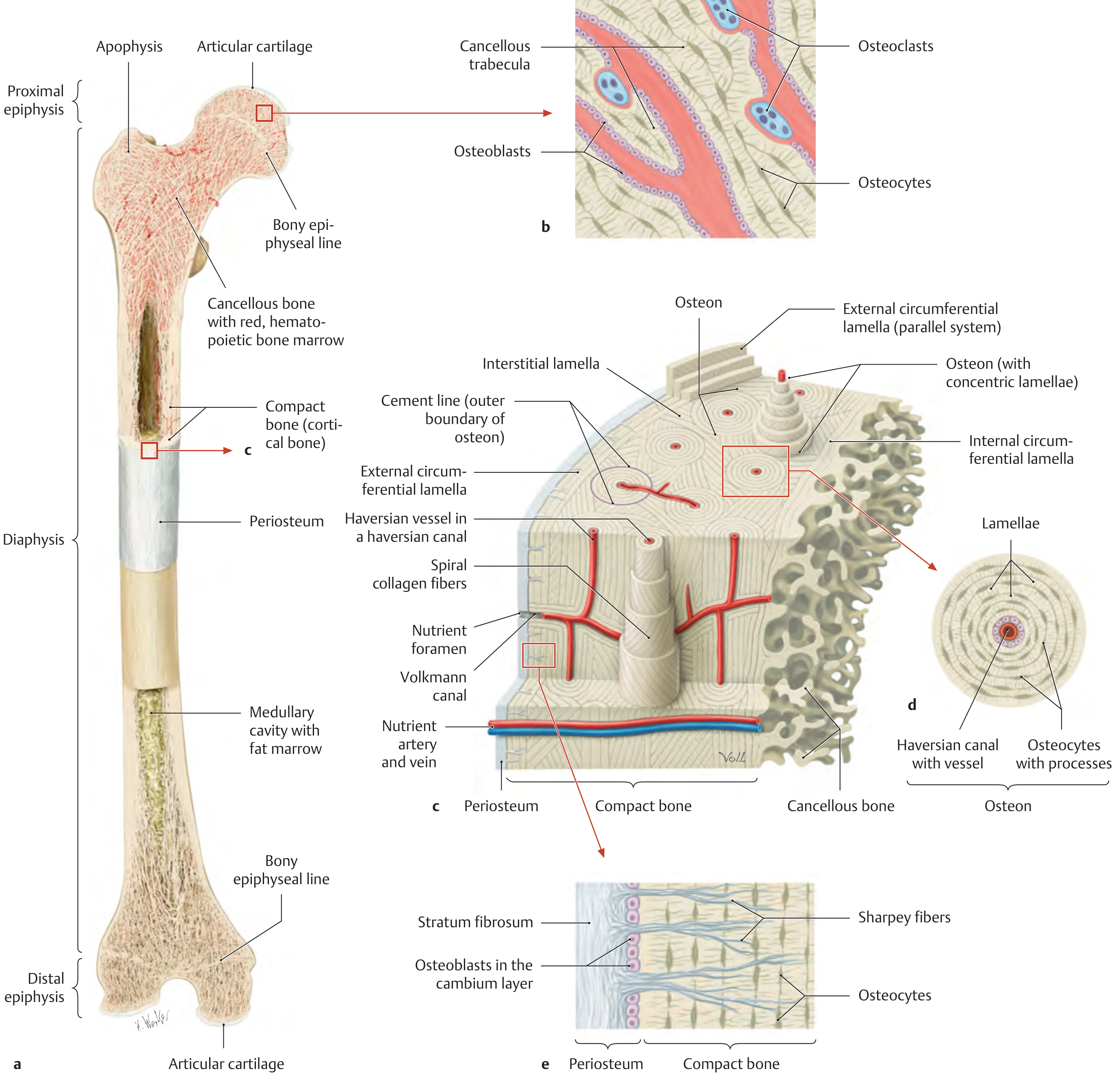
This diagram summarizes the entire macro-to-micro hierarchy: (a) whole femur, (b) cancellous trabeculae with cells, (c) 3D compact bone with osteons and canal systems, (d) cross-section of a single osteon, (e) periosteum layers.
Layers from Outside to Inside in the Diaphysis
PERIOSTEUM
↓
OUTER CIRCUMFERENTIAL LAMELLAE (subperiosteal)
↓
HAVERSIAN SYSTEMS (osteons) — the bulk of compact bone
↓
INNER CIRCUMFERENTIAL LAMELLAE (perimedullary)
↓
ENDOSTEUM
↓
MEDULLARY CAVITY (yellow marrow / fat in adults)
1. Periosteum
A double-layered sleeve of dense connective tissue covering the outer surface:
- Stratum fibrosum (outer layer) - dense collagenous fibrous tissue; the stratum of mechanical attachment
- Stratum germinativum / cambium layer (inner layer) - contains osteoprogenitor cells and active osteoblasts; responsible for appositional (periosteal) bone growth and fracture repair
The periosteum is anchored to underlying bone by Sharpey fibers - bundles of type I collagen that penetrate obliquely into the outer circumferential lamellae.
2. Cortical (Compact) Bone
- Forms the outer wall of the diaphysis; appears dense and ivory-white on gross examination
- Porosity is only ~5-10% (vs. ~75-95% for cancellous bone)
- The structural units are osteons (Haversian systems), cylinders approximately 1 cm long and 250-350 µm in diameter, oriented parallel to the long axis of the bone
- Haversian canals run longitudinally and are interconnected by Volkmann canals (transverse/oblique) and also communicate with periosteal and medullary vessels via the nutrient foramen
3. Medullary Cavity
- Central hollow space within the diaphysis
- In adults, contains yellow bone marrow (primarily adipose tissue)
- Lined internally by endosteum (described below)
- Metaphyseal/epiphyseal regions contain red bone marrow (haematopoietic)
4. Endosteum
- A thin single cell layer lining all internal bone surfaces: medullary cavity, trabecular surfaces, Haversian canal walls, and Volkmann canal walls
- Contains osteoprogenitor cells (endosteal cells), osteoblasts, and osteoclasts
- The endosteal surface is the main site of bone remodeling and marrow-bone cell exchange
Part 2 — Microscopic Structure
Adult cortical bone is lamellar bone - organized into layers of parallel collagen fibers that alternate in orientation between successive lamellae (like plywood), giving it exceptional strength. This contrasts with woven (immature) bone, which has randomly oriented collagen and is found only in the fetus, rapidly growing areas, fracture callus, and Paget disease.
Four Components of Compact Lamellar Bone
| Component | Description | Function |
|---|---|---|
| Osteons (Haversian systems) | Main structural units; concentric lamellae around a central canal | Primary structural and nutritive unit |
| Interstitial lamellae | Irregular remnants of old osteons | Fills space between osteons |
| Outer circumferential lamellae | Parallel plates immediately under periosteum | Strength; structural integration with periosteum |
| Inner circumferential lamellae | Parallel plates around medullary cavity | Structural integrity at marrow interface |
The Osteon (Haversian System) - In Detail
Histology: Ground bone cross-section (Junqueira's)
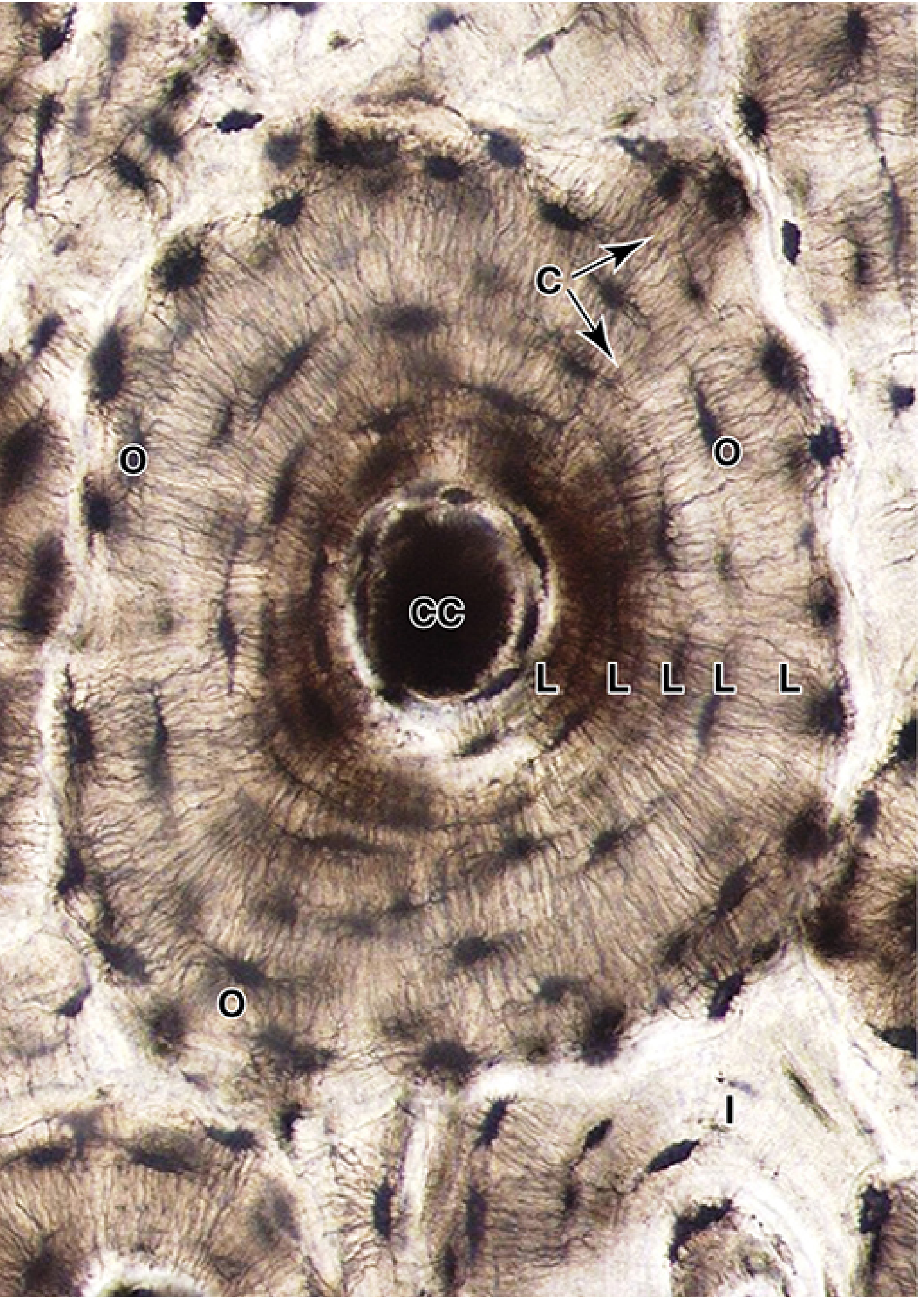
Dimensions
- Diameter: 100-250 µm (Junqueira) / 250-350 µm for the full osteon including circumferential zone (THIEME)
- Length: approximately 1 cm along bone axis, sometimes branching (bifurcated)
- Number of concentric lamellae per osteon: 5-20
Components of the osteon from center outward:
-
Haversian canal (central canal)
- Contains 1-2 small blood vessels (capillaries/postcapillary venules), unmyelinated nerve fibers, and endosteum
- Diameter: ~20-50 µm
- All cells of the osteon are nourished via diffusion from these vessels
-
Concentric lamellae
- Rings of mineralized bone matrix deposited concentrically around the Haversian canal
- Each lamella is ~3-7 µm thick
- The collagen fibers in each lamella run parallel to each other but at an angle to adjacent lamellae (alternating orientations)
- This plywood-like arrangement is responsible for the resistance to torsional, bending, and compressive forces
-
Lacunae
- Small lens-shaped spaces between lamellae (and within them)
- Each contains one osteocyte
- Interconnected by canaliculi
-
Canaliculi
- Fine tunnels radiating from each lacuna in all directions
- Contain the dendritic (cytoplasmic) processes of osteocytes
- Connect adjacent osteocytes via gap junctions
- Provide the nutrient-waste exchange pathway between the Haversian canal and the furthest osteocytes
-
Cement line (reversal line)
- Outer boundary of each osteon; marks where osteoclast resorption stopped and osteoblast deposition began
- Rich in non-collagenous proteins (osteopontin, osteocalcin) and mineralized collagen
- Acts as a crack-arresting interface - prevents fracture propagation across osteon boundaries
Polarized Light Appearance of Lamellar Bone
Under polarized light, the alternating collagen fiber orientations in successive lamellae produce alternating bright and dark bands - a hallmark of lamellar bone that distinguishes it from woven bone:
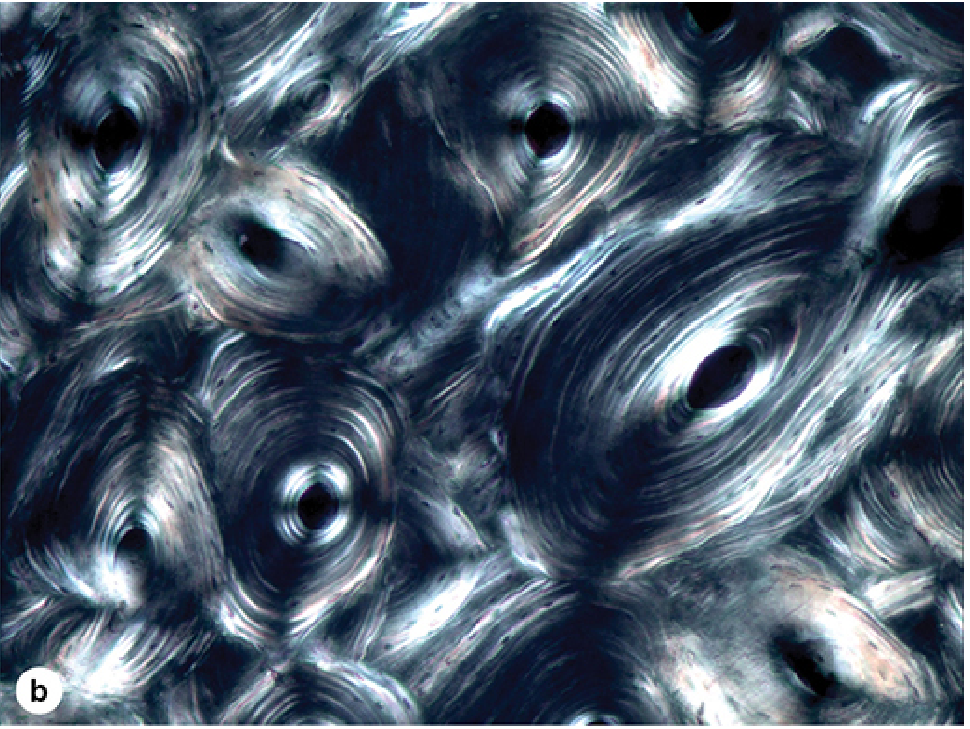
- Bright (birefringent) bands = lamellae with collagen fibers oriented perpendicular to the polarizer
- Dark bands = lamellae with collagen fibers parallel to the polarizer
- This alternation indicates that fibers in successive lamellae have different orientations - the structural basis of the bone's strength
Interstitial Lamellae and Generations of Osteons
As bone remodels throughout life, successive generations of osteons are formed. Each new osteon partially destroys its predecessor, leaving behind angular remnants of old lamellae called interstitial lamellae:
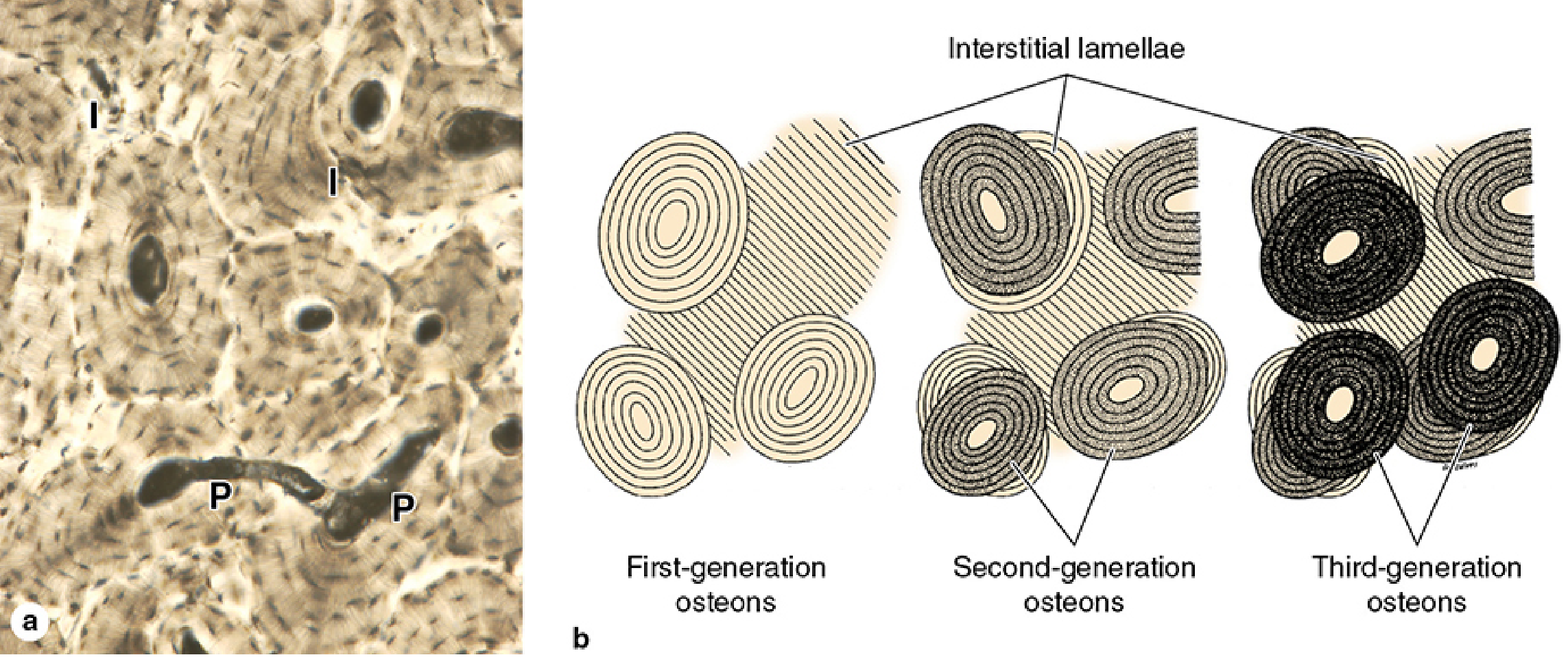
- Most compact bone in adult humans = secondary bone (remodeled osteons)
- Interstitial lamellae are the remnants of the first-, second-, and third-generation osteons
- Successive generations are visible by their different degrees of mineralization (darker = more mineralized = older)
Canal Systems of Cortical Bone
| Canal | Orientation | Contents | Lamellae? |
|---|---|---|---|
| Haversian (osteonal) canal | Longitudinal (parallel to bone axis) | Blood vessels, nerves, endosteum | Yes - concentric |
| Volkmann (perforating) canal | Transverse / oblique | Blood vessels | No - perforates through existing lamellae |
Volkmann canals connect:
- Adjacent Haversian canals to each other
- Haversian canals to periosteal vessels
- Haversian canals to the medullary cavity
The key distinguishing feature: Haversian canals are surrounded by concentric lamellae; Volkmann canals are not.
The Cells of Cortical Bone
| Cell | Origin | Location | Function |
|---|---|---|---|
| Osteoprogenitor cells | Mesenchymal stem cells (via RUNX2/CBFA1) | Periosteum (cambium), endosteum, marrow | Stem cell precursors; differentiate into osteoblasts |
| Osteoblasts | Osteoprogenitor cells | Periosteal cambium, endosteum, Haversian canal walls | Synthesize and secrete osteoid (unmineralized matrix); direct mineralization via matrix vesicles |
| Osteocytes | Osteoblasts (trapped in lacunae) | Within lacunae throughout bone | Maintain matrix; mechanosensing; communicate via canalicular network |
| Osteoclasts | Hematopoietic precursors (monocyte fusion); RANK-RANKL signaling | Howship's lacunae on resorbing bone surfaces | Bone resorption - secrete H⁺ (acid) and lysosomal enzymes |
Extracellular Matrix (ECM) Composition
Adult cortical bone matrix is approximately:
- 65-70% inorganic (mineral): Hydroxyapatite Ca₁₀(PO₄)₆(OH)₂ crystals deposited on and between collagen fibers; provides compressive strength and hardness
- 30-35% organic (osteoid): predominantly Type I collagen (~90% of organic matrix); the remaining ~10% = non-collagenous proteins
| Non-collagenous protein | Role |
|---|---|
| Osteocalcin (bone-Gla protein) | Binds Ca²⁺; regulates mineralization; marker of bone turnover |
| Osteopontin | Adhesion molecule; part of cement line; osteoclast attachment |
| Osteonectin | Binds collagen and mineral; initiates mineralization |
| Bone sialoprotein | Nucleates hydroxyapatite crystal formation |
| Proteoglycans (decorin, biglycan) | Regulate collagen fibril diameter and matrix organization |
Cortical Bone Remodeling (The Cutting Cone / BMU)
Remodeling in cortical bone proceeds via a Basic Multicellular Unit (BMU):
- Activation - Osteoclasts are recruited to a site (triggered by mechanical strain, microdamage, hormonal signals)
- Resorption - Osteoclasts tunnel through cortical bone forming a cylindrical "cutting cone" (~200 µm diameter)
- Reversal - Transition from resorption to formation; mononuclear cells smooth the surface; cement line is deposited
- Formation - Osteoblasts follow the advancing tunnel ("closing cone"), deposit concentric lamellae inward, trapping themselves as osteocytes
- The residual central canal = new Haversian canal with its blood supply
In healthy adults, 5-10% of bone turns over annually via this mechanism.
Summary Table
| Feature | Detail |
|---|---|
| Porosity | ~5-10% |
| Fundamental structural unit | Osteon (Haversian system) |
| Osteon diameter | 100-350 µm |
| Osteon length | ~1 cm; may bifurcate |
| Lamellae per osteon | 5-20 concentric rings |
| Collagen arrangement | Alternating angles in successive lamellae (plywood pattern) |
| Haversian canal | Longitudinal; contains vessels + nerves; surrounded by lamellae |
| Volkmann canal | Transverse/oblique; connects Haversian canals; no own lamellae |
| Interstitial lamellae | Remnants of remodeled (destroyed) old osteons |
| Outer circumferential lamellae | Just beneath periosteum; anchored by Sharpey fibers |
| Inner circumferential lamellae | Around medullary cavity / endosteum |
| Cement line | Outer osteon boundary; rich in non-collagenous proteins |
| Annual turnover rate | 5-10% |
Sources:
- Junqueira's Basic Histology - Text and Atlas, 17th Ed - Types & Organisation of Bone, Lamellar Bone sections
- Histology: A Text and Atlas with Correlated Cell and Molecular Biology - General Structure of Bone Tissue
- THIEME Atlas of Anatomy: General Anatomy and Musculoskeletal System - Structure of a Typical Tubular Bone (femur)
- Forensic Anthropology: A Comprehensive Introduction, 2nd Ed - Bone Structure
- Rheumatology, 2-Volume Set (2022, Elsevier) - Microscopic level and Effects of Remodeling on Cortical Bone
This is a shared conversation. Sign in to Orris to start your own chat.