
Where are emmiserry veins present ?
- Mastoid emissary vein - passes through the mastoid foramen, connecting the sigmoid sinus to the posterior auricular/occipital veins
- Parietal emissary vein - passes through the parietal foramen, connecting the superior sagittal sinus to scalp veins
- Occipital emissary vein - connects the confluence of sinuses to the occipital veins
- Condylar emissary vein - passes through the posterior condylar canal, connecting the sigmoid sinus to suboccipital veins
- Sphenoidal emissary vein (vein of Vesalius) - passes through the foramen of Vesalius (sphenoidal emissary foramen), connecting the cavernous sinus to the pterygoid plexus
emissary veins skull cranial sinuses diagram anatomy
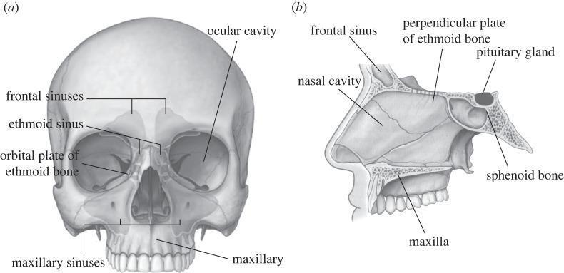
This medical anatomical diagram displays the human skull in two views, focusing on the paranasal sinuses and internal cranial structures. View (a) is an anterior (frontal) perspective of the skull with semi-transparent overlays illustrating the paranasal sinuses. Labeled structures include the frontal sinuses located superior to the orbits, the ethmoid sinus situated medially between the eyes, the orbital plate of the ethmoid bone forming the medial orbital wall, the ocular cavity (orbit), and the large maxillary sinuses within the maxilla. View (b) provides a mid-sagittal section of the facial and mid-cranial region. It highlights the spatial relationship between the nasal cavity and surrounding structures, including the frontal sinus, the perpendicular plate of the ethmoid bone (part of the nasal septum), and the maxilla. Posteriorly, it shows the sphenoid bone containing the sella turcica, where the pituitary gland is seated. This illustration is designed for medical education regarding craniofacial anatomy, specifically for procedures such as Functional Endoscopic Sinus Surgery (FESS) and maxillofacial osteotomies.
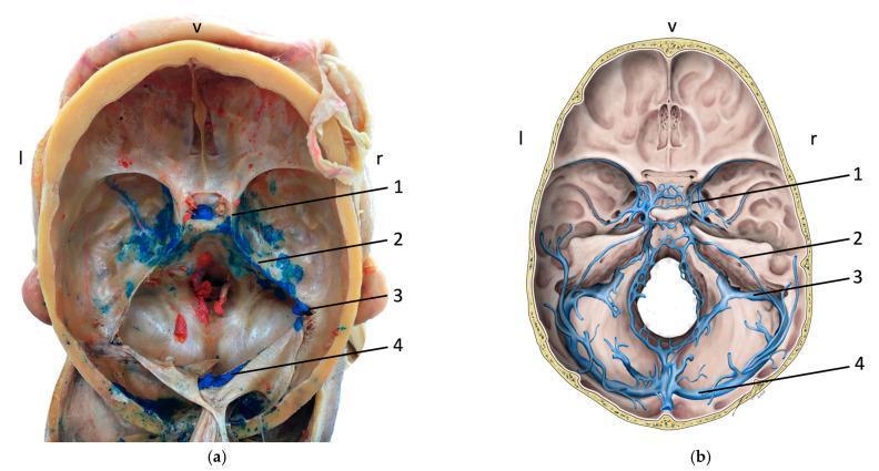
This medical image displays a comparative study of the cranial base dural venous sinuses through an anatomical cadaveric specimen and a corresponding schematic diagram. Panel (a) shows an internal view of a human skull base following calvaria and brain removal. The dural sinuses are highlighted by blue latex dispersion injected via the facial vein, demonstrating venous connections. Panel (b) provides an idealized anatomical illustration of the same structures. Key numbered structures in both panels include: (1) Cavernous sinus, located ventrally and centrally; (2) Superior petrosal sinus, extending posterolaterally along the petrous part of the temporal bone; (3) Sigmoid sinus, following an S-shaped course toward the jugular foramen; and (4) Transverse sinus, positioned most posteriorly within the tentorium cerebelli. Orientation markers indicate ventral (v), left (l), and right (r) directions. This content serves to teach the complex intracranial venous anatomy and the clinical significance of venous anastomoses between the extracranial facial veins and the intracranial dural sinus system, which is relevant for understanding the spread of infections or thrombi.
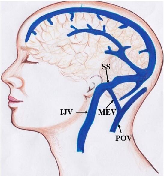
Anatomical diagram depicting the venous drainage of the human head in a lateral view. The illustration highlights the relationship between intracranial dural sinuses and extracranial veins. Key structures identified include the Sigmoid Sinus (SS), which transitions into the Internal Jugular Vein (IJV) as it descends anteriorly. A significant anatomical feature shown is the Mastoid Emissary Vein (MEV), which provides a valveless communication pathway between the intracranial sigmoid sinus and the extracranial Posterior Auricular Vein (POV). The POV is situated posteriorly to the ear and runs roughly parallel to the upper portion of the IJV. The diagram effectively illustrates the concept of emissary veins as collateral drainage routes and their role in intracranial pressure regulation and potential surgical risk during posterior fossa or mastoid procedures. The focus is on neurovascular anatomy, specifically the communication between the dural venous system and superficial scalp veins.
Emissary Veins - Location and Connections
Complete List of Emissary Veins
| Emissary Vein | Foramen/Opening | Intracranial Connection | Extracranial Connection |
|---|---|---|---|
| Parietal emissary vein | Parietal foramen | Superior sagittal sinus | Superficial temporal vein |
| Mastoid emissary vein | Mastoid foramen (junction of temporal & occipital bones) | Sigmoid sinus | Occipital / posterior auricular vein |
| Condylar emissary vein | Posterior condylar canal | Sigmoid sinus | External vertebral venous plexus |
| Occipital emissary vein | Near external occipital protuberance | Confluence of sinuses | Occipital vein |
| Vein of Vesalius (sphenoidal emissary vein) | Foramen of Vesalius (sphenoidal emissary foramen) | Cavernous sinus | Pterygoid venous plexus |
| Emissary vein of foramen cecum | Foramen cecum | Superior sagittal sinus | Nasal cavity veins |
| Venous plexus of hypoglossal canal | Hypoglossal canal | Sigmoid sinus / jugular bulb | Suboccipital plexus |
| Venous plexus of foramen ovale | Foramen ovale | Cavernous sinus | Pterygoid plexus |
| Internal carotid venous plexus | Carotid canal | Cavernous sinus | Internal carotid plexus |
Clinical Significance
- Spread of infection from scalp/face to intracranial sinuses (e.g., meningitis, cavernous sinus thrombosis)
- Bleeding risk during mastoid or posterior fossa surgery (mastoid emissary vein is the most surgically relevant)
- Collateral drainage when a dural sinus is obstructed - for example, the mastoid emissary vein can provide collateral flow around an obstructed sigmoid sinus
Emissary veins in heat hematoma forensic medicine
emissary veins heat hematoma forensic medicine postmortem fire death
Heat Hematoma and Emissary Veins - Forensic Medicine
What is a Heat Hematoma?
Role of Emissary Veins - The Mechanism
- Intense heat causes blood within the diploic layer of the skull bones to boil and vaporize
- This pressurized blood is extruded outward through the emissary veins and diploic veins into the extradural (epidural) space
- The blood then clots and forms the characteristic heat hematoma
- Heat causes shrinkage of the meninges and brain
- This shrinkage may aspirate blood from the venous sinuses into the extradural space
"It may be due to blood being boiled from the diploic layer of bone through emissary veins and venous sinus which rupture or shrinkage of meninges and brain, which may aspirate blood from the venous sinuses."
"Blood and marrow are expressed from the skull bones by heat that accumulates between dura and the bone... The diploic veins and longitudinal sinus is involved."
Characteristics of Heat Hematoma
| Feature | Heat Hematoma |
|---|---|
| Nature | Postmortem artifact |
| Location | Extradural (epidural) space |
| Most common site | Parietotemporal region (sometimes frontal or occipital) |
| Colour | Light chocolate / brown; pink if blood contains CO |
| Consistency | Soft, friable |
| Appearance | Honeycomb (due to steam bubbles from boiling blood) |
| Thickness | 1.5 - 15 mm |
| Volume | ~100-120 mL |
| COHb level | Same as peripheral blood |
| Adjacent brain | Shrunken, firm, yellow-brown ("cooked"), dura leathery |
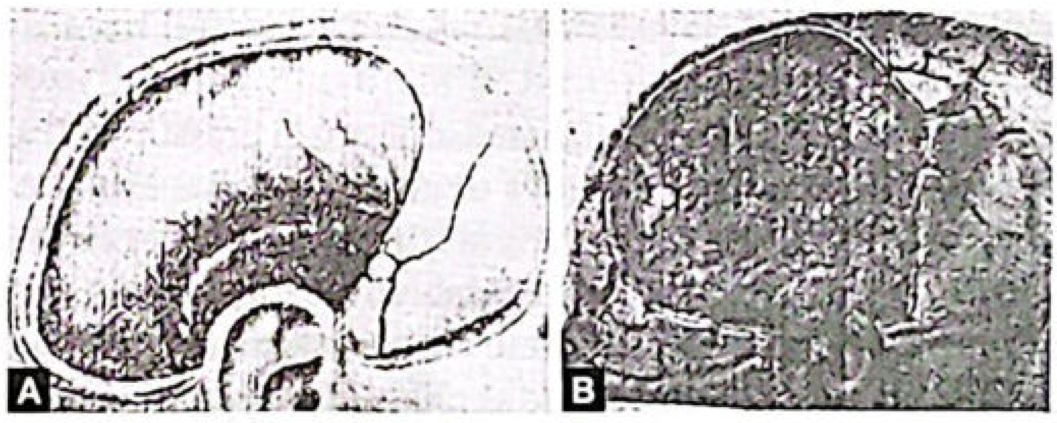
Key Forensic Distinction: Heat Hematoma vs. Traumatic Extradural Hemorrhage
| Feature | Heat Hematoma | Traumatic Extradural Hematoma |
|---|---|---|
| Origin | Postmortem artifact from heat | Antemortem - blunt trauma |
| Colour | Pink/brown, cherry-red if COHb | Dark red |
| Appearance | Honeycomb (bubbly, foamy) | Smooth clot |
| COHb in hematoma | Same as peripheral blood | Little or no COHb |
| Associated skull fracture | Thermal fractures (no suture involvement) | Fracture over temporal bone (middle meningeal artery) |
| Typical site | Parietotemporal, follows charring distribution | Temporoparietal (fixed - MMA territory) |
| Dura | Shrunken, leathery | Normal |
| Signs of blunt force | Absent | Present |
| CT appearance | Low density, crescent-shaped (may cross midline) | High density, lens/biconvex shape |
- Equal to peripheral blood = heat hematoma (blood was in circulation when fire started, became saturated with CO, then was expressed by heat)
- Little or no COHb = true traumatic extradural bleed that occurred before the fire
Forensic Significance
- Artifact recognition - Heat hematoma must not be mistaken for evidence of antemortem head trauma or homicide
- Fire as a means of concealing homicide - Distinguishing heat hematoma from true EDH is critical in cases where fire may have been used to conceal a murder
- COHb analysis serves as the key chemical test to differentiate the two
- The valveless nature of emissary veins means heat-boiled blood from the diploe can be driven bidirectionally into the extradural space with minimal resistance
Is emissary veins subdural or extra dural?
Emissary Veins - Extradural or Subdural?
Anatomical Position of Emissary Veins
SCALP
  ↕  (extracranial veins)
SKULL BONE  ← emissary veins run here (through foramina/diploic channels)
  ↕  (intracranial sinuses)
EXTRADURAL SPACE  ← between bone and dura
DURA MATER
SUBDURAL SPACE  ← between dura and arachnoid
ARACHNOID
SUBARACHNOID SPACE
PIA MATER
BRAIN
In the Context of Heat Hematoma (Forensic)
| Space | Contents in fire death |
|---|---|
| Extradural | Heat hematoma (from emissary veins + diploic veins + venous sinuses) |
| Subdural | Only from injury, NOT from heat/emissary veins |
Simple Summary
| Question | Answer |
|---|---|
| Where do emissary veins physically run? | Through the skull bone (transmural) |
| What space does heat hematoma form in? | Extradural (epidural) space |
| Can emissary veins cause subdural hematoma? | No - not by the heat mechanism |