
What are the things I must know about microbiology as a clinical pharmacist
microbiology essentials for clinical pharmacists antimicrobials resistance
antimicrobial resistance mechanisms bacteria diagram
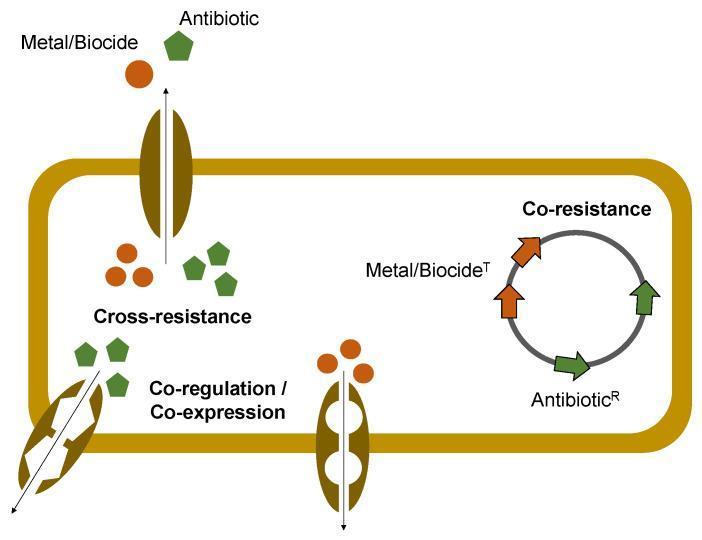
This pathophysiology diagram illustrates the three primary mechanisms of bacterial co-selection for antimicrobial resistance (AMR) triggered by metals and biocides. A bacterial cell is shown with three distinct genetic and functional pathways: 1) Cross-resistance, depicted by a single transmembrane efflux pump protein transporting both 'Metal/Biocide' (orange circles) and 'Antibiotic' (green pentagons), suggesting a shared resistance mechanism. 2) Co-resistance, represented by a circular plasmid or genetic element containing linked genes for 'Metal/Biocide T' (tolerance) and 'Antibiotic R' (resistance), showing how selective pressure for one leads to the retention of the other. 3) Co-regulation / Co-expression, shown by linked transmembrane transporters that are jointly activated by a common regulatory pathway. The diagram highlights how environmental stressors, such as biocides or heavy metals, can inadvertently select for antibiotic-resistant bacteria through these physiological and genetic linkages.
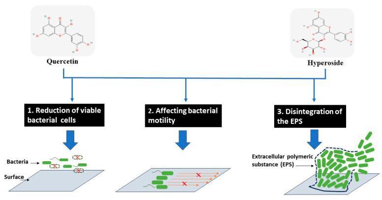
This medical pathophysiology diagram illustrates the antimicrobial and antibiofilm modes of action for two polyphenolic compounds, Quercetin and Hyperoside, against bacterial cells. The diagram is organized into three primary mechanisms of action: 1. Reduction of viable bacterial cells, depicted by rod-shaped bacteria on a surface with several cells marked by red 'X's indicating cell death or non-viability. 2. Affecting bacterial motility, shown by bacterial cells with flagella whose movement paths are interrupted by red 'X's, representing the inhibition of swimming or swarming movements. 3. Disintegration of the Extracellular Polymeric Substance (EPS), visualized by a disrupted dashed boundary around a bacterial cluster, signifying the breakdown of the protective biofilm matrix. The top of the diagram displays the chemical structures of Quercetin (a flavonoid) and Hyperoside (quercetin-3-O-galactoside). This visual summarizes how these flavonoids interfere with bacterial colonization, surface attachment, and the structural integrity of biofilms, which is clinically relevant for developing strategies to overcome antibiotic resistance and foodborne pathogens.
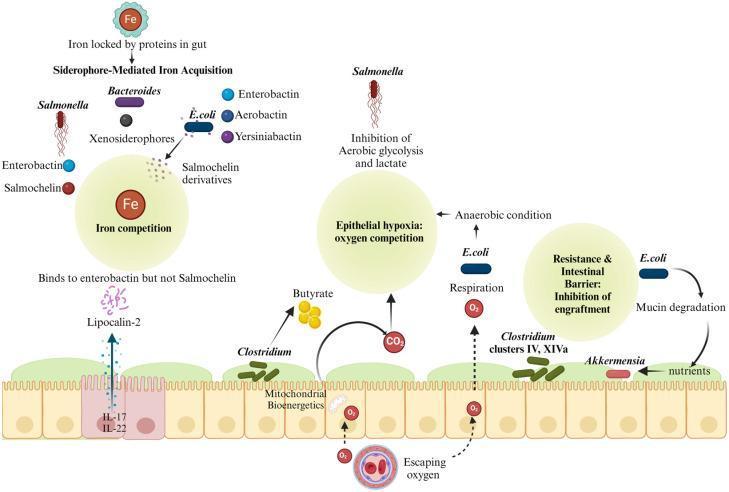
This medical illustration depicts the mechanisms of colonization resistance and ecocompetition within the human gut microbiome between commensal bacteria and pathogenic Salmonella. The diagram is divided into three functional areas: siderophore-mediated iron acquisition, epithelial hypoxia/oxygen competition, and mucosal barrier resistance. In the iron acquisition section, the diagram shows Salmonella producing salmochelin to bypass the host antimicrobial protein Lipocalin-2 (which binds enterobactin), while commensal E. coli and Bacteroides compete for iron using various siderophores like aerobactin and yersiniabactin. Centrally, the diagram illustrates how Clostridium-derived butyrate fuels mitochondrial bioenergetics in colonocytes, consuming oxygen and maintaining epithelial hypoxia. This anaerobic state, supplemented by E. coli respiration, inhibits the aerobic glycolysis and growth of Salmonella. Finally, the intestinal barrier section shows commensal E. coli degrading mucin to support beneficial bacteria like Akkermensia, collectively inhibiting Salmonella engraftment and translocation. The schematic emphasizes the symbiotic role of the microbiota in maintaining intestinal homeostasis and nutrient sequestration to prevent pathogen proliferation.
gram positive gram negative bacteria cell wall structure comparison
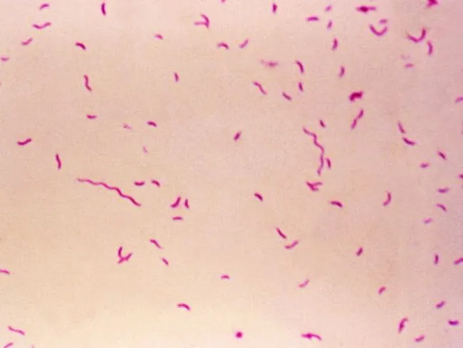
Bright-field light microscopy of a Gram-stained bacterial smear reveals numerous small, slender, curved Gram-negative rods with a characteristic gull-wing appearance consistent with Campylobacter species. The image captures bacteria oriented individually and in small clusters against a pale pink counterstain, illustrating Bacillary morphology typical of enteric pathogens. Gram staining shows purple/blue rods (crystal violet retained by cell wall) with a light pink background from the counterstain (safranin), enabling discrimination from Gram-positive organisms. The organisms appear slender and curved, with a single polar flagellum suggested by motility or alignment in the smear. Specimen type is a bacterial smear obtained from a gastrointestinal sample (feces or culture isolate), prepared for diagnostic microbiology. The imaging modality is bright-field microscopy at high magnification (approximately 1000x with oil immersion), following Gram staining to highlight bacterial cell wall structure. Clinically, detection of Campylobacter species supports infectious gastroenteritis; in the IPSID (immunoproliferative small intestinal disease) context, Campylobacter involvement has been proposed as a pathogenic trigger mirroring Helicobacter pylori's role in gastric MALT lymphoma. Differential diagnoses include Helicobacter, Vibrio, and other curved Gram-negative bacteria. This image serves educational and diagnostic utility for microbiology, clinical pathology, gastroenterology, and infectious disease research.
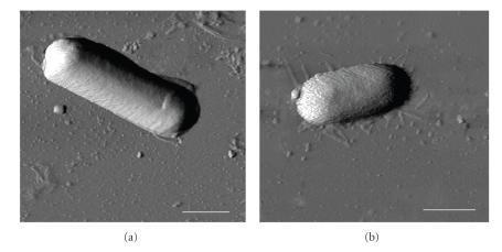
This diagnostic imaging set displays Atomic Force Microscopy (AFM) phase images of two distinct bacterial species, Bacillus cereus (a) and Escherichia coli (b), deposited on a mica surface. The images facilitate a morphological comparison between Gram-positive and Gram-negative bacteria at the nanometer scale. Panel (a) shows B. cereus as a distinctly elongated, rod-shaped cell with a relatively uniform, smooth surface texture and well-defined edges. In contrast, panel (b) depicts E. coli with a more compact, oval or capsule-shaped morphology. The E. coli cell exhibits a significantly rougher surface topography with visible granularity compared to the smoother envelope of B. cereus. The background depicts the mica substrate with faint thread-like structures visible around the E. coli cell, likely representing extracellular filaments or surface dehydration artifacts. A 1 μm scale bar is provided in both panels. These images are used in microbiology to study bacterial cell wall structural integrity, surface roughness (Rq), and biophysical responses to environmental conditions like relative humidity (RH).
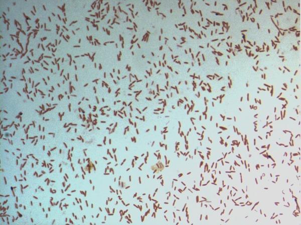
This diagnostic image shows a light microscopy view of a bacterial culture following Gram staining. The specimen displays numerous small, rod-shaped (bacilli) bacteria distributed across the field. The microorganisms exhibit a consistent pink-to-red hue, identifying them as Gram-negative. This staining characteristic indicates a cell wall structure with a thin peptidoglycan layer and an outer membrane that does not retain the crystal violet stain. The arrangement of the bacilli is predominantly individual or scattered, with occasional small clusters and pairs. Specifically identified as D. fastidiosa strain JC13T, this image serves as a primary microbiological reference for the morphological and staining properties of this novel genus within the family Erysipelotrichaceae. The visual demonstrates key diagnostic features including cell morphology, arrangement, and Gram reaction, which are essential for clinical microbiology classification and initial pathogen identification.
Microbiology Essentials for Clinical Pharmacists
1. Bacterial Classification - The Foundation of Drug Selection
Gram Stain: Your First Clue
| Feature | Gram-Positive | Gram-Negative |
|---|---|---|
| Cell wall | Thick peptidoglycan, no outer membrane | Thin peptidoglycan + lipopolysaccharide (LPS) outer membrane |
| Gram stain color | Purple (retains crystal violet) | Pink/red (counterstained with safranin) |
| Outer membrane | Absent | Present - acts as a drug permeability barrier |
| Examples | S. aureus, Streptococci, Enterococci | E. coli, Klebsiella, Pseudomonas, Acinetobacter |
Morphology Categories
- Cocci: spherical bacteria (e.g., Staphylococcus, Streptococcus, Enterococcus)
- Bacilli: rod-shaped (e.g., E. coli, Klebsiella, Pseudomonas)
- Spirochetes: helical (Treponema pallidum, Borrelia burgdorferi)
- Atypical bacteria: lack cell walls or have unusual structures (Mycoplasma, Chlamydia, Rickettsia) - these are intrinsically resistant to beta-lactams
Key Clinical Pathogens You Must Know by Organism Class
- Staphylococcus aureus (including MRSA) - skin, bone, endocarditis, bacteremia
- Streptococcus pneumoniae - pneumonia, meningitis, otitis media
- Streptococcus pyogenes (Group A Strep) - pharyngitis, necrotizing fasciitis
- Enterococcus faecalis / faecium (including VRE) - UTI, endocarditis
- E. coli - UTI, intra-abdominal, bacteremia
- Klebsiella pneumoniae - pneumonia, UTI, liver abscess
- Proteus mirabilis - UTI, wound
- Enterobacter spp. - HAP, VAP, urinary
- Pseudomonas aeruginosa - VAP, wound infections, sepsis in immunocompromised
- Acinetobacter baumannii - ICU-acquired pneumonia, often pan-resistant
- Clostridioides difficile - antibiotic-associated diarrhea/colitis
- Listeria monocytogenes - meningitis in immunocompromised, neonates, elderly
- Mycoplasma pneumoniae, Chlamydophila pneumoniae, Legionella pneumophila - community-acquired pneumonia; only covered by macrolides, fluoroquinolones, tetracyclines
2. Antimicrobial Classes and Mechanisms of Action
| Class | Mechanism | Spectrum | Key Drugs |
|---|---|---|---|
| Beta-lactams (penicillins, cephalosporins, carbapenems, monobactams) | Inhibit cell wall synthesis (bind PBPs) | Broad; carbapenem = widest | Ampicillin, piperacillin-tazobactam, ceftriaxone, meropenem |
| Glycopeptides | Inhibit cell wall synthesis (bind D-Ala-D-Ala) | Gram-positive only | Vancomycin, teicoplanin |
| Aminoglycosides | Inhibit protein synthesis (30S ribosome) | Gram-negative + synergy for gram-positive | Gentamicin, amikacin, tobramycin |
| Fluoroquinolones | Inhibit DNA gyrase/topoisomerase IV | Broad (gram-negative + atypicals) | Ciprofloxacin, levofloxacin, moxifloxacin |
| Macrolides | Inhibit protein synthesis (50S ribosome) | Gram-positive + atypicals | Azithromycin, clarithromycin |
| Tetracyclines | Inhibit protein synthesis (30S ribosome) | Broad including atypicals | Doxycycline, tigecycline, minocycline |
| Oxazolidinones | Inhibit protein synthesis (50S, initiation) | Gram-positive (incl. MRSA, VRE) | Linezolid, tedizolid |
| Lipopeptides | Disrupt cell membrane | Gram-positive (incl. MRSA, VRE) | Daptomycin |
| Polymyxins | Disrupt outer membrane | Gram-negative (last resort) | Colistin, polymyxin B |
| Nitroimidazoles | DNA strand breakage | Anaerobes, protozoa | Metronidazole |
| Azoles / Echinocandins / Polyenes | Antifungal mechanisms (ergosterol, glucan synthesis, membrane binding) | Fungal pathogens | Fluconazole, caspofungin, amphotericin B |
3. Antimicrobial Resistance Mechanisms - The Most Critical Topic
The Four Main Mechanisms:
- Porin loss or mutation: Gram-negative bacteria use protein channels (porins) to allow drug entry. Loss of OprD in Pseudomonas confers resistance to imipenem specifically.
- Efflux pumps: Energy-dependent transporters expel antibiotics. Five major families: MFS, MATE, SMR, RND, ABC transporters. MexA-MexB-OprM in Pseudomonas raises MICs for beta-lactams and aminoglycosides broadly.
- Beta-lactamases: Over 3,000 variants exist. Categories critical for pharmacists:
- Broad-spectrum (TEM, SHV) - inhibited by clavulanate, sulbactam, tazobactam
- ESBLs (e.g., CTX-M): Hydrolyze third-generation cephalosporins. Treat with carbapenems or ceftazidime-avibactam
- AmpC beta-lactamases: Not inhibited by classic inhibitors; cefepime or carbapenems required
- Carbapenemases (KPC, NDM, VIM, IMP, OXA): Hydrolyze carbapenems - the most feared resistance. MDR organisms require novel agents (ceftazidime-avibactam, meropenem-vaborbactam, cefiderocol)
- Aminoglycoside-modifying enzymes: AAC (acetyltransferases), ANT (adenylyltransferases), APH (phosphotransferases) - inactivate aminoglycosides before they reach the ribosome
- mecA gene in MRSA: produces PBP2a, a modified penicillin-binding protein with low affinity for all beta-lactams - this is why all beta-lactams fail in MRSA regardless of disk sensitivity
- VanA/VanB in VRE: modifies the D-Ala-D-Ala terminus to D-Ala-D-Lac, eliminating vancomycin binding
- Quinolone resistance: Mutations in DNA gyrase (GyrA) and topoisomerase IV (ParC) reduce fluoroquinolone binding
- Overproduction of target enzymes
- Biofilm formation (physical protection, tolerates extremely high antibiotic concentrations)
Multidrug-Resistant (MDR) Organisms to Know:
- MRSA (Methicillin-Resistant S. aureus) - treat with vancomycin, daptomycin, linezolid, ceftaroline
- VRE (Vancomycin-Resistant Enterococcus) - treat with linezolid, daptomycin, tigecycline
- ESBL-producing Enterobacterales - treat with carbapenems (or ceftazidime-avibactam if carbapenem-sparing needed)
- CRE/CPE (Carbapenem-Resistant Enterobacterales) - ceftazidime-avibactam, meropenem-vaborbactam, cefiderocol, colistin (last resort)
- CRAB/CRPA (Acinetobacter and Pseudomonas) - often require combination therapy
4. Laboratory Tests You Must Be Able to Interpret
Culture and Sensitivity (C&S) Reports
- Gram stain: Gives you morphology and gram-reaction within 1 hour
- Culture: Final organism identification takes 24-72 hours
- Susceptibility Testing: Results reported as S (susceptible), I (intermediate/susceptible-dose-dependent), or R (resistant)
MIC and Breakpoints - From [Goodman & Gilman]:
- Higher MIC within the susceptible range = need higher doses or prolonged infusion strategies
- MIC 1 dilution below breakpoint = consider dose optimization before treating as susceptible
Rapid Diagnostics
- Molecular/PCR tests: Detect resistance genes directly from samples in under 2 hours (e.g., mecA gene for MRSA, rifampin resistance in M. tuberculosis)
- Blood cultures: Minimum 2 sets before antibiotics; positive results change management in ~20-30% of bacteremia cases
- Beta-D-glucan / Galactomannan: Fungal biomarkers for invasive candidiasis and aspergillosis
- Procalcitonin (PCT): Supports antibiotic de-escalation decisions in sepsis
5. PK/PD Principles for Antimicrobial Dosing
| PK/PD Type | Predictor of Efficacy | Examples | Clinical Implication |
|---|---|---|---|
| Time-dependent | % Time > MIC | Beta-lactams | Extended/continuous infusion improves outcomes |
| Concentration-dependent | Cmax/MIC | Aminoglycosides, fluoroquinolones | Once-daily dosing maximizes kill, reduces toxicity |
| AUC-dependent | AUC/MIC (fAUC) | Vancomycin, fluoroquinolones | Target AUC/MIC ≥ 400-600 for vancomycin (guideline-recommended) |
Special Dosing Scenarios:
- Renal impairment: Dose-adjust aminoglycosides, vancomycin, beta-lactams (except nafcillin), fluconazole, antiviral agents
- Hepatic impairment: Dose-adjust metronidazole, clindamycin, chloramphenicol, antifungals
- Obesity: Weight-based dosing adjustments for aminoglycosides (use adjusted body weight), vancomycin, daptomycin
- CNS infections: Use agents with high CNS penetration (meropenem, ampicillin for meningitis); avoid those that don't cross the blood-brain barrier (cefazolin)
- Critically ill patients: Volume of distribution is altered - loading doses matter more; augmented renal clearance can sub-therapeutically dose renally-cleared drugs
6. Common Infection Syndromes and Empiric Coverage
| Infection | Common Pathogens | Empiric Drug(s) |
|---|---|---|
| Community-acquired pneumonia (mild) | S. pneumoniae, Mycoplasma, Chlamydophila | Amoxicillin ± macrolide, or respiratory fluoroquinolone |
| CAP (severe, ICU) | Above + Legionella, S. aureus | Beta-lactam + macrolide or fluoroquinolone |
| Healthcare-associated pneumonia (HAP/VAP) | Pseudomonas, MRSA, Enterobacterales | Antipseudomonal beta-lactam + MRSA coverage |
| Uncomplicated UTI | E. coli, Klebsiella | Nitrofurantoin, TMP-SMX (check local resistance) |
| Complicated UTI/pyelonephritis | Same + Proteus | Ceftriaxone, ciprofloxacin, or pip-tazo |
| Intra-abdominal infection | Polymicrobial (GNR + anaerobes) | Pip-tazo, or ceftriaxone + metronidazole |
| Sepsis (unknown source) | Broad empiric | Pip-tazo or carbapenem; add MRSA coverage if risk factors |
| Skin/soft tissue (non-purulent) | Streptococcus | Beta-lactam (e.g., cefazolin) |
| Skin/soft tissue (purulent) | S. aureus incl. MRSA | TMP-SMX, doxycycline; IV vancomycin if severe |
| C. difficile infection (CDI) | Clostridioides difficile | Fidaxomicin (preferred) or vancomycin PO |
| Candida bloodstream | Candida spp. | Echinocandin (caspofungin, micafungin) empirically |
7. Antimicrobial Stewardship - Your Core Professional Role
- Prospective audit and feedback: Review antimicrobial prescriptions, flag inappropriate use, recommend de-escalation once culture data is available
- Formulary restriction and preauthorization: Gate-keep broad-spectrum and last-resort agents (carbapenems, linezolid, daptomycin)
- Applying the 5 D's: Right Drug, correct Dose, right Delivery route, appropriate Duration, timely De-escalation to pathogen-directed therapy
- IV-to-oral switch: Identify patients stable enough to switch from IV to oral antibiotics (saves costs, reduces CLABSI risk)
- Therapeutic drug monitoring (TDM): Particularly for vancomycin (target AUC 400-600), aminoglycosides (target troughs and peaks), voriconazole (target trough 1-5.5 mg/L)
8. Key Infection-Specific Drug Knowledge
Fungal Infections
- Candida: Echinocandins first-line for candidemia; fluconazole step-down after susceptibility confirmed and clinical stability
- Aspergillus: Voriconazole or isavuconazole first-line for invasive disease
- Cryptococcus (in HIV): Amphotericin B + flucytosine induction, then fluconazole consolidation
- Drug interactions alert: Azoles inhibit CYP3A4/2C9 (fluconazole > voriconazole > posaconazole > isavuconazole); monitor for drug-drug interactions with immunosuppressants, anticoagulants, statins
Viral Infections
- HIV: Understand antiretroviral classes (NRTIs, NNRTIs, PIs, INSTIs) and resistance genotyping
- Herpes viruses: Acyclovir (HSV/VZV), ganciclovir (CMV - dose-adjust in renal impairment, myelosuppressive)
- Influenza: Oseltamivir (start within 48h); neuraminidase resistance (H275Y mutation) can develop
- Hepatitis B/C: Direct-acting antivirals - high cure rates, but significant drug interactions (especially HCV drugs)
Parasites
- Malaria: Know chloroquine resistance patterns; artemisinin-based combinations for P. falciparum
- Toxoplasma: Pyrimethamine + sulfadiazine (+ leucovorin for bone marrow protection)
9. Infection Control and Prevention Concepts
- Standard precautions: Apply to all patients; hand hygiene is the single most effective intervention
- Transmission-based precautions:
- Contact (MRSA, VRE, C. difficile) - gown and gloves; note that C. diff spores are NOT killed by alcohol gel (use soap and water)
- Droplet (Influenza, Meningococcal, RSV)
- Airborne (TB, measles, varicella) - N95 required
- Surgical prophylaxis: First- or second-generation cephalosporins (cefazolin is the standard); administer within 60 minutes of incision; discontinue within 24 hours
- Biofilm: Staphylococcus epidermidis and S. aureus form biofilm on prosthetic devices and catheters - requires removal of hardware plus prolonged therapy; antibiotics alone rarely cure biofilm infections
10. Key Microbiology Concepts for Specific Drug Decisions
| Scenario | What to Know |
|---|---|
| Beta-lactam allergy | Cross-reactivity between penicillins and cephalosporins is low (~1-2%); carbapenems also very low. Side-chain similarity matters more than core structure. Avoid all if anaphylaxis/severe reaction |
| MRSA bacteremia | Do NOT use daptomycin for pneumonia (inactivated by surfactant); vancomycin or ceftaroline for lung involvement |
| ESBL infection | Do NOT use cephalosporins even if susceptible by disk - inoculum effect; use carbapenems |
| Enterobacter / Serratia with AmpC | Do NOT use third-generation cephalosporins (inducible AmpC) - treat with cefepime or carbapenems |
| C. difficile infection | Stop offending antibiotics; do NOT use systemic antibiotics that reach the colon in active CDI; fidaxomicin preferred for recurrence prevention |
| Daptomycin for VRE | Check prior vancomycin exposure; can cause daptomycin resistance through membrane adaptation |
Summary: The Clinical Pharmacist's Microbiology Checklist
- Classify the organism from the report: gram stain morphology, genus/species
- Know the resistance phenotype: MRSA? ESBL? CPE? What enzymes are present?
- Read the MIC, not just S/I/R: A high MIC in the susceptible range demands dose optimization
- Match PK/PD to the bug: Use extended infusion for beta-lactams against high-MIC gram-negatives
- De-escalate when culture data is back - don't stay on empiric broad coverage indefinitely
- Check source control: No antibiotic works if an abscess isn't drained or a line isn't removed
- Verify duration: Most infections have defined courses; prolonged courses select for resistance and cause harm
- Drug interactions: Azoles, linezolid (serotonin syndrome), rifampin (CYP inducer) - always reconcile