
Monomophic Ventricular Tachykardia management
"monomorphic ventricular tachycardia"[MeSH Terms] AND management
monomorphic ventricular tachycardia ECG wide complex
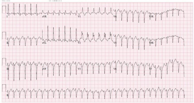
A 12-lead electrocardiogram (ECG) demonstrating a regular wide complex tachycardia (WCT). The rhythm is characterized by a rapid ventricular rate and broad QRS complexes, indicating delayed ventricular depolarization. Key morphological findings include a Right Bundle Branch Block (RBBB) pattern, notably visible as an RSR' or notched morphology in the right precordial leads (V1, V2). There is significant left axis deviation (LAD), with predominantly negative QRS complexes in the inferior leads (II, III, and aVF) and positive complexes in leads I and aVL. This combination of RBBB morphology and left axis deviation in the context of wide complex tachycardia is a classic presentation of idiopathic left ventricular tachycardia, also known as Belhassen tachycardia or verapamil-sensitive fascicular ventricular tachycardia. The rhythm appears monomorphic, and the baseline shows no clearly identifiable P waves, suggesting atrioventricular dissociation or retrograde conduction common in ventricular arrhythmias.
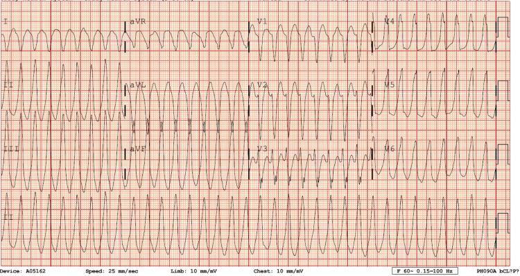
This diagnostic image is a 12-lead electrocardiogram (ECG) demonstrating a regular, monomorphic wide-complex tachycardia (WCT). The ventricular rate is approximately 250 beats per minute, characterized by widened QRS complexes that maintain a consistent morphology across each lead. P waves are not clearly discernible, and there is no obvious evidence of atrioventricular (AV) dissociation on this tracing, making it visually challenging to differentiate between ventricular tachycardia (VT) and supraventricular tachycardia (SVT) with aberrant conduction. Notably, the limb leads aVR and aVL exhibit signs of potential lead reversal, complicating axis determination. The rhythm is monomorphic, suggesting a uniform ventricular depolarization pathway. This ECG is a critical educational example used to illustrate the emergency department presentation of tachyarrhythmias, often requiring clinical correlation and potentially electrical cardioversion. It serves as a teaching tool for advanced cardiac life support (ACLS) and cardiology-level interpretation of life-threatening arrhythmias.
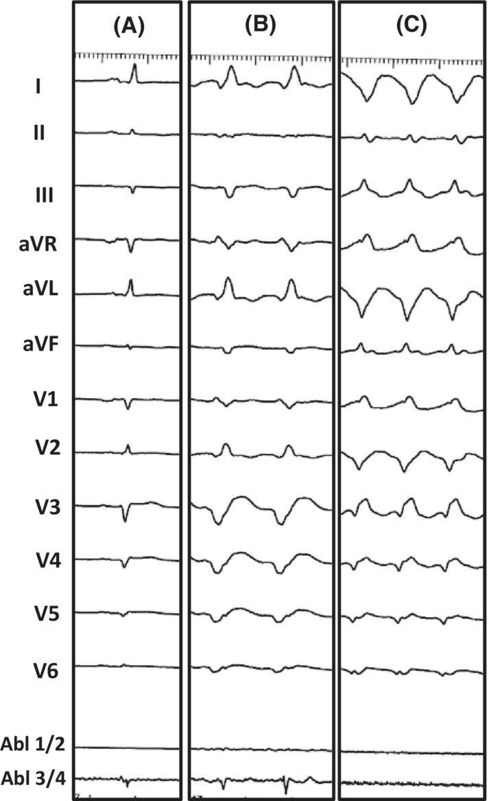
This diagnostic image consists of three vertical panels (A, B, and C) displaying 12-lead electrocardiogram (ECG) tracings alongside intracardiac ablation catheter signals (Abl 1/2, Abl 3/4). Panel A represents the patient's baseline ECG, showing narrow QRS complexes and normal sinus rhythm. Panel B illustrates Ventricular Tachycardia (VT) Morphology 1, characterized by a wide-complex tachycardia with a right bundle branch block (RBBB) morphology in V1 and a predominantly positive axis in lead I. Panel C displays VT Morphology 2, which shows a different wide-complex tachycardia pattern with a more discordant QRS morphology across the precordial leads and higher frequency compared to Panel B. The image serves as an educational tool for comparing baseline cardiac rhythms with different morphologies of monomorphic ventricular tachycardia in the context of electrophysiology studies and catheter ablation. It demonstrates clinical features such as QRS widening, secondary ST-T wave changes, and morphology shifts indicative of different ventricular exit sites during a VT storm.
Monomorphic Ventricular Tachycardia (MVT) - Management
Definition & ECG Recognition
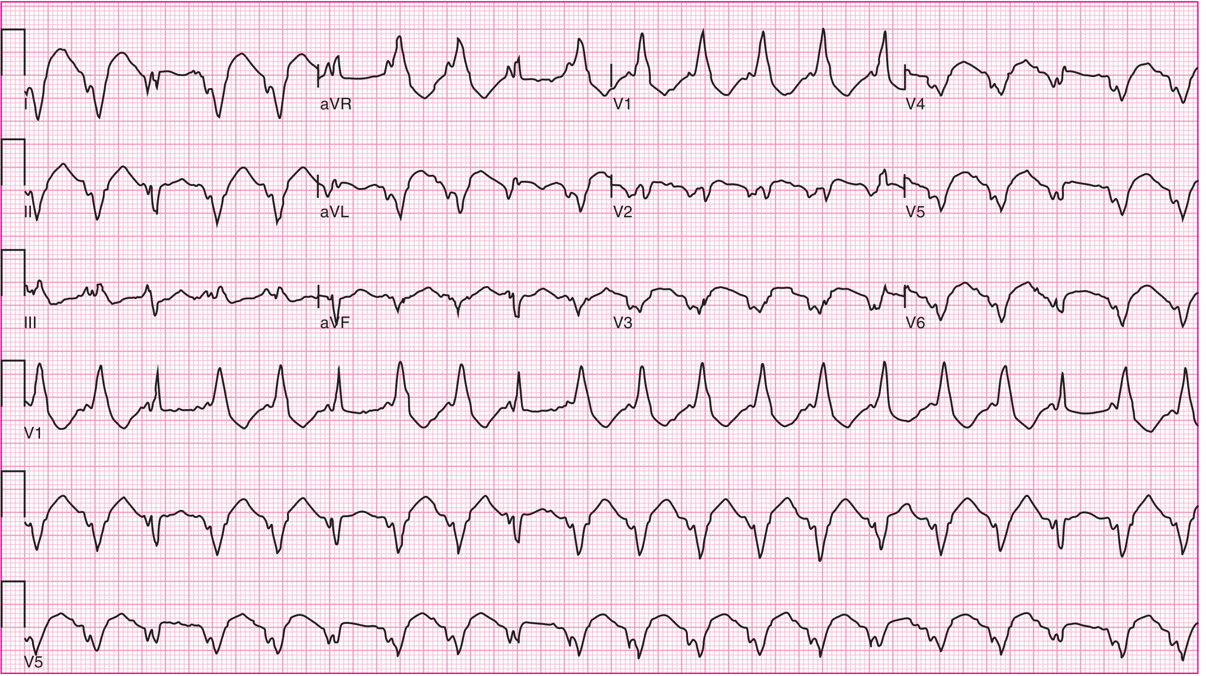
- Regular wide QRS tachycardia (rate typically 120-300 bpm)
- AV dissociation (P waves march independently from QRS)
- Fusion beats (narrow QRS intermixed - pathognomonic)
- Capture beats
Acute Management
Step 1: Assess Hemodynamics Immediately
| Status | Action |
|---|---|
| Hemodynamically UNSTABLE (hypotension, impaired consciousness, pulmonary edema) | Immediate synchronized DC cardioversion (after sedation if patient is conscious) |
| Hemodynamically STABLE | Obtain 12-lead ECG; proceed to pharmacologic termination |
- Defibrillation pads/external defibrillator must be connected immediately - VT can degenerate to VF at any time, including in response to drugs.
- If diagnosis uncertain in stable patient: IV adenosine bolus under ECG monitoring (transiently blocks AV/VA conduction to clarify rhythm).
Step 2: Pharmacologic Termination (Stable MVT)
| Drug | Dose | Notes |
|---|---|---|
| Procainamide (1st choice) | 10 mg/kg (up to 1000 mg) IV over 20 min | Superior to amiodarone for acute termination in randomized trial; avoid in end-stage renal disease (metabolite NAPA accumulates → QT prolongation → polymorphic VT) |
| Amiodarone | 150 mg IV over 10 min | More effective at slower rates; better for preventing recurrence after SR restored; vasodilator - up to 30% hypotension |
| Lidocaine | 50 mg IV bolus | Less effective than amiodarone; more effective at faster rates; good for acute ischemia-related VT; does NOT prevent recurrences |
| Beta-blockers (IV) | e.g., metoprolol or esmolol | Particularly useful for idiopathic VTs without structural disease |
| Verapamil / Diltiazem IV | Only if idiopathic LV fascicular (Belhassen) VT is certain | CONTRAINDICATED in structural heart disease VT - risk of hemodynamic collapse |
Key point: IV calcium channel blockers are contraindicated in MVT unless idiopathic fascicular VT is confirmed in a patient WITHOUT structural heart disease. - Braunwald's Heart Disease
Step 3: Correct Reversible Precipitants
- Hypokalemia / hypomagnesemia
- Hypoxia / acidosis
- Elevated sympathetic tone (pain, anxiety, acute illness)
- Acute heart failure
- Myocardial ischemia (check ECG + troponins - though acute MI is rarely the cause of sustained MVT)
- Proarrhythmic drugs, beta-agonists, milrinone
Electrical Storm
- ACLS - cardioversion for each episode
- IV Amiodarone
- Reduce sympathetic tone (most important):
- Sedation → escalate to general anesthesia if needed
- Beta-blockade: propranolol (non-selective) shown superior to metoprolol in one study
- Check ICD function (if implanted): confirm appropriate sensing, pacing, rule out proarrhythmic medications
- Stellate ganglion block or high thoracic epidural anesthesia
- Emergent catheter ablation if refractory
- Mechanical circulatory support: IABP, axial pump, percutaneous VAD, or ECMO if hemodynamically compromised
- Consider LVAD or transplant workup if refractory
Long-Term / Definitive Management
Evaluation After First Presentation
- 12-lead ECG in sinus rhythm (look for prior MI, cardiomyopathy pattern)
- Echocardiography or cardiac MRI - assess LV/RV function and scar
- Coronary angiography if ischemic substrate suspected
- Genetic workup if no clear ischemic cause (~40% of non-ischemic cardiomyopathies are genetic)
- Rule out ARVC, cardiac sarcoidosis (especially for RV-origin VT)
Long-Term Management by Substrate
1. Structural Heart Disease (Ischemic or Non-Ischemic)
- Clearly indicated for secondary prevention (sustained VT or hemodynamically significant VT)
- ICDs reduce annual mortality from 12.3% to 8.8% and lower arrhythmic deaths by ~50% vs antiarrhythmic drugs alone - Harrison's 22E
- Antiarrhythmic drugs alone (without ICD) do not improve survival
- Recommended for patients failing or intolerant of antiarrhythmic drugs (strongest indication)
- The VANISH trial showed catheter ablation superior to escalating antiarrhythmic drug doses
- BERLIN VT trial: preventive ablation not shown superior to deferred strategy, but fewer VT recurrences
- Considered when long-term amiodarone therapy (with its toxicities) is not desired
- Emergent catheter ablation for incessant VT / electrical storm refractory to drugs
- Role: reduce ICD therapies (shocks + ATP), improve quality of life
- Amiodarone: most effective but significant long-term toxicity
- Sotalol: alternative
- Beta-blockers: guideline-directed medical therapy for all patients with HF/CAD + VT
- ACE inhibitors: standard heart failure therapy, important for VT substrate reduction
2. Idiopathic VT (No Structural Heart Disease)
- Cardiac arrest from idiopathic VT transforming to VF is exceedingly rare
- Catheter ablation is preferred - potentially curative
- Beta-blockers (metoprolol 50 mg BID, atenolol 50 mg OD): safe first-line for symptomatic PVCs/NSVT but not very effective
- ICD generally not indicated unless sustained VT causes syncope
- Fascicular (Belhassen) VT: verapamil-sensitive; IV verapamil terminates acutely; catheter ablation is curative long-term
- If PVC/NSVT burden is very high → risk of tachycardia-induced cardiomyopathy → aggressive ablation warranted even without symptoms
Summary Algorithm
MVT Diagnosed
     │
     ├─ Hemodynamically UNSTABLE?
     │        └─ YES → Synchronized DC Cardioversion (sedate if conscious)
     │
     └─ STABLE?
              └─ IV Procainamide (preferred) or Amiodarone or Lidocaine
                       │
                       ├─ Terminated? → Correct precipitants
                       │                 Evaluate for structural heart disease
                       │                 Long-term: ICD ± Ablation ± AAD
                       │
                       └─ Refractory / Electrical Storm?
                                 → IV Amiodarone + Beta-blocker
                                 → Reduce sympathetic tone (sedation/GA)
                                 → Emergent catheter ablation
                                 → Mechanical support (IABP/ECMO) if needed
Recent Evidence (2024-2025)
- A 2025 meta-analysis (PMID 39542872) found that ischemia testing and coronary revascularization does not reduce mortality or VT recurrence in MVT without acute coronary syndrome - supporting the guideline position that revascularization is not routinely indicated for MVT substrate control.
- A 2024 systematic review of VT trials (PMID 38821759) highlights catheter ablation as the most actively investigated intervention, reflecting its growing role over antiarrhythmic drugs.