
Thyroid from surgery
What aspect of thyroid surgery are you interested in?
surgical anatomy of the thyroid gland
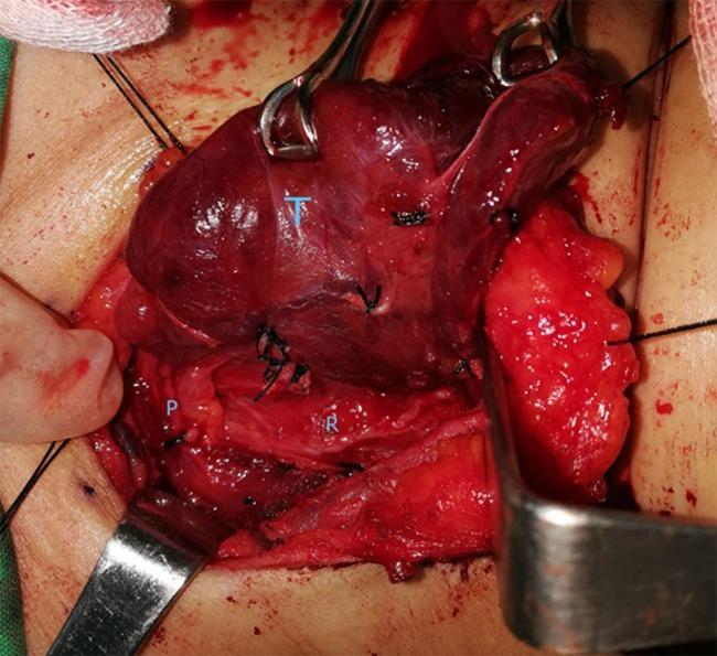
This clinical photograph displays an intraoperative view of the neck during a thyroidectomy procedure, focusing on the surgical anatomy of the thyroid bed before the application of methylene blue. The primary anatomical structures are labeled for educational identification: the thyroid gland (T) is visible as a superiorly located, reddish-brown vascular mass held by surgical forceps; the recurrent laryngeal nerve (R) is identified as a thin, pale, linear structure in the tracheoesophageal groove inferior to the thyroid lobe; and a parathyroid gland (P) is shown as a small, yellowish-tan nodule situated adjacent to the nerve and posterior thyroid capsule. The surgical field includes various instruments, such as retractors and forceps used for tissue manipulation, and black silk sutures are visible throughout the dissection site. This image serves as a clinical anatomical guide for identifying critical structures that must be preserved during endocrine surgery to prevent vocal cord paralysis or hypocalcemia. It illustrates the standard surgical topography and the natural appearance of these tissues in an active surgical field.
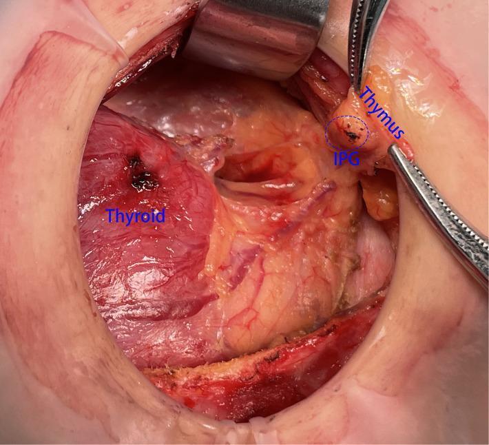
This clinical photograph shows an intraoperative view of a thyroidectomy or neck dissection, highlighting the surgical anatomy of the thyroid gland, thymus, and inferior parathyroid gland (IPG). The thyroid gland is a large, reddish, vascularized structure visible on the left side. Surgical forceps are used to retract the thymus, an elongated, yellowish-tan tissue located superior and lateral to the thyroid. The IPG is visible as a small, distinct ovoid nodule closely associated with the superior aspect of the thymic capsule, circled and labeled in blue. This visual demonstrates the 'in situ reservation' technique, where the IPG and thymus are preserved as an organic unit to maintain parathyroid blood supply during central lymph node dissection. The surgical field displays exposed soft tissue, retractor placement, and visible vasculature, providing educational context for endocrine surgeons and residents regarding the preservation of parathyroid function.
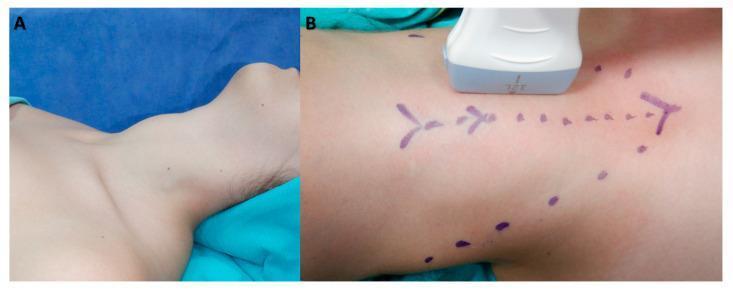
This composite of clinical photographs illustrates the preoperative preparation and intraoperative evaluation for thyroid surgery. Panel A shows a patient in the supine position under general anesthesia, with the neck extended to provide optimal exposure of the anterior cervical region. The visible anatomy includes the lower mandible, the submental and anterior neck areas, and the superior thoracic inlet. Panel B demonstrates the application of an 8–12 MHz linear ultrasound transducer on the skin surface for the ultrasonographic evaluation of the thyroid gland. Purple surgical ink markings on the neck denote the midline and key anatomical landmarks or intended incision pathways, including a dotted line and directional arrows. This sequence highlights the importance of patient positioning and the use of intraoperative ultrasonography for precise volumetric measurement and surgical planning in endocrine surgery, such as total thyroidectomy.
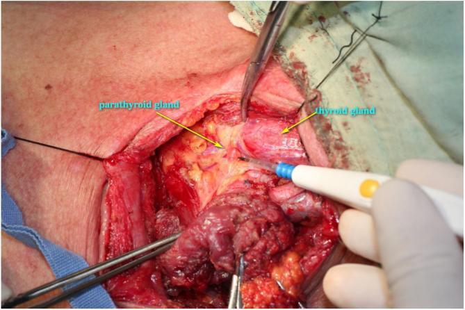
This clinical photograph displays an intraoperative view of a deep surgical dissection in the left lateral neck during a fistula repair procedure. The strap muscles have been retracted or partially removed to expose the visceral compartment of the neck. Key anatomical landmarks are identified with labels: the thyroid gland is visible as a reddish, vascularized lobular structure, and a parathyroid gland is indicated as a smaller, yellowish-tan nodule located on the posterior aspect of the thyroid. The surgical field also demonstrates the recurrent laryngeal nerve pathway and surrounding adipose tissue. Surgical instruments are in use, including an electrocautery probe held by a gloved hand and hemostatic clamps for tissue retraction. The image serves as an educational reference for surgical anatomy of the thyroid and parathyroid glands, nerve preservation techniques, and the management of cervical fistulae. This material is relevant for surgical training in otorhinolaryngology and endocrine surgery.
Surgical Anatomy of the Thyroid Gland
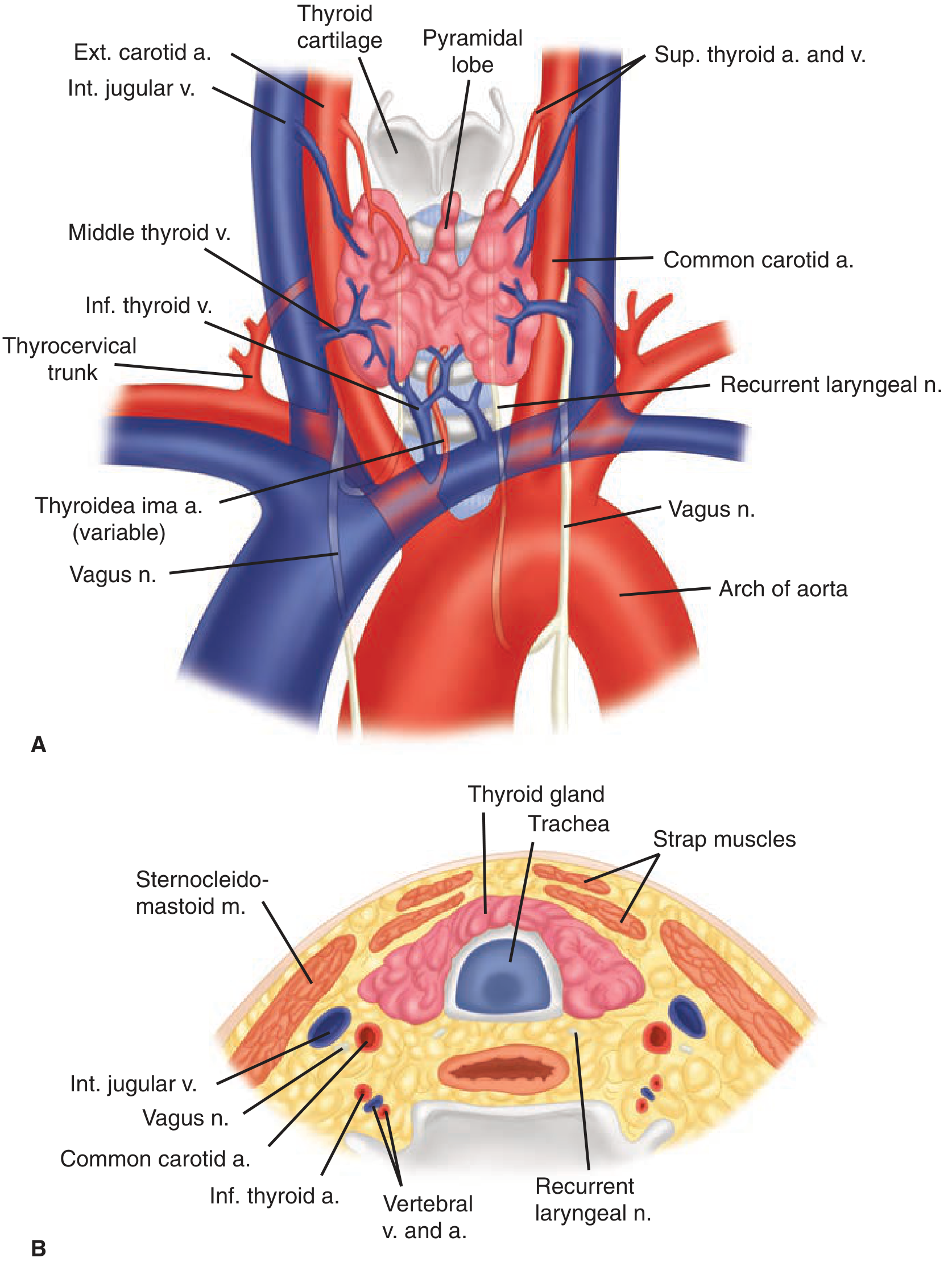
Gross Anatomy and Position
- Anterior: sternothyroid and sternohyoid strap muscles
- Lateral: medial aspects of the carotid sheath (common carotid artery, internal jugular vein, vagus nerve)
- Posterior: trachea and esophagus (especially left side)
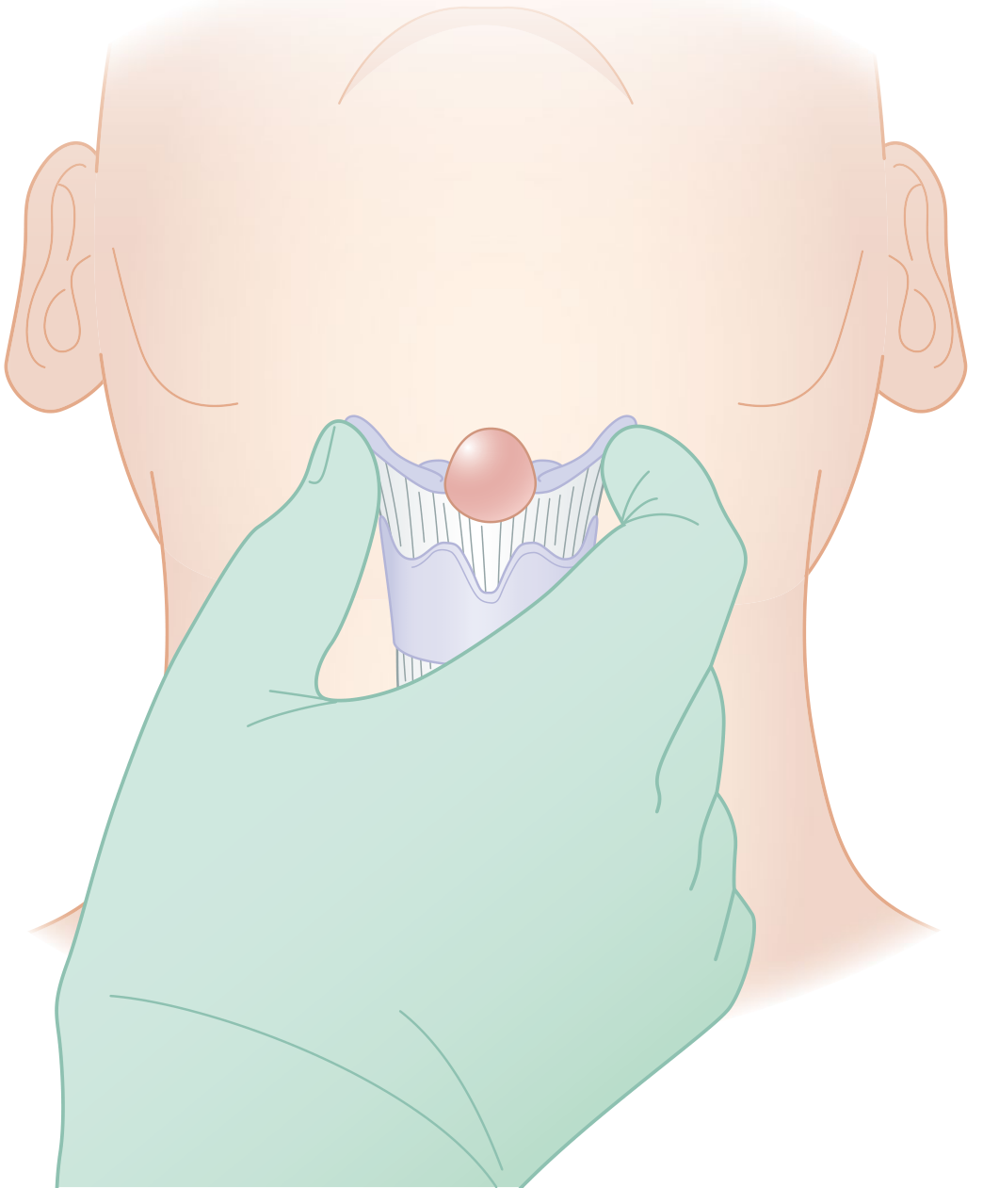
Ligament of Berry - Critical Surgical Point
- The recurrent laryngeal nerve (RLN) and its terminal branches are intimately associated here
- Branches of the RLN traverse this ligament in ~25% of individuals and are particularly vulnerable to injury
- The tubercle of Zuckerkandl (the posterolateral extension of the thyroid lobe at the level of the cricoid) must be mobilized to identify the RLN before dividing the ligament. - Schwartz's Principles of Surgery, p. 1655
Blood Supply
Arterial
| Artery | Origin | Course |
|---|---|---|
| Superior thyroid a. | External carotid a. | Descends along inferior pharyngeal constrictor; divides into anterior and posterior branches at the superior pole |
| Inferior thyroid a. | Thyrocervical trunk (from subclavian a.) | Courses medially deep to carotid sheath; enters the thyroid at its midpoint; crosses the RLN |
| Thyroidea ima a. | Directly from aorta or innominate | Present in 1-10% of individuals; enters the isthmus or replaces a missing inferior thyroid artery; may cause significant blood loss if unrecognized |
Venous Drainage
- Superior thyroid vein: drains with the superior thyroid artery → internal jugular vein
- Middle thyroid vein: least consistent; drains → internal jugular vein. May be torn during medial mobilization if unrecognized.
- Inferior thyroid veins: form a plexus → brachiocephalic (innominate) veins
Nerves - Most Critical Structures in Thyroid Surgery
1. Recurrent Laryngeal Nerve (RLN)
- Left RLN: arises from vagus at the aortic arch, loops around the ligamentum arteriosum, ascends medially in the tracheoesophageal groove
- Right RLN: arises from vagus at its crossing with the right subclavian artery; more oblique course; slightly more variable
- Unilateral: ipsilateral vocal cord palsy - paramedian position = weak but nearly normal voice; abducted position = hoarse voice + ineffective cough
- Bilateral: airway obstruction requiring emergency tracheostomy or loss of voice
2. External Branch of the Superior Laryngeal Nerve (EBSLN)
- Lies on the inferior pharyngeal constrictor and descends alongside the superior thyroid vessels before innervating the cricothyroid muscle
- The Cernea classification describes its relationship to the superior thyroid vessels:
- Type 1: crosses ≥1 cm above the superior pole (safest)
- Type 2a: crosses <1 cm above the pole (occurs in ~20% - at higher risk)
- Type 2b: crosses below the superior pole (highest risk)
- Therefore, the superior pole vessels must never be ligated en masse - they should be individually divided low on the thyroid capsule to protect this nerve
- Injury causes loss of ability to tense the ipsilateral vocal cord - difficulty projecting, voice fatigue, inability to hit high notes
3. Internal Branch of the Superior Laryngeal Nerve
- Sensory to the supraglottic larynx; injury may result in aspiration (rare in standard thyroid surgery)
Parathyroid Glands - Anatomical Relationship
- Superior parathyroids: lie posterior (deep) to the plane of the RLN, classically near the cricothyroid junction ~1 cm superior to where the RLN crosses the inferior thyroid artery
- Inferior parathyroids: lie anterior (superficial) to the plane of the RLN; more variable in position and more prone to inadvertent devascularization
- Blood supply to both: primarily from inferior thyroid artery branches (~80%); ~20% of superior glands receive supply from the superior thyroid artery
Lymphatics
- Prelaryngeal, pretracheal, and bilateral paratracheal lymph nodes
- Bounded: hyoid bone (superior) → innominate artery (inferior) → carotid sheaths (lateral)
- Delphian lymph nodes = prelaryngeal nodes just above the isthmus; when positive for metastases, they signal adverse tumor characteristics
Intraoperative Nerve Monitoring
Embryologic Anomalies Relevant to Surgery
| Anomaly | Surgical Implication |
|---|---|
| Thyroglossal duct cyst | Requires Sistrunk procedure (with midportion of hyoid bone) |
| Lingual thyroid | May be the only thyroid tissue - check before removing |
| Ectopic thyroid | Can harbor cancer; lateral neck thyroid tissue = nodal metastasis, not ectopic tissue |
| Non-recurrent laryngeal nerve | Associated with aberrant right subclavian artery - detectable on preoperative CT |
- Identify the RLN before ligating any inferior thyroid artery branches
- Mobilize the tubercle of Zuckerkandl to expose the RLN near the ligament of Berry
- Individually ligate superior pole vessels close to the capsule to protect the EBSLN
- Dissect along the thyroid capsule to preserve parathyroid blood supply
- Be alert for non-recurrent RLN if preoperative imaging shows aberrant subclavian artery
Thyroglossal duct cyst
thyroglossal duct cyst neck midline
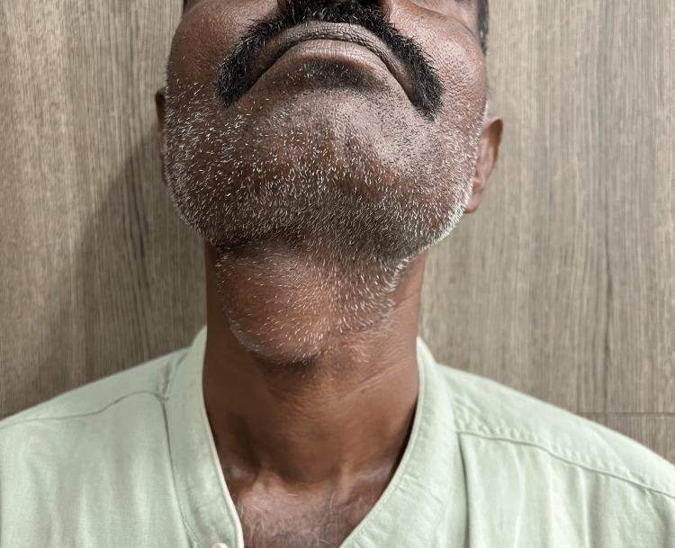
A clinical photograph of an adult male depicting a prominent midline neck mass, consistent with a thyroglossal duct cyst. The patient's neck is slightly extended, revealing a well-circumscribed, ovoid, and globular swelling located in the anterior midline of the neck, specifically at the level of the hyoid bone and inferior to the submental region. The overlying skin is intact and appears unremarkable, without signs of erythema, inflammation, ulceration, or fistulous tracts. This visual presentation is characteristic of a congenital thyroglossal duct anomaly, which typically manifests as a painless, mobile midline mass that often moves superiorly with tongue protrusion or swallowing. The image serves as a clinical example for Otolaryngology and General Surgery education, emphasizing the anatomical localization and physical characteristics necessary for the differential diagnosis of midline neck swellings, such as dermoid cysts or ectopic thyroid tissue.
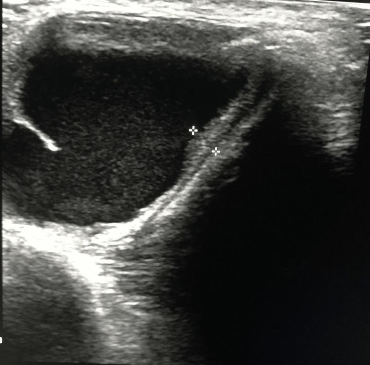
This diagnostic ultrasound image in the transverse plane depicts a large, well-circumscribed thyroglossal duct cyst in the anterior midline of the neck. The cyst is primarily anechoic to hypoechoic, indicating a clear fluid-filled interior, and exhibits smooth, well-defined borders. A key anatomical feature is the presence of an associated thyroglossal tract extending from the cyst wall, which is demarcated by two electronic calipers (plus-sign markers). This tract appears as a linear, moderately echogenic structure extending superiorly toward the hyoid bone region. A small, focal echogenic projection or internal debris is visible along the left inner margin of the cyst. The image demonstrates typical sonographic findings used to diagnose thyroglossal duct anomalies and evaluate their relationship to surrounding midline neck structures, which is essential for pre-surgical planning of a Sistrunk procedure.
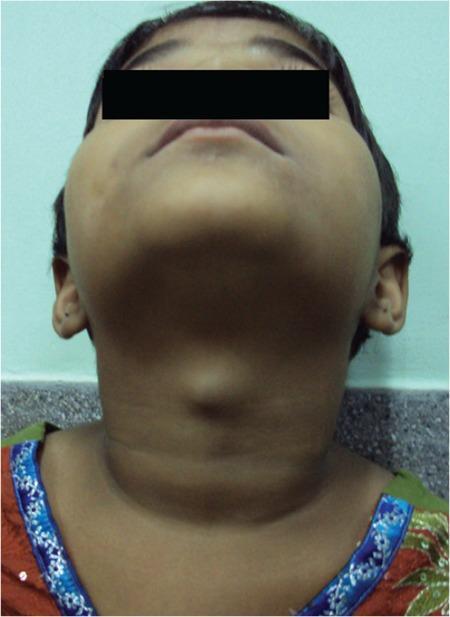
Clinical photograph of a pediatric patient's neck, viewed from an inferior-anterior perspective (looking up toward the chin). The image demonstrates a prominent, well-circumscribed, midline cystic swelling in the anterior neck, located superior to the thyroid cartilage. The overlying skin appears normal with no visible erythema, inflammation, or ulceration. The morphology is characteristic of a thyroglossal duct cyst, a common congenital abnormality occurring along the descent pathway of the thyroid gland. In this clinical context, such a mass often contains ectopic functional thyroid tissue and typically moves upward upon protrusion of the tongue or swallowing. The visual findings illustrate a classic presentation of a midline neck mass in childhood, serving as an educational example for differential diagnosis in pediatric otolaryngology and endocrinology, specifically regarding ectopic thyroid tissue and thyroglossal duct remnants.
Thyroglossal Duct Cyst (TDC)
Embryology
Epidemiology
- The most common congenital cystic neck mass
- Second most common benign neck mass overall (after benign lymphadenopathy)
- Predominantly a pediatric condition, though can present in adults
- Classic teaching: "An upper midline cystic neck lesion in a toddler is a TDC until proven otherwise" - Sabiston, p. 117
Clinical Presentation
| Feature | Details |
|---|---|
| Location | Midline (75%) or up to 2 cm off-midline (25%) |
| Position | At or just below hyoid bone (65% infrahyoid, 15% at hyoid level) |
| Character | Painless, soft, smooth, well-circumscribed cystic mass |
| Mobility | Moves cranially with swallowing or tongue protrusion - pathognomonic sign |
| Size | Typically 1-4 cm |
| Symptoms | Usually asymptomatic; can become infected → pain, redness, swelling |
| Rare complications | Dysphagia, airway obstruction |
Clinical Photos
Pathology (Robbins)
- Cysts are 1-4 cm, lined by:
- Stratified squamous epithelium when near the base of tongue
- Pseudostratified columnar epithelium in lower (cervical) locations
- Transitional patterns also occur
- The fibrous cyst wall often contains lymphoid aggregates and thyroid remnants
- Malignant transformation of the lining epithelium is exceedingly rare - Robbins Pathologic Basis of Disease, p. 694
Diagnosis
Ultrasound (first-line)
- Appearance varies:
- Classic: homogeneous anechoic cyst with posterior wall enhancement
- Pseudosolid: due to proteinaceous secretions
- Heterogeneous: due to prior hemorrhage or infection
- Key imaging points:
- Relationship to the hyoid bone
- Presence of normal thyroid tissue inferiorly (rule out lingual thyroid as only thyroid tissue before resecting)
- Any solid component within the cyst (1% risk of carcinoma)
CT / MRI
- MRI: invariably high T2 signal; T1 variable (depends on protein content)
- CT: useful for anatomical planning pre-operatively
FNA
- Can be performed to rule out malignancy or other neck masses in atypical presentations - Sabiston, p. 1343
Critical pre-operative step: Always confirm a normally located thyroid gland is present before surgery, especially in children. In lingual thyroid, the ectopic tongue-base thyroid may be the only functioning thyroid tissue.
Treatment: The Sistrunk Procedure
- The cyst itself
- The complete thyroglossal duct tract
- The central portion (body) of the hyoid bone - en bloc
- The tract superior to the hyoid bone extending to the base of the tongue / foramen cecum
- Simple cystectomy alone → recurrence in 40-50% of cases
- The Sistrunk procedure reduces recurrence to <5%
- Sabiston Textbook of Surgery, p. 117
Malignancy in TDC
- Occurs in approximately 1-1.5% of thyroglossal duct cysts
- Papillary thyroid carcinoma (PTC) is the most common type
- Most cases: Sistrunk procedure alone is adequate
- More extensive surgery or radioactive iodine is considered in high-risk patients:
- Age >55
- Suspicious lymph nodes
- Tumor >4 cm
- Soft tissue extension
- Cummings Otolaryngology, p. 2237
Differential Diagnosis of Midline Neck Mass
| Condition | Distinguishing Feature |
|---|---|
| TDC | Moves with tongue protrusion; at/below hyoid |
| Dermoid cyst | Does NOT move with tongue protrusion; doughy consistency |
| Lingual thyroid | At base of tongue; may be only thyroid tissue |
| Ectopic thyroid in neck | Central compartment; solid on imaging |
| Submental lymph node | Firm; may be reactive/tender |
| Lipoma | Soft, mobile, subcutaneous |