
Polyarthritis disease wise in detail information about investigation and examination
polyarthritis joint examination rheumatoid arthritis hand deformity
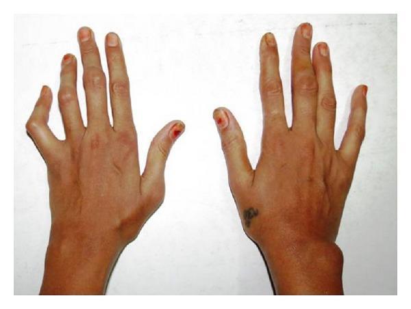
Clinical photograph of the dorsal aspect of bilateral hands and wrists in a patient with Rheumatoid Arthritis (RA). The image demonstrates characteristic chronic inflammatory joint deformities. On the left hand, there is a prominent swan-neck deformity of the fifth digit, characterized by hyperextension of the proximal interphalangeal (PIP) joint and flexion of the distal interphalangeal (DIP) joint. Swelling is visible across the interphalangeal joints of the third and fourth digits. The right hand shows a significant, localized soft tissue swelling on the lateral/ulnar aspect of the wrist joint, consistent with synovial hypertrophy or a rheumatoid nodule. General architectural changes include joint stiffness and minor deviations of the fingers. This visual illustrates the progressive peripheral joint involvement and resulting anatomical deformities typical of systemic inflammatory polyarthritis, emphasizing the clinical signs used in diagnosing the severity of RA.
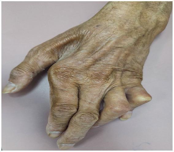
This clinical photograph displays a dorsal view of a human hand exhibiting advanced chronic polyarthritis and joint deformities associated with leprosy. Key visual features include significant ulnar deviation of the digits at the metacarpophalangeal joints and a prominent Boutonniere deformity of the fourth digit, characterized by flexion at the proximal interphalangeal (PIP) joint. The joints appear swollen and misaligned, suggesting a presentation that mimics rheumatoid arthritis. The skin is notably thin, wrinkled, and atrophic with visible underlying vasculature and patchy hyperpigmentation, consistent with age-related changes or long-term chronic disease. The nails show mild scaling and thickening. This image serves as an educational example of how late-stage leprosy can present with severe musculoskeletal manifestations and joint destruction, requiring differentiation from primary rheumatologic conditions.
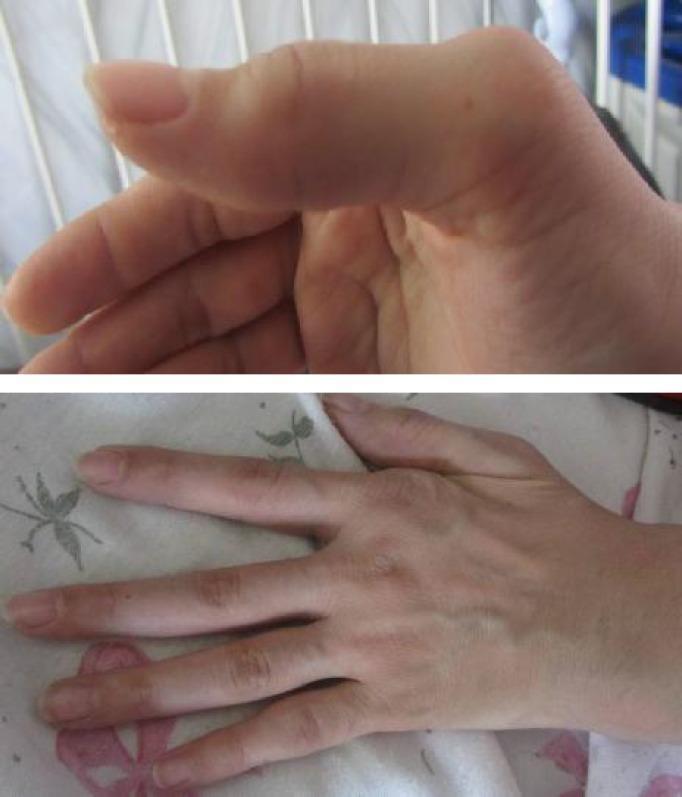
Clinical photographs of a hand demonstrating chronic structural deformities characteristic of rheumatoid arthritis. The upper image provides a lateral view focusing on a digit showing a prominent deformity at the proximal interphalangeal (PIP) joint and enlargement of the metacarpophalangeal (MCP) joint. The lower image provides a dorsal view of the hand, illustrating significant ulnar deviation of the fingers at the MCP joints, which is a classic hallmark of progressive inflammatory arthritis. There is visible soft tissue swelling and joint enlargement at the 2nd metacarpal and multiple PIP joints. The images exemplify the long-term musculoskeletal manifestations of active rheumatoid arthritis, showcasing joint misalignment and chronic inflammatory changes in the small joints of the hand. This content is suitable for teaching clinical signs of autoimmune rheumatological diseases and physical examination findings in hand pathology.
rheumatoid arthritis radiograph erosions investigation
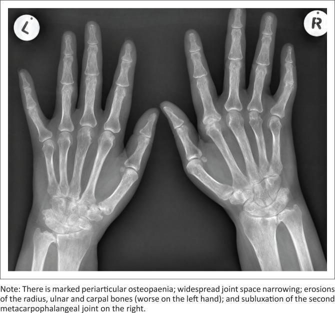
A frontal (posteroanterior) radiograph of both hands and wrists, demonstrating classic radiographic features of Rheumatoid Arthritis. The image shows bilateral, relatively symmetrical involvement. Key findings include marked periarticular osteopenia, most notable around the metacarpophalangeal (MCP) and proximal interphalangeal (PIP) joints. Extensive cortical erosions are visible within the carpal bones, distal radius, and ulna, with more advanced destruction appearing on the left side. There is widespread, uniform narrowing of the joint spaces throughout the carpal, MCP, and PIP joints. Additionally, subluxation is noted at the second MCP joint of the right hand, indicating significant ligamentous and articular instability. The distribution and nature of these findings (erosions, osteopenia, symmetry, and joint narrowing) are highly characteristic of chronic inflammatory erosive arthritis. This diagnostic image serves as an educational example for identifying structural damage and joint malalignment in systemic rheumatologic conditions.
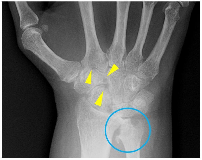
This diagnostic image is a posteroanterior (PA) radiograph of the wrist and proximal hand in a 57-year-old female patient with rheumatoid arthritis. The imaging demonstrates classic features of chronic inflammatory arthritis. Scattered marginal erosions are visible within the carpal bones, specifically highlighted by yellow arrowheads, indicating loss of cortical integrity in the 'bare areas' of the joints. A blue circle highlights the distal radius and ulna, which exhibit moderate peri-articular demineralization. This demineralization is characterized by reduced radiopacity and a sparse, less distinct trabecular pattern compared to healthy bone. The visual findings illustrate the progression of rheumatoid arthritis, where synovial inflammation leads to hyperemia and subsequent bone resorption and erosion before significant joint space narrowing is observed. These features are clinically significant for the assessment of disease activity and structural damage in rheumatological disorders.
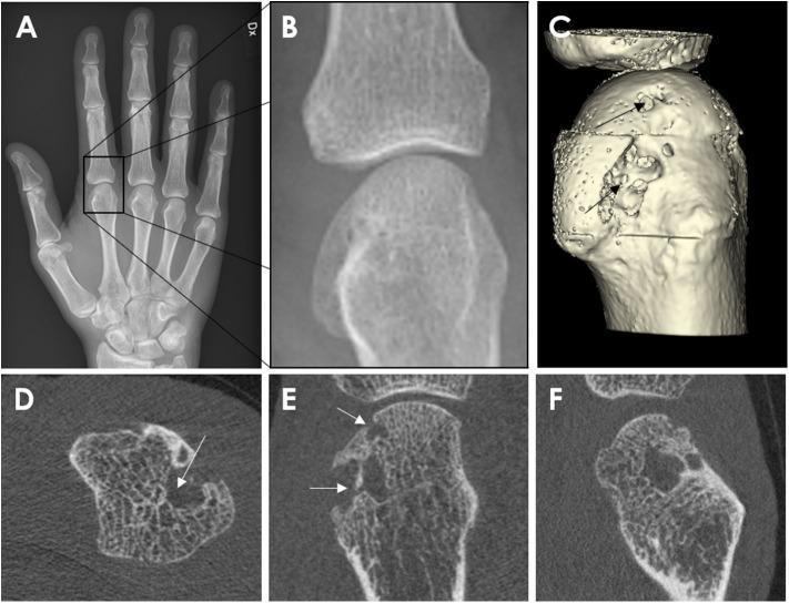
This composite diagnostic image demonstrates the utility of different imaging modalities in identifying bone erosions in a 62-year-old male with rheumatoid arthritis. Panel A is an anterior-posterior (AP) radiograph of the right hand, with a magnified view (B) focusing on the second metacarpophalangeal (MCP) joint. Panels C-F utilize High-Resolution Peripheral Quantitative Computed Tomography (HR-pQCT). Panel C shows a 3D surface reconstruction of the metacarpal head, where arrows highlight deep, pitted cortical erosions and irregular surface morphology. Panels D (axial), E (coronal), and F (sagittal) provide cross-sectional HR-pQCT views, revealing significant discontinuity in the cortical shell and the subsequent loss of underlying trabecular bone structure. These images illustrate the superior sensitivity of HR-pQCT over conventional radiography in detecting and quantifying juxta-articular erosions, which appear as non-linear breaks in the cortical bone in multiple perpendicular planes. The visual data emphasizes the pathological bone remodeling and microstructural degradation characteristic of advanced inflammatory arthritis.
Polyarthritis: Disease-Wise Examination & Investigations
General Approach to Any Polyarthritis
History (Seven Dimensions)
| Feature | Significance |
|---|---|
| Onset (acute vs. gradual) | Acute = septic/crystal/viral; Gradual = RA, CTD |
| Pattern (symmetric vs. asymmetric) | Symmetric = RA, SLE; Asymmetric = SpA, PsA |
| Joint distribution | Small = RA; Large + axial = SpA |
| Morning stiffness >60 min | Inflammatory arthritis (RA, SpA) |
| Gel phenomenon (short-lived stiffness) | Osteoarthritis |
| Axial involvement | Spondyloarthritis |
| Fever, rash, GI, genital, ocular sx | Systemic diseases (SLE, reactive, Behcet) |
| Travel history | Chikungunya, Lyme, brucellosis |
| Sexual history | Gonococcal arthritis, reactive arthritis |
| Family history | RA, psoriasis, AS |
Physical Examination Principles
- Swelling: soft (synovial) vs. hard (osteophyte/OA)
- Redness: strongly suggests septic arthritis or gout (rarely in RA/SLE)
- Warmth: better indicator than redness in chronic inflammatory disease
- Tenderness: palpation of joint line vs. periarticular
- Squeeze test: lateral compression of MCP/MTP - more specific for inflammatory arthritis but low sensitivity
- Range of motion: active vs. passive restriction
1. Rheumatoid Arthritis (RA)
Clinical Examination
- Symmetric synovitis of MCPs, PIPs, wrists, MTPs (spares DIPs)
- "Squeeze test" positive at MCP/MTP joints
- Wrist: Carpal tunnel, extensor tenosynovitis, piano-key sign (ulnar head instability)
- Late deformities: ulnar deviation, swan-neck, boutonniere, Z-thumb
- Forefoot: MTP subluxation, hammer toes, calluses under metatarsal heads
- Atlantoaxial subluxation (C1-C2) - check for myelopathic signs
- Rheumatoid nodules (extensor surfaces, olecranon)
- Sicca (secondary Sjogren)
- Scleritis, episcleritis
- Pleural/pericardial rub
- Peripheral neuropathy, mononeuritis multiplex (rare)
- Splenomegaly + neutropenia = Felty's syndrome
Investigations
- RF (IgM): positive in ~80% established RA; also positive in HCV, SLE, Sjogren - not specific alone
- Anti-CCP (ACPA): sensitivity ~70%, specificity ~95% - most useful confirmatory test; can be positive years before symptoms
- ANA: positive in ~30% RA (low titer), not diagnostic
- ESR, CRP: elevated; useful for monitoring disease activity (DAS28 score)
- CBC: normocytic normochromic anemia (ACD), thrombocytosis in active disease, neutropenia (Felty)
- LFT/RFT: baseline before DMARDs
- WBC 5,000-50,000/mm³ (class II, inflammatory)
- Turbid, poor viscosity, low glucose
- No crystals; culture negative
- X-ray hands/feet (bilateral): periarticular osteopenia, symmetric joint space narrowing, marginal erosions (hallmark), ulnar deviation, subluxation
- Ultrasound: detects synovitis and erosions earlier than X-ray; power Doppler shows active vascularity
- MRI: most sensitive for early erosions and synovitis, bone marrow edema
- Joint involvement (0-5)
- Serology - RF/ACPA (0-3)
- Acute phase reactants - CRP/ESR (0-1)
- Duration ≥6 weeks (0-1)
2. Systemic Lupus Erythematosus (SLE)
Clinical Examination
- Arthritis: non-erosive, non-deforming polyarthritis - MCPs, PIPs, wrists, knees
- Jaccoud arthropathy: reducible deformities (no erosions on X-ray)
- Malar (butterfly) rash - spares nasolabial folds
- Photosensitivity, discoid rash, oral ulcers
- Serositis signs: pleuritic rub, pericardial friction rub
- Lymphadenopathy, hepatosplenomegaly
- Neurological: psychosis, seizures, mononeuritis
- Raynaud's phenomenon
- Livedo reticularis, hair loss
Investigations
- ANA: sensitivity >95% - best screening test; if negative, SLE unlikely
- Anti-dsDNA: high specificity (~95%); correlates with disease activity and nephritis
- Anti-Smith (anti-Sm): highest specificity (~99%) but low sensitivity (~30%)
- Anti-Ro/SSA, Anti-La/SSB: neonatal lupus, subacute cutaneous lupus
- Anti-phospholipid antibodies: aCL, anti-β2GP1, lupus anticoagulant (LA)
- Complement C3, C4: low in active disease (consumption); useful for monitoring
- Hemolytic anemia (Coombs positive), leukopenia (<4,000), lymphopenia (<1,000), thrombocytopenia (<100,000) - any of these = diagnostic criteria
- Proteinuria >0.5 g/24h, red cell casts, active urinary sediment = lupus nephritis
- 24h urine protein or spot PCR
- ESR elevated (often very high), CRP usually normal or mildly elevated (except in serositis/infection)
- LFT, RFT
- Skin/renal biopsy when indicated
3. Psoriatic Arthritis (PsA)
Clinical Examination
- 5 clinical patterns: oligoarthritis (most common), symmetric polyarthritis (RA-like), DIP predominant, arthritis mutilans, axial disease
- Dactylitis ("sausage digit"): diffuse swelling of entire digit (highly characteristic)
- Enthesitis: tenderness at Achilles insertion, plantar fascia, tibial tuberosity
- DIP joint involvement (unlike RA)
- Nail changes (80%): pitting (>20 pits = strongly suggestive), onycholysis, subungual hyperkeratosis, oil drop sign
- Psoriatic skin plaques: check scalp, navel, natal cleft, ears
- Asymmetric joint involvement common
- Relative absence of rheumatoid nodules
Investigations
- RF: negative (seronegative arthritis)
- ACPA: negative
- ESR, CRP: elevated in active disease
- ANA: may be mildly positive in some
- Skin biopsy if psoriasis diagnosis uncertain
- HLA-B27: positive in ~50% with axial involvement
- X-ray: characteristic "pencil-in-cup" deformity (arthritis mutilans), periosteal new bone formation (fluffy periostitis), DIP erosions, ankylosis; NO periarticular osteopenia (unlike RA)
- MRI/Ultrasound: enthesitis, dactylitis, axial disease
- CASPAR criteria for classification (sensitivity 91%, specificity 99%)
4. Ankylosing Spondylitis / Axial Spondyloarthritis (AS/axSpA)
Clinical Examination
- Predominantly axial but peripheral arthritis in ~30-50% (lower limb, large joint, asymmetric)
- Modified Schober's test: measure lumbar flexion (<5 cm increase = restricted)
- Tragus-to-wall distance: >15 cm = reduced cervical flexion
- Chest expansion: <2.5 cm (4th intercostal space) = restricted
- FABER test (Flexion, Abduction, External Rotation): sacroiliac joint tenderness
- Sacroiliac joint tenderness on direct pressure
- Loss of lumbar lordosis, thoracic kyphosis
- Enthesitis: Achilles, plantar fascia, costochondral junctions
- Extra-articular: anterior uveitis (acute, unilateral, painful red eye), aortitis (AR murmur), apical lung fibrosis, cauda equina syndrome
Investigations
- HLA-B27: positive in ~90% AS (Caucasian); much lower specificity in populations with high prevalence
- ESR, CRP: elevated in active disease; may be normal in axial-only disease
- RF, ACPA: negative (seronegative)
- ANA: negative
- X-ray pelvis/SI joints: sacroiliitis grading (Grade 0-IV) - Grade II bilateral or Grade III unilateral = modified NY criteria
- X-ray spine: squaring of vertebral bodies, "bamboo spine" (late), Romanus lesions, Anderson lesions
- MRI SI joints: bone marrow edema (STIR sequence) = active sacroiliitis - key for early/non-radiographic axSpA (nr-axSpA)
- BASMI, BASDAI: clinical activity scores
5. Reactive Arthritis (formerly Reiter's Syndrome)
Clinical Examination
- Classic triad: urethritis + conjunctivitis + arthritis ("can't see, can't pee, can't climb a tree")
- Asymmetric oligoarthritis/polyarthritis of lower limb large joints (knees, ankles)
- Dactylitis, enthesitis (Achilles)
- Keratoderma blennorrhagicum: pustular hyperkeratotic rash on palms/soles (virtually pathognomonic)
- Circinate balanitis: painless penile lesion
- Oral ulcers (painless)
- Preceding infection: urogenital (Chlamydia) or gastrointestinal (Salmonella, Shigella, Campylobacter, Yersinia)
- Conjunctivitis, anterior uveitis
Investigations
- Urethral/cervical swab: Chlamydia trachomatis PCR
- Stool culture: enteric pathogens if GI trigger suspected
- Serology: Yersinia, Salmonella, Campylobacter antibodies
- HLA-B27: positive in ~50-80% (associated with severity and chronicity)
- RF, ACPA: negative
- ESR, CRP: elevated
- Synovial fluid: inflammatory (WBC 10,000-50,000), no crystals, culture negative
- Urinalysis: sterile pyuria (in urogenital form)
- X-ray: periostitis, calcaneal spur, sacroiliitis (in chronic/recurrent cases)
6. Viral Arthritis (Parvovirus, Chikungunya, Hepatitis B/C, Rubella)
Clinical Examination
- Parvovirus B19: symmetric RA-like polyarthritis (MCPs, PIPs, wrists); associated with "slapped cheek" rash; self-limiting (<6 weeks)
- Chikungunya: acute onset with high fever + rash + severe polyarthralgia/arthritis; can persist for months/years; mimics seronegative RA
- Hepatitis C: erosive polyarthritis + mixed cryoglobulinemia; positive RF (up to 50%)
- Hepatitis B: prodromal immune complex polyarthritis + urticaria before jaundice
- Rubella: symmetric small joint polyarthritis in women; post-vaccination arthritis
Investigations
- Parvovirus B19: IgM antibody (acute), IgG (past infection); PCR in immunocompromised
- Chikungunya: RT-PCR (acute <7 days), IgM/IgG ELISA
- Hepatitis B/C serology: HBsAg, anti-HBc, anti-HCV, HCV RNA
- Rubella IgM/IgG
- ESR, CRP: elevated
- RF: may be positive in HCV-associated arthritis
- Cryoglobulins: HCV arthritis
- Synovial fluid: inflammatory, sterile
7. Gout / Pseudogout (Crystal Arthropathy)
Clinical Examination
- Gout: asymmetric, acute attacks; classic first MTP (podagra), ankles, knees; tophi on ears, olecranon, Achilles
- Pseudogout (CPPD): large joints (knee most common), wrists; older patients; can be polyarticular in acute calcium pyrophosphate arthritis ("pseudorheumatoid")
- Skin overlying joint: erythematous, warm, shiny (acute attack)
- Tophi: subcutaneous collections of urate crystals
Investigations
- Synovial fluid aspiration (GOLD STANDARD):
- Gout: needle-shaped, negatively birefringent crystals (yellow when parallel to slow ray under polarized light)
- Pseudogout: rhomboid-shaped, positively birefringent crystals (blue when parallel)
- Serum uric acid: often normal during acute attack; elevated baseline confirms hyperuricemia
- ESR, CRP: markedly elevated in acute attack
- RF: negative
- X-ray:
- Gout: punched-out erosions with overhanging edge ("rat bite"), soft tissue tophi, preserved joint space (unlike RA)
- Pseudogout: chondrocalcinosis (calcification of fibrocartilage - menisci, triangular fibrocartilage of wrist)
- DECT (Dual Energy CT): detects urate deposits non-invasively; excellent sensitivity
8. Septic Arthritis (Bacterial Polyarthritis)
Clinical Examination
- Usually monoarticular but can be polyarticular in disseminated gonococcal infection (DGI) and immunosuppressed
- DGI: migratory tenosynovitis + polyarthralgia → settles in 1-2 joints; petechial/pustular rash on skin
- Hot, red, swollen joint(s); severe pain
- Fever, rigors, systemic sepsis
- Risk factors: IV drug use, prosthetic joints, diabetes, immunosuppression
Investigations
- Synovial fluid aspiration (URGENT):
- WBC >50,000/mm³ with >90% neutrophils (septic until proven otherwise)
- Gram stain: low sensitivity (~60% gram-positive, ~30% gram-negative)
- Culture and sensitivity (hold up to 7 days)
- Blood cultures: positive in ~50%
- CBC: leukocytosis, neutrophilia
- ESR, CRP: markedly elevated
- Gonococcal: urethral/cervical/rectal/pharyngeal swab + culture; blood cultures often negative in DGI; nucleic acid amplification test (NAAT)
- X-ray: normal early; joint space widening (effusion)
- Ultrasound: confirms effusion, guides aspiration
- MRI: osteomyelitis extension
9. Adult-Onset Still's Disease (AOSD)
Clinical Examination
- Classic triad: quotidian (daily) high fever + salmon-pink evanescent rash (appears with fever) + arthritis
- Polyarthritis: knees, wrists, ankles; can be destructive
- Lymphadenopathy, splenomegaly, hepatomegaly
- Sore throat (often first symptom)
- Serositis (pleuritis, pericarditis)
- Macrophage activation syndrome (MAS) - rare but life-threatening
Investigations
- Serum ferritin: markedly elevated (>10,000 ng/mL = highly suggestive); glycosylated ferritin <20% (normal >50%)
- ESR, CRP, WBC: markedly elevated
- CBC: leukocytosis (WBC >10,000), neutrophilia, anemia
- LFT: elevated transaminases
- RF, ANA, ACPA: negative (seronegative)
- Exclusions: blood cultures (rule out sepsis), viral serology (EBV, CMV, parvovirus), malignancy workup
- Yamaguchi criteria for classification (sensitivity 96%)
10. Sjogren's Syndrome
Clinical Examination
- Primary Sjogren: sicca complex = dry eyes (keratoconjunctivitis sicca) + dry mouth (xerostomia) + arthritis
- Symmetric, non-erosive polyarthritis (RA-like distribution)
- Parotid gland enlargement
- Schirmer's test: <5 mm in 5 min = abnormal
- Rose Bengal/Lissamine green staining: corneal damage
- Vaginal dryness, skin dryness
Investigations
- Anti-Ro/SSA: most sensitive (~70-90%); present in primary Sjogren + SLE
- Anti-La/SSB: more specific for primary Sjogren
- RF: positive in ~70-80%
- ANA: positive (speckled pattern)
- Minor salivary gland biopsy (lip biopsy): focal lymphocytic sialoadenitis with focus score ≥1 = hallmark
- Schirmer test, slit lamp exam
- ESR, CRP, hypergammaglobulinemia
- Urinalysis: renal tubular acidosis
11. Polymyalgia Rheumatica (PMR)
Clinical Examination
- Age >50 (almost always); bilateral shoulder and pelvic girdle pain/stiffness
- Morning stiffness >45 min
- Restricted shoulder abduction on examination
- No true peripheral joint swelling (though mild synovitis can occur at wrists/knees)
- Look for temporal artery tenderness, scalp tenderness, jaw claudication (GCA association)
- Muscle strength: normal (distinguishes from myositis)
Investigations
- ESR >40 mm/hr (often >100): characteristic
- CRP: markedly elevated
- CBC: normocytic anemia, thrombocytosis
- ALP, GGT: mildly elevated
- RF, ANA: negative
- CK: normal (distinguishes from inflammatory myopathy)
- X-ray shoulder/pelvis: often normal
- Ultrasound/MRI: subdeltoid bursitis, bicipital tenosynovitis, glenohumeral synovitis - present in >80%
- Temporal artery biopsy if GCA suspected (giant cell arteritis occurs in 10-40% of PMR)
Summary Table: Key Differentiating Investigations
| Disease | RF | ACPA | ANA | Anti-dsDNA | HLA-B27 | X-ray Hallmark | Synovial Fluid |
|---|---|---|---|---|---|---|---|
| RA | +ve (80%) | +ve (70-80%) | Low titer | - | - | Marginal erosions, periarticular osteopenia | Inflammatory, no crystals |
| SLE | +ve | - | +ve >95% | +ve (60%) | - | Normal/Jaccoud | Inflammatory |
| PsA | -ve | -ve | - | - | +ve (axial) | Pencil-in-cup, periostitis | Inflammatory |
| AS | -ve | -ve | -ve | - | +ve (90%) | Bamboo spine, sacroiliitis | Inflammatory |
| Reactive | -ve | -ve | -ve | - | +ve (50-80%) | Periostitis, calcaneal spur | Inflammatory, sterile |
| Gout | -ve | -ve | - | - | - | Punched-out erosions | Negatively birefringent crystals |
| CPPD | -ve | -ve | - | - | - | Chondrocalcinosis | Positively birefringent crystals |
| Septic | -ve | -ve | - | - | - | Normal early | >50,000 WBC, organisms |
| AOSD | -ve | -ve | -ve | - | - | Normal | Inflammatory |
| Viral | Variable | -ve | - | - | - | Normal | Inflammatory, sterile |
Stepwise Approach to First-Line Investigations in Any Polyarthritis
- CBC - anemia, leukocytosis, thrombocytopenia
- ESR + CRP - inflammatory activity
- RF + ACPA - RA serology
- ANA panel - ANA, anti-dsDNA, anti-Sm, anti-Ro/La (if SLE suspected)
- Uric acid - gout screening
- Urine analysis + PCR - lupus nephritis
- Hepatitis B/C serology - viral arthritis, secondary RF positivity
- Synovial fluid aspiration - crystals, WBC, culture (ESSENTIAL if acute monoarthritis/oligoarthritis)
- X-ray of affected joints - erosions, chondrocalcinosis, sacroiliitis
- HLA-B27 - if spondyloarthritis suspected
- Ferritin - AOSD
- Complement (C3, C4) - SLE