
Explain meningeal haemorrhage
Reading File
Now I'll search the medical library for meningeal haemorrhage using parallel queries.
Finding Sources
Finding Sources
Finding Sources
Good results found. Now let me read the detailed content from the most comprehensive sources in parallel.
Reading File
Reading File
Reading File
Excellent. Now let me read the subdural and subarachnoid content, plus fetch key images in parallel.
Reading File
Reading File
Loading Image
Now let me read more on subdural and subarachnoid haemorrhage content in parallel.
Reading File
Reading File
Reading File
Let me now read the subdural haemorrhage content from Parikh's and the subarachnoid section in parallel.
Reading File
Finding Sources
I now have comprehensive content for a thorough answer. Let me also fetch a subdural image to illustrate.
Reading File
Finding Sources
Reading File
I now have enough comprehensive information from multiple authoritative textbooks to deliver a thorough answer. Let me compile it.
Meningeal Haemorrhage
Meningeal haemorrhage refers to bleeding that occurs in relation to the meninges - the three membranes (dura mater, arachnoid mater, pia mater) that envelop the brain and spinal cord. Depending on the anatomical compartment involved, it is classified into three main types: extradural (epidural), subdural, and subarachnoid haemorrhage.
Anatomical Background
The meninges create distinct potential spaces between the skull and brain:
- Extradural space - between the inner skull and dura mater
- Subdural space - between the dura and arachnoid mater
- Subarachnoid space - between the arachnoid and pia mater (normally filled with CSF)
1. Extradural (Epidural) Haemorrhage
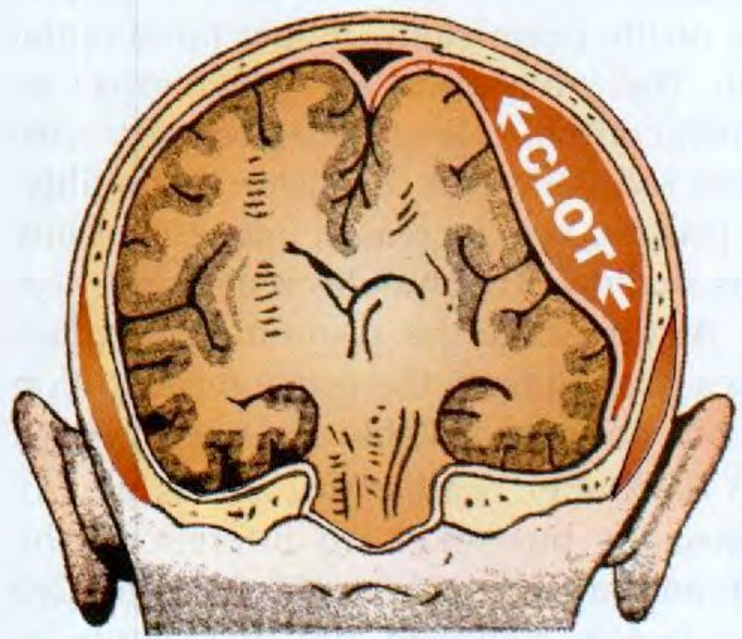
Definition and Frequency
Bleeding between the inner surface of the skull and the dura mater. It is the least common of the three types - Dikshit's Forensic Medicine cites Rowbotham's figure of only 3% of head injuries having epidural haemorrhage.
Mechanism and Source
- Almost always traumatic and strongly associated with skull fracture (except in infants/children, where the dura is more firmly adherent)
- Classical cause: rupture of the middle meningeal artery, most often its anterior branch, at the point it exits the bony canal at the pterion (temporal bone)
- Can also arise from posterior branch of the middle meningeal artery, anterior meningeal vessels (fracture of anterior fossa), internal maxillary artery, or dural venous sinuses
- Acute epidural haemorrhage: arterial in origin (middle meningeal artery rupture)
- Subacute epidural haemorrhage: from torn dural sinuses, middle meningeal veins, or diploic veins; symptoms appear 3+ days after injury
- High-pressure arterial blood progressively strips the dura from the skull, accumulating a haematoma. A minimum of ~35 ml is needed before clinical signs appear, though 100 ml is typically associated with fatalities
Classic Clinical Course (Lucid Interval)
- Initial concussion - loss of consciousness immediately after trauma
- Lucid interval - patient regains consciousness; the dura is slowly stripped by accumulating blood, but ICP remains normal as CSF is displaced into the spinal canal
- Confusion and irritability as ICP rises
- Deteriorating consciousness progressing to coma
- Contralateral motor signs: twitching then paralysis progressing face → arm → leg (as clot spreads over motor cortex from below upwards)
- Ipsilateral pupil: initially constricts then dilates (3rd nerve compression from uncal herniation through tentorial hiatus)
- Eventually bilateral fixed dilated pupils and decerebrate rigidity (midbrain cone)
Note: The classical lucid interval picture is actually not common in acute cases. The more frequent presentation is severe head injury with skull fracture and stupor progressing to deep coma.
Localising the Side
- Side of skull fracture
- Boggy swelling beneath the temporal muscle (ipsilateral)
- Side of initial pupil dilatation
Key Features - S Das, Manual on Clinical Surgery:
- Supratentorial haemorrhage causes uncal herniation through the tentorial hiatus → midbrain compression
- Infratentorial haemorrhage (far less common): elevated BP, slow pulse, irregular respiration, ataxia, nystagmus, lower cranial nerve palsies
Medicolegal Significance
- Good prognosis with prompt surgical drainage (burr hole or craniotomy), but a contralateral haematoma must always be excluded
- Patient may be discharged during the lucid interval and die at home - risk of medical negligence charge
- Clinical picture may resemble drunkenness, leading to missed diagnosis (patient may die in police custody)
- Heat haematoma - a forensic artefact that mimics extradural haemorrhage: blood extruded from diploe/venous sinuses into extradural space during fire; honeycombed, brown, friable, pink due to high carboxyhaemoglobin; distinguishable by comparing COHb levels in haematoma vs peripheral blood
2. Subdural Haemorrhage
Definition and Frequency
Bleeding into the space between the dura and arachnoid membrane. It is 6 times more common than extradural haemorrhage and is one of the most common head injuries ending fatally.
Mechanism and Source
- Results from rapid deceleration of the head, tearing bridging (communicating) veins that connect cortical veins to the dural venous sinuses (especially the superior sagittal sinus)
- These thin, unsupported veins traverse both the subarachnoid and subdural spaces and are particularly vulnerable to rotational movement
- Less often due to cortical vein laceration or injury to dural sinuses
- Not necessarily associated with skull fracture
- Blood accumulates at lower venous pressure than in EDH, so onset is slower
Location
Most common over the convexities of the hemispheres (greatest freedom of brain movement), and less common in the posterior fossa.
Classification
| Type | Timeline | Mechanism | Features |
|---|---|---|---|
| Acute | < 3 days | Venous bleeding, often from severe injury | Lucid interval may be absent; rapid neurological decline |
| Subacute | 3 days - 3 weeks | Smaller bridging vein rupture | Gradual onset; associated with minor contusions |
| Chronic | Weeks to months | Ruptured perforating dural veins in atrophic brain | Common in elderly, alcoholics; mimics dementia |
Chronic subdural haematoma deserves special mention:
- Occurs in elderly and chronic alcoholics with cerebral atrophy - the brain can oscillate within the now-oversized skull, stretching bridging veins
- Often follows trivial or forgotten trauma
- Symptoms: mild confusion, forgetfulness, emotional disorder - easily misdiagnosed as schizophrenia (in young) or dementia (in elderly)
- The haematoma does not resolve because the subdural space lacks mesothelial lining; it becomes encased by inner and outer fibrous membranes
- Susceptible to re-expansion by recurrent bleeding (membrane vessels are thin and poorly endothelialised) and osmotic fluid absorption
- Unsuspected chronic subdural haematoma is a known cause of sudden unexplained death in alcoholics
Gross Appearance by Age
| Age | Appearance |
|---|---|
| Recent (weeks) | Tan/brown, gelatinous membrane |
| Older (months) | Firm, tough bilateral membrane resembling hot-water bottle rubber |
Histological Dating (Dikshit's Forensic Medicine)
| Time | Histological Changes |
|---|---|
| Within 36 hours | Fibroblasts appear at margin of clot |
| By 4 days | Neo-membrane adjacent to dura, few cells thick |
| 5-8 days | Membrane well established; fibroblasts migrate into clot; haemosiderin-laden phagocytes (stain with Perl's Prussian blue reaction) |
| By 8 days | Membrane 12-14 cells thick, visible to naked eye |
| By 11 days | Fibroblast strands subdivide the clot |
| By 15 days | Inner membrane present; outer membrane ½ to ⅓ dural thickness |
| 1-3 months | Membrane loses fibroblastic nuclei, becomes hyaline |
| 6-12 months | Thick, fibrous, resembles dura |
3. Subarachnoid Haemorrhage (SAH)
Definition
Bleeding into the subarachnoid space (between arachnoid and pia mater), where it mixes with CSF. Blood in the subarachnoid space is diluted, less likely to clot, and is more mobile. Haemolysis turns CSF xanthochromic (yellow) within hours to days; within weeks, the blood is absorbed, leaving residual yellow-brown staining of the pia and arachnoid.
Causes
Natural (non-traumatic):
- Rupture of a berry (saccular) aneurysm - most common non-traumatic cause; aneurysms typically occur at bifurcations of the Circle of Willis or its major branches, due to developmental defects in the vessel media
- Arteriosclerotic changes in vessel media, associated with hypertension (in older persons)
- Leaking intracerebral haemorrhage
- Disease states: purpuric states, leukaemia, angioma
Traumatic:
- Cerebral contusions or lacerations
- Explosive blast
- Asphyxia by strangulation
- Traumatic asphyxia
- Damage to vertebral arteries (e.g., fracture of upper cervical vertebrae, blows to the neck)
- Prolonged hyperextension of the head (e.g., during bronchoscopy - may result in SAH over the base of the brain, raising concern for malpractice)
Large collections of blood in the subarachnoid space at the base of the brain are more typical of natural disease; traumatic basal SAH usually involves laceration of the internal carotid, vertebral, or basilar arteries and is likely immediately fatal.
Comparison: Extradural vs. Subdural Haemorrhage
| Feature | Extradural | Subdural |
|---|---|---|
| Incidence | Less common (3% of head injuries) | ~6x more common |
| Source | Middle meningeal artery (arterial) | Bridging veins (venous) |
| Association with fracture | Usually present | Not necessarily |
| Lucid interval | Typically present, clear | Shorter or absent (blood accumulates faster) |
| Speed of compression signs | Later onset | Earlier onset |
| Paralysis pattern | Face → arm → leg (contralateral); then ipsilateral herniation sign | Earlier ipsilateral hemiplegia if unilateral |
| X-ray | May show fracture over middle meningeal groove | Fracture not typical |
| Bilateral | Rare | ~50% bilateral |
| Resolution | Rapid without sequelae; fibroblastic proliferation on dura, no re-bleeding | Does not resolve easily; forms membranes; can re-expand |
Treatment
- Extradural haemorrhage: Surgical emergency - burr hole or craniotomy for evacuation. Must always exclude a contralateral haematoma.
- Subdural haemorrhage: Surgical drainage for symptomatic acute/subacute cases; chronic SDH may be managed conservatively or with burr hole drainage.
- Subarachnoid haemorrhage: Supportive management, aneurysm clipping or coiling for berry aneurysm rupture, nimodipine to prevent vasospasm.
Sources: S Das, A Manual on Clinical Surgery, 13th Ed.; PC Dikshit, Textbook of Forensic Medicine and Toxicology; Parikh's Textbook of Medical Jurisprudence, Forensic Medicine and Toxicology; Grainger & Allison's Diagnostic Radiology
Space occupying lesions within the shull
Reading File
This is a broad medical topic - I'll run parallel searches across headings and paragraphs in the library.
Finding Sources
Finding Sources
Finding Sources
Excellent finds. Now I'll read the most comprehensive dedicated sections in parallel - S Das (surgical perspective), Bailey & Love (raised ICP), and Schwartz's (detailed ICP/SOL).
Reading File
Reading File
Reading File
Good content. Now let me read more of S Das (types of SOL and cone formation), Bailey & Love (investigation and management), and fetch key images.
Reading File
Reading File
Reading File
Let me now get the key sections on types of intracranial tumours, cone formation, management, and also the Robbins pathology content on herniation.
Reading File
Reading File
Finding Sources
Excellent. Now let me fetch the herniation diagram and read the Bailey & Love section on specific SOL types and management.
Loading Image
Reading File
Finding Sources
Let me read the rest of the common brain tumours section and get the Pye's content on SOL types.
Reading File
Reading File
I now have comprehensive content from multiple authoritative textbooks. Let me compile the full answer.
Space-Occupying Lesions (SOL) Within the Skull
A space-occupying lesion (SOL) is any mass within the rigid cranial vault that displaces normal brain tissue and, by progressively increasing intracranial volume, raises intracranial pressure (ICP). The skull cannot expand, so even a small mass will eventually exhaust compensatory mechanisms and compress the brain.
The Monro-Kellie Doctrine
The skull is a rigid, closed compartment with three normal contents:
- Brain tissue (~80%)
- Blood (~10%)
- CSF (~10%)
Normal ICP is 4-14 mmHg. Sustained ICP above 20 mmHg causes brain injury. When a new lesion is added (tumour, abscess, haematoma, etc.), volume is initially compensated by displacement of CSF into the spinal canal and reduction in cerebral blood volume. Once this compensation is exhausted, pressure rises steeply on the pressure-volume curve and herniation becomes imminent. - Schwartz's Principles of Surgery, 11th Ed.
Classification of Intracranial SOLs
1. Neoplasms (Tumours)
Primary Brain Tumours
| Tumour | Features |
|---|---|
| Glioblastoma multiforme (GBM) | Most malignant; adult males; IDH wild-type; WHO Grade IV; median survival ~12 months |
| Astrocytoma | Any age; frontal lobes in adults; cerebral hemispheres in young |
| Medulloblastoma | Childhood (5-15 years); boys > girls; posterior fossa; midline |
| Meningioma | Usually benign; arises from meninges; causes mass effect + vasogenic oedema; may show local scalp thickening with engorged veins or an audible bruit |
| Acoustic neurofibroma (Vestibular schwannoma) | Rare before age 30; cerebellopontine angle; presents with hearing loss, tinnitus, balance disturbance |
| Pituitary adenoma | Basophil adenoma = Cushing's; acidophil adenoma = acromegaly/gigantism; bitemporal hemianopia from optic chiasm compression |
| Oligodendroglioma | Characteristic IDH mutation + 1p/19q co-deletion |
Molecular classification (WHO 2016): Gliomas are now classified by both histology and molecular markers - IDH mutation status and 1p/19q co-deletion define distinct tumour entities with different prognoses and treatment responses. - Bailey & Love's Short Practice of Surgery, 28th Ed.
Secondary (Metastatic) Tumours
Cerebral metastases are the most common intracranial tumours, diagnosed in ~25% of cancer patients. Common primaries include:
- Lung (most common), Breast, Melanoma, Renal cell carcinoma, Colorectal cancer
They typically present acutely with fast-growing mass effect and surrounding oedema.
2. Haematomas
| Type | Space | Source | Key Feature |
|---|---|---|---|
| Extradural haematoma | Skull - dura | Middle meningeal artery | Lucid interval; temporal fracture |
| Subdural haematoma | Dura - arachnoid | Bridging veins | More common; venous; can be chronic |
| Intracerebral haematoma | Brain parenchyma | Hypertensive vessels; trauma; AVM | Associated with deep grey matter |
3. Cerebral Abscess
A focal collection of pus within the brain parenchyma, presenting as an SOL. Sources include:
- Direct spread from frontal sinusitis, otitis media/mastoiditis, dental infections
- Haematogenous spread (infective endocarditis, pulmonary AVM)
- Post-traumatic (compound skull fractures)
Patients present with the classic SOL triad (raised ICP + seizures + focal deficit) plus signs of infection. Must be excluded before giving corticosteroids for mass effect. - Bailey & Love's
4. Other SOLs
- Chronic subdural haematoma (can behave as a slow-growing mass)
- Cerebral oedema (diffuse or focal)
- Hydrocephalus (increased CSF volume with ventricular enlargement)
- Arachnoid cysts
- Parasitic cysts (e.g., neurocysticercosis)
Clinical Features
A chronic SOL produces symptoms after an initial period of silent growth, then presents with three categories of symptoms. - S Das, Manual on Clinical Surgery, 13th Ed.
(a) Focal/Localising Symptoms
- Epileptic fits arising for the first time in an adult should always be suspected as tumour until proven otherwise; hallucinations of taste/smell suggest uncinate process involvement
- Twitching/paresis starting in both feet spreading to one side: parasagittal meningioma of the opposite side
- Incoordination ipsilateral to tumour + nystagmus: cerebellar hemisphere tumour
- Personality change, memory loss, poor concentration: frontal lobe tumour
- Progressive focal deficit (as opposed to sudden vascular onset) is highly suggestive of tumour
Tumour location and expected deficits:
| Location | Deficit |
|---|---|
| Pituitary | Bitemporal hemianopia; gaze palsies |
| Cerebellopontine angle | Hearing loss; tinnitus; balance disturbance |
| Anterior skull base (olfactory groove meningioma) | Anosmia; ipsilateral optic atrophy + contralateral papilloedema = Foster Kennedy syndrome |
| Occipital | Contralateral visual field defect |
| Frontal | Personality change; executive dysfunction |
(b) Symptoms of Raised ICP
The speed of onset depends on tumour location:
- Midline and posterior fossa tumours: symptoms appear earliest (they partially obstruct CSF outflow causing internal hydrocephalus)
- Temporal and parietal lobe tumours: moderate delay
- Frontal lobe tumours: symptoms appear latest (push ventricles back rather than obstructing them)
Key symptoms:
- Headache - the most constant symptom; classically worst in the early morning on getting out of bed and on coughing/straining (raised hydrostatic pressure); occipital headache radiating down the neck = subtentorial growth; bitemporal headache = pituitary tumour; unilateral headache = ipsilateral tumour
- Vomiting - projectile, usually morning, before breakfast, not preceded by nausea; aggravated by coughing and straining
- Dimness of vision / papilloedema - optic disc swelling with blurred margins on fundoscopy; occurs early in subtentorial and inferior frontal/temporal tumours
- Bradycardia - due to raised ICP (Cushing's reflex - also includes hypertension and irregular respirations)
- Diplopia - from VIth nerve (abducent) palsy, a "false localising sign" as it is stretched over the petrous ridge with raised ICP
- Drowsiness and slowed mentation
In infants (before skull suture fusion): raised ICP presents with increasing head circumference, prominent scalp veins, bulging fontanelle, and sunsetting sign (loss of upgaze = Parinaud's syndrome). - Bailey & Love's
(c) Symptoms of Cone (Herniation) Formation
These appear as the final stage, when brain tissue is forced through rigid dural openings:
- Drowsiness progressing to coma
- Slow pulse rate
- Neck stiffness
- Paroxysmal (plateau) headache
- Pupillary dilatation (ipsilateral - from 3rd nerve compression)
Herniation Syndromes
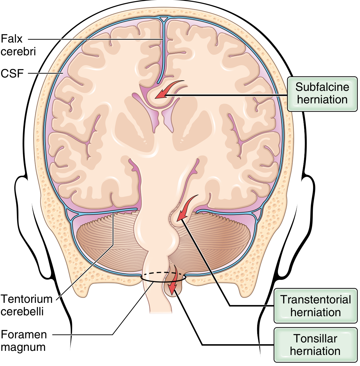
1. Subfalcine (Cingulate) Herniation
- Unilateral/asymmetric hemisphere expansion pushes the cingulate gyrus under the falx cerebri
- Can compress the anterior cerebral artery (ACA), causing medial frontal/parietal infarcts
2. Transtentorial (Uncal) Herniation
- Medial temporal lobe (uncus) is compressed against the free margin of the tentorium cerebelli
- Compresses CN III → ipsilateral pupil dilation, ptosis, ophthalmoplegia
- Compresses posterior cerebral artery (PCA) → contralateral homonymous hemianopia
- With large herniation, the contralateral cerebral peduncle is compressed against the opposite tentorial edge → Kernohan's notch phenomenon (ipsilateral hemiparesis - a "false localising sign")
- Duret haemorrhages: secondary flame-shaped midline/paramedian brainstem bleeds from distortion/tearing of penetrating vessels supplying the upper brainstem; indicate severe, often fatal herniation
3. Tonsillar Herniation
- Cerebellar tonsils are displaced downward through the foramen magnum
- Life-threatening - compresses the medullary respiratory and cardiac centres
- Results in bradycardia, respiratory arrest, and death
- Robbins, Cotran & Kumar Pathologic Basis of Disease; Schwartz's Principles of Surgery
Investigation
| Investigation | Use |
|---|---|
| CT scan (first line) | Identifies mass lesions, bleeds, oedema, hydrocephalus; guides treatment |
| MRI | Superior for elective assessment of most tumour types, posterior fossa, and surgical planning (third ventriculostomy anatomy) |
| ICP monitoring | Gold standard - external ventricular drain or intraparenchymal monitor; P1 (arterial) > P2 (tidal, elevated in poor compliance) > P3 (venous) |
| Lumbar puncture | CONTRAINDICATED until CT has excluded SOL (risk of coning by downward herniation) |
| MR Spectroscopy / PET | Tumour characterisation and metabolic activity |
| Angiography | Vascular lesions (AVMs, aneurysms) |
| Biopsy / stereotactic biopsy | Tissue diagnosis |
Management
Immediate/Emergency
- Airway and ventilation - controlled hyperventilation lowers PaCO₂ → cerebral vasoconstriction → reduces ICP acutely
- Head elevation to 30° - reduces hydrostatic pressure at cranial vault
- Mannitol 0.5-1 g/kg IV - osmotic diuresis draws free water from brain; effect delayed ~20 minutes; lasts 4-6 hours
- Hypertonic saline - alternative osmotherapy
- Emergency CT + neurosurgical referral
Specific
| Lesion | Treatment |
|---|---|
| Extradural/Subdural haematoma | Urgent burr hole/craniotomy and evacuation |
| Brain tumour (raised ICP) | High-dose dexamethasone (reduces peritumoral oedema); external ventricular drain for obstructive hydrocephalus (especially posterior fossa) |
| Glioblastoma | Maximal surgical resection (5-ALA fluorescence guidance + neuronavigation) + radiotherapy + temozolomide chemotherapy |
| Meningioma | Surgical excision; radiosurgery for recurrence |
| Cerebral metastases | Steroids; whole brain radiotherapy or stereotactic radiosurgery; surgical excision for single accessible lesion |
| Brain abscess | IV antibiotics + surgical drainage (aspiration or excision); steroids are withheld until abscess is excluded |
| Hydrocephalus | External ventricular drain; endoscopic third ventriculostomy; ventriculoperitoneal shunt |
Anticonvulsants are used for seizure management but prophylactic anticonvulsants are not recommended in brain tumour patients without prior seizures. - Bailey & Love's
Key Points Summary
- A chronic SOL produces symptoms after a silent growth phase, appearing first as focal/localising signs, then raised ICP symptoms, and finally herniation signs
- The triad of brain tumour presentation: raised ICP + seizures + focal neurological deficit
- Epilepsy of new onset in an adult must be treated as tumour until excluded
- Lumbar puncture is contraindicated until SOL is excluded by imaging
- Herniation (coning) is the final common pathway to death: subfalcine → transtentorial → tonsillar
- Metastases are the most common intracranial tumours overall; lung and breast are the most frequent primaries
Sources: S Das, A Manual on Clinical Surgery, 13th Ed. | Bailey & Love's Short Practice of Surgery, 28th Ed. | Schwartz's Principles of Surgery, 11th Ed. | Robbins, Cotran & Kumar Pathologic Basis of Disease
Explain neurons and neuroglia from snell book
Reading File
First, let me find Snell's book in the library and locate the neurons/neuroglia sections.
Finding Sources
Finding Sources
Snell's isn't listed by that name. Let me search the full catalog for neuroanatomy books and search for neurons/neuroglia content.
Finding Sources
Finding Sources
Snell's Clinical Neuroanatomy is not available in the library. However, there are excellent neuroscience and neuroanatomy books. Let me search specifically for dedicated neuron and neuroglia sections in the available books, particularly the Neuroscience (Bear) and Neuroanatomy books.
Finding Sources
Finding Sources
Finding Sources
Finding Sources
Snell's book is not in the library. The best equivalent sources are Histology (Eroschenko/Ross) with dedicated THE NEURON and NEUROGLIA sections, and Neuroscience: Exploring the Brain. Let me read these comprehensive sections in parallel.
Reading File
Reading File
Reading File
Excellent. Now let me read the detailed neuron content from histology book and neuroscience book in parallel.
Reading File
Reading File
Reading File
Now let me read the central neuroglia section and fetch key neuron/neuroglia images in parallel.
Reading File
Reading File
Loading Image
Excellent content and a great diagram. Let me now read more on astrocytes, oligodendrocytes, microglia, ependymal cells, and the neuroglia from the Neuroscience book.
Reading File
Reading File
Let me now read the glia section from Neuroscience Exploring the Brain, and the oligodendrocyte/microglia detail from histology.
Reading File
Reading File
Let me fetch the glial cell distribution diagram and read the glia section from the Neuroscience book.
Loading Image
Reading File
Reading File
I now have comprehensive material on neurons and neuroglia. Let me read the glia section from Neuroscience Exploring the Brain and microglia detail from histology for completeness.
Reading File
Reading File
I now have comprehensive content from multiple authoritative textbooks. Let me read the Neuroscience glia section quickly and then compile the full answer.
Reading File
I have all the information needed. Let me read the glia/astrocyte section from Neuroscience Exploring the Brain quickly.
Reading File
I now have all necessary content to compose a thorough, well-illustrated answer. Let me note upfront that Snell's Clinical Neuroanatomy is not in the library, and I'll draw on the closest authoritative equivalents.
Note on Sources: Snell's Clinical Neuroanatomy is not available in this textbook library. The explanation below draws from the three most authoritative equivalents available: Histology: A Text and Atlas (Ross & Pawlina, 8th Ed.), Neuroscience: Exploring the Brain (Bear, Connors & Paradiso, 5th Ed.), and Medical Physiology (Boron & Boulpaep). The content closely parallels Snell's own coverage of these topics.
Neurons and Neuroglia
Part I: The Neuron
Definition and Overview
The neuron is the structural and functional unit of the nervous system. The human nervous system contains more than 10 billion neurons. Although neurons show the greatest variation in size and shape of any group of cells in the body, all neurons share the same fundamental functional role: rapid communication via electrical and chemical signals.
The adult human brain contains approximately 85 billion neurons and a similar number of glial cells. Neurons are responsible for the most unique features of the brain - sensing environmental changes, rapidly communicating them to other neurons, and coordinating the body's responses. - Neuroscience: Exploring the Brain, 5th Ed.
Neuron Doctrine
The Neuron Doctrine states that each neuron is a discrete, independent cell that is not cytoplasmatically continuous with other neurons. This was established by Santiago Ramón y Cajal using the Golgi silver stain method, and was finally proven by electron microscopy in the 1950s, which showed that neurites of different neurons are separated by a narrow gap (the synaptic cleft) of ~50 nm. - Neuroscience: Exploring the Brain
Structure of the Neuron
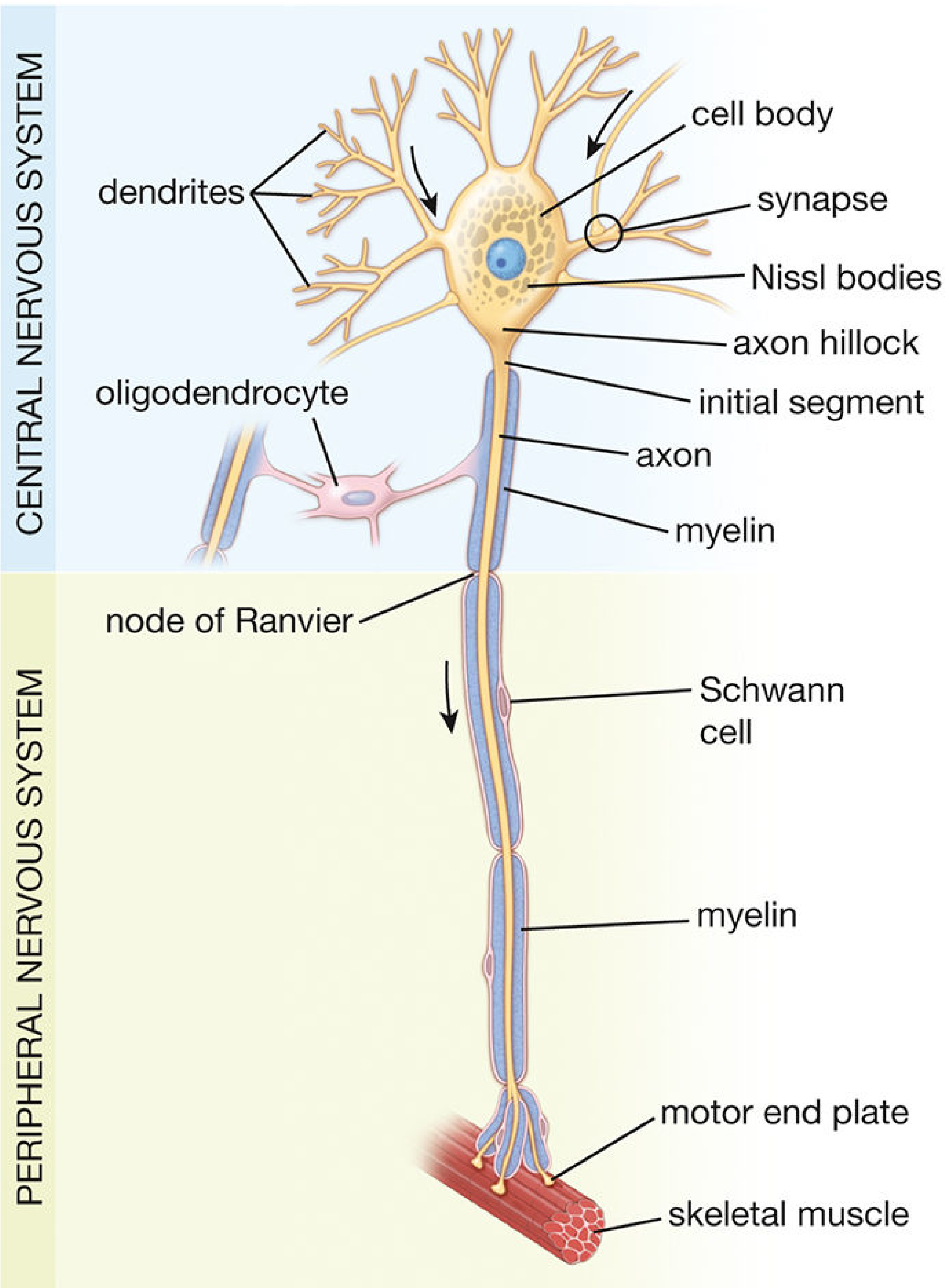
The functional components of every neuron include: cell body (soma/perikaryon), dendrites, axon, and synaptic junctions.
1. Cell Body (Soma / Perikaryon)
The cell body contains the nucleus and all the organelles that maintain the cell.
| Organelle | Function in Neurons |
|---|---|
| Nucleus | Large, pale-staining, contains euchromatin (active transcription); single prominent nucleolus |
| Nissl bodies (rough ER + polyribosomes) | Protein synthesis; absent from axon hillock and axon |
| Smooth ER & Golgi apparatus | Post-translational modification, packaging of proteins for transport |
| Mitochondria | ATP production; highly abundant due to neuron's high energy demands |
| Neuronal membrane | ~5 nm thick; studded with ion channels, pumps, and receptors; composition varies across soma, dendrites, and axon |
| Cytoskeleton | Three components (see below) |
Cytoskeleton components:
- Microtubules (25 nm diameter): composed of tubulin dimers; serve as tracks for axonal transport (kinesin for anterograde, dynein for retrograde)
- Neurofilaments (10 nm): intermediate filaments unique to neurons; provide structural support and regulate axon calibre; accumulation seen in Alzheimer's disease (neurofibrillary tangles of hyperphosphorylated tau)
- Microfilaments / actin filaments (8 nm): anchored to the inner membrane; regulate synaptic structure and dendritic spine morphology
2. Dendrites
Shorter, branching processes that receive incoming signals and transmit impulses toward the cell body. Key features:
- Most neurons have many dendrites, which dramatically increase the receptive surface area
- Contain dendritic spines - small protrusions that form the postsynaptic site of the majority of excitatory synapses
- Dendritic spines contain a postsynaptic density with clusters of neurotransmitter receptors (mainly glutamate receptors for fast excitatory transmission), voltage-gated Na⁺ and K⁺ channels, and an actin cytoskeleton
- Unlike axons, dendrites can contain polyribosomes (allowing local protein synthesis near synapses, critical for synaptic plasticity)
- Dendritic spine morphology is crucial for learning and memory; abnormal spines (long, thin, immature) are associated with intellectual disability and autism - Neuroscience: Exploring the Brain
3. Axon
The axon is specialized for transmitting impulses away from the cell body to distant targets. Key features:
- Each neuron has only one axon (but may have many collateral branches)
- Begins at the axon hillock, which tapers into the initial segment - the site where action potentials are initiated (highest concentration of voltage-gated Na⁺ channels)
- No rough ER in the axon; proteins must be synthesised in the cell body and transported down
- Membrane composition is fundamentally different from the soma membrane
- Terminates in axon terminals (boutons terminaux / synaptic knobs) which form synapses with the next neuron or effector cell
- Axonal Transport:
- Anterograde (soma → terminal): Kinesin motors carry vesicles, organelles, and newly synthesised proteins down the axon
- Retrograde (terminal → soma): Dynein motors return used membrane, organelles, and trophic signals back; this pathway is exploited by herpes virus and tetanus toxin to reach the CNS
4. Synapse
The point of communication between two neurons (or a neuron and an effector). The narrow synaptic cleft (~50 nm) separates the presynaptic terminal from the postsynaptic membrane. Neurotransmitters are released from vesicles in the presynaptic terminal, diffuse across the cleft, and bind to receptors on the postsynaptic membrane.
Classification of Neurons
A. By Number of Processes (Morphological)
| Type | Description | Example |
|---|---|---|
| Multipolar | One axon + many dendrites; most common | Motor neurons, interneurons |
| Bipolar | One axon + one dendrite | Retinal bipolar cells, cochlear ganglion |
| Unipolar (pseudounipolar) | Single process that bifurcates; both branches transmit impulses | Dorsal root ganglion sensory neurons |
B. By Function
| Type | Role | Fibre type |
|---|---|---|
| Sensory (Afferent) | Convey impulses from receptors to CNS | Somatic afferent (pain, temp, touch, proprioception); Visceral afferent |
| Motor (Efferent) | Convey impulses from CNS to effectors | Somatic efferent (skeletal muscle); Visceral efferent (smooth muscle, cardiac, glands) |
| Interneurons (Intercalated) | Form the integrative and communicating network between sensory and motor neurons | >99.9% of all neurons |
C. By Axon Length
- Golgi Type I (Projection neurons): Long axons extending to distant brain regions (e.g., pyramidal cells)
- Golgi Type II (Local circuit neurons): Short axons that do not extend beyond the local area (e.g., cortical stellate cells)
Impulse Conduction
An action potential is an electrochemical event initiated at the axon hillock when sufficient excitatory input depolarises the membrane to threshold.
Mechanism:
- Voltage-gated Na⁺ channels at the initial segment open → Na⁺ influx → depolarisation (resting potential of -70 mV rises to +30 mV)
- Na⁺ channels inactivate; voltage-gated K⁺ channels open → K⁺ efflux → repolarisation
- Local current flows to adjacent membrane, propagating the action potential
Saltatory (Jumping) Conduction in Myelinated Axons:
- Myelin acts as an insulator; voltage reversal (depolarisation) can only occur at the nodes of Ranvier (gaps in the myelin sheath with high densities of voltage-gated channels)
- The impulse "jumps" from node to node - this is saltatory conduction (Latin: saltus = to jump)
- Saltatory conduction is much faster than continuous conduction in unmyelinated fibres
- Conduction velocity increases with axon diameter and myelin thickness
Part II: Neuroglia (Glial Cells)
Neuroglia constitute approximately half the volume of the brain and outnumber neurons. The term "glia" is derived from the Greek word for glue, reflecting the original (now outdated) view that their main function is support. Modern understanding recognises that glial functions are far richer than mere structural support. - Neuroscience: Exploring the Brain
Only the nuclei of glial cells are visible in routine H&E-stained sections. Special heavy metal staining (e.g., gold sublimate) or immunocytochemistry (e.g., anti-GFAP for astrocytes) is required to visualise the full cell shape. - Histology: A Text and Atlas
Distribution of All Glial Cell Types in Brain
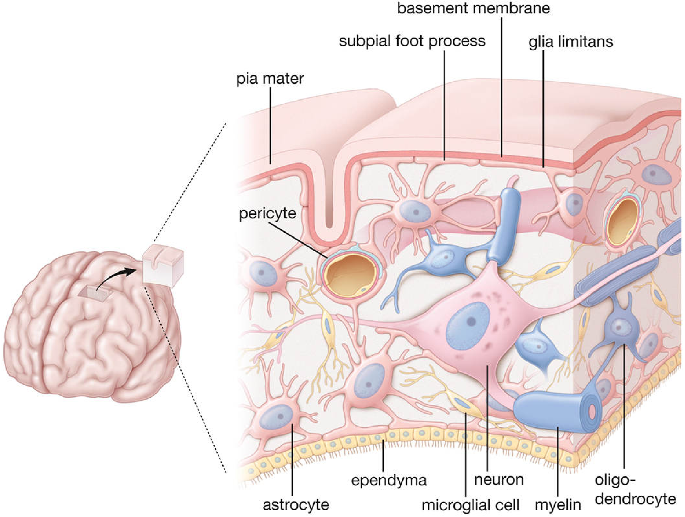
Central Neuroglia (CNS)
There are four types of central neuroglia:
1. Astrocytes
The largest of the neuroglial cells. They form an extensive interconnected network and communicate with neurons to support and modulate their activities.
Two subtypes:
| Type | Location | Morphology | Function |
|---|---|---|---|
| Protoplasmic | Gray matter | Numerous short, branching processes | Interact with up to 2 million synapses per cell; major role in synaptic modulation |
| Fibrous | White matter | Fewer, longer, straighter processes; fewer branches | Run along axons; contact nodes of Ranvier |
Both types contain prominent intermediate filaments of Glial Fibrillary Acidic Protein (GFAP) - the specific immunohistochemical marker for astrocytes. More abundant in fibrous astrocytes.
Functions of Astrocytes:
-
Structural scaffolding: During development, radial glial cells (derived from astrocytes) guide neuronal migration; some astrocytes span the entire brain thickness
-
Blood-brain barrier (BBB) maintenance: Astrocyte endfeet (perivascular feet) wrap around all brain capillaries and are integral to the tight junctions forming the BBB; regulate glucose uptake, store it as glycogen, and supply neurons with lactate
-
Potassium spatial buffering: Astrocyte membranes have abundant K⁺ pumps and channels; they absorb excess extracellular K⁺ released during neural activity, maintaining the microenvironment and preventing excessive neuronal excitability
-
Neurotransmitter regulation: Astrocytes take up and remove excess glutamate and other neurotransmitters from synaptic clefts; confine neurotransmitters to the synaptic space
-
Synapse formation and pruning: Signal neurons to initiate synapse growth (during development) and actively phagocytose excess synapses (synaptic pruning) to establish precise neural circuitry
-
Glia limitans: Protoplasmic astrocytes extend subpial feet to the pia mater, forming the glia limitans - a relatively impermeable barrier surrounding the CNS
-
Reactive gliosis: After injury, astrocytes proliferate and form a glial scar that walls off the damaged area but also impedes axonal regeneration
-
Phagocytosis: Reactive astrocytes can phagocytose myelin debris when microglia are overwhelmed
Tumours from fibrous astrocytes = Astrocytomas - account for ~80% of adult primary brain tumours; GFAP-positive on immunostaining. - Histology: A Text and Atlas
2. Oligodendrocytes
Small cells with few processes compared to astrocytes, aligned in rows between axons. Their sole primary function is producing and maintaining the myelin sheath in the CNS.
How they myelinate:
- Each oligodendrocyte gives off several tongue-like processes; each process wraps itself around a segment of a nearby axon, forming one internode of myelin
- A single oligodendrocyte can myelinate up to 40 different axons simultaneously (unlike Schwann cells, which myelinate only one axon segment each)
- The nucleus-containing cell body may be at some distance from the axons it myelinates
CNS vs PNS Myelin proteins:
- CNS myelin: Proteolipid protein (PLP), Myelin oligodendrocyte glycoprotein (MOG), Oligodendrocyte myelin glycoprotein (OMgp)
- PNS myelin: P0, PMP22 (expressed by Schwann cells)
Clinical relevance: In multiple sclerosis (MS), the immune system attacks oligodendrocytes and their myelin, causing demyelinating plaques → slowed or blocked conduction in affected axons.
3. Microglia
Inconspicuous cells with small, dark, elongated nuclei and relatively few processes. They are the resident immune cells of the CNS.
Origin: Unlike other glial cells (which derive from the neural tube neuroectoderm), microglia originate from mesoderm (bone marrow-derived monocyte precursors that migrate into the CNS during embryonic development). This makes them functionally analogous to macrophages.
Functions:
- Phagocytosis: Primary phagocytic cells of the brain; remove cellular debris, dead neurons, pathogens, and excess synapses
- Immune surveillance: Constantly survey the brain parenchyma with motile processes; activated by injury, infection, or disease
- Antigen presentation: Can present antigens during CNS inflammation
- Neuroprotection and neurotoxicity: Can be neuroprotective (clearing debris) or contribute to neuronal damage if chronically activated
Histological identification: Elongated, dark nuclei visible on H&E; best demonstrated with special stains or microglial markers (Iba-1); in diffuse microgliosis, large numbers become visible in routine preparations.
4. Ependymal Cells
Columnar-to-cuboidal cells that line the ventricles of the brain and the central canal of the spinal cord, forming an epithelium-like layer.
Features:
- Single layer of cells; lack an external lamina (unlike true epithelia)
- Apical surface: cilia (for CSF circulation) + microvilli (for CSF absorption)
- Bound by junctional complexes at their apical surfaces
- Basal surface: numerous infoldings that interdigitate with adjacent astrocyte processes
Specialised variants:
- Choroid plexus cells: Modified ependymal cells associated with capillary loops; responsible for producing CSF by active transport and secretion
- Tanycytes: Specialised ependymal cells in the floor of the third ventricle; lack cilia; have a long basal process projecting into brain parenchyma; involved in transporting substances between CSF and hypothalamic portal circulation; sensitive to glucose levels (possible role in energy balance regulation)
Peripheral Neuroglia (PNS)
5. Schwann Cells
The major glial cells of the PNS; analogous to oligodendrocytes in function but structurally distinct. Origin: neural crest cells.
Three phenotypes:
| Phenotype | Role |
|---|---|
| Myelinating Schwann cell | Produces myelin for large-diameter PNS axons; each cell myelinates only ONE axon segment |
| Non-myelinating (Remak) Schwann cell | Envelops multiple small-diameter unmyelinated axons in grooves (Remak bundles); mostly autonomic post-ganglionic fibres |
| Repair Schwann cell | After nerve injury, myelinating and Remak cells dedifferentiate; form bands of Büngner - regeneration tracks that guide axon sprouts back to their targets; secrete trophic factors and cytokines; mediate myelin autophagy |
Key differences: Oligodendrocyte vs Schwann cell
| Feature | Oligodendrocyte (CNS) | Schwann Cell (PNS) |
|---|---|---|
| Number of axons myelinated | Up to 40 | Only 1 |
| Myelin proteins | PLP, MOG, OMgp | P0, PMP22 |
| Regeneration after injury | Poor | Good (Bands of Büngner) |
| Cell of origin | Neuroectoderm | Neural crest |
6. Satellite Cells
- Surround neuronal cell bodies in peripheral ganglia (analogous to astrocytes in the CNS)
- Provide metabolic support and regulate the microenvironment around ganglion cells
7. Other Peripheral Neuroglia
- Terminal (perisynaptic) Schwann cells (teloglia): Cover axon terminals at the neuromuscular junction; regulate synaptic transmission
- Enteric glial cells: Associated with ganglia in the wall of the alimentary canal (enteric nervous system)
- Müller cells: Radial glial cells of the retina; support retinal neurons and photoreceptors
Summary Comparison Table
| Feature | Neurons | Astrocytes | Oligodendrocytes | Microglia | Ependymal Cells | Schwann Cells |
|---|---|---|---|---|---|---|
| Location | CNS + PNS | CNS | CNS | CNS | CNS (ventricles) | PNS |
| Origin | Neuroectoderm | Neuroectoderm | Neuroectoderm | Mesoderm | Neuroectoderm | Neural crest |
| Primary function | Signal transmission | Support, BBB, K⁺ buffering, synapse regulation | CNS myelination | Immune surveillance, phagocytosis | CSF lining & production | PNS myelination, repair |
| Marker | - | GFAP | MBP, PLP, MOG | Iba-1 | - | S100, P0 |
| H&E appearance | Large cell body visible | Only nucleus visible | Small nucleus in rows | Small elongated dark nucleus | Columnar, ciliated | Nucleus seen at Schwann cell-axon junction |
Sources: Histology: A Text and Atlas with Correlated Cell and Molecular Biology, Ross & Pawlina, 8th Ed. | Neuroscience: Exploring the Brain, Bear, Connors & Paradiso, 5th Ed. | Medical Physiology, Boron & Boulpaep
What is difference between neuron and nerve
A neuron is a single cell - the individual nerve cell with its cell body, dendrites, and axon. A nerve is a bundle of many axons (from multiple neurons) bound together by connective tissue sheaths, running through the peripheral nervous system. Think of a neuron as a single wire, and a nerve as a cable containing many wires.
Searching sources...
Finding Sources
Finding Sources
Reading File
Reading File
Finding Sources
Reading File
Reading File
Loading Image
All needed. Here is the full, textbook-sourced answer.
Neuron vs. Nerve
These two terms are frequently confused. They refer to completely different levels of organisation in the nervous system.
The Neuron - The Individual Cell
A neuron is a single cell - the fundamental structural and functional unit of the nervous system. It consists of:
- Cell body (soma/perikaryon) - contains the nucleus and organelles
- Dendrites - multiple branching processes that receive incoming signals
- Axon - a single long process that transmits impulses away from the cell body
- Synaptic terminals - where the neuron communicates with the next cell
A neuron is entirely a microscopic entity. Its cell body typically ranges from 5 to 100 µm in diameter, and while its axon can be very long (up to 1 metre for a motor neuron to the foot), the axon itself is still just one cellular process of one cell.
"A peripheral nerve is a bundle of nerve fibers held together by connective tissue." - Histology: A Text and Atlas (Ross & Pawlina)
The Nerve - A Bundle of Axons
A nerve is a macroscopic, organ-level structure found in the peripheral nervous system (PNS). It is built from the axons of many neurons, packaged together with connective tissue sheaths, blood vessels, and Schwann cells. The nerve itself is not a single cell - it is a collection of hundreds to thousands of axon fibres from many different neurons.
Structure of a Peripheral Nerve
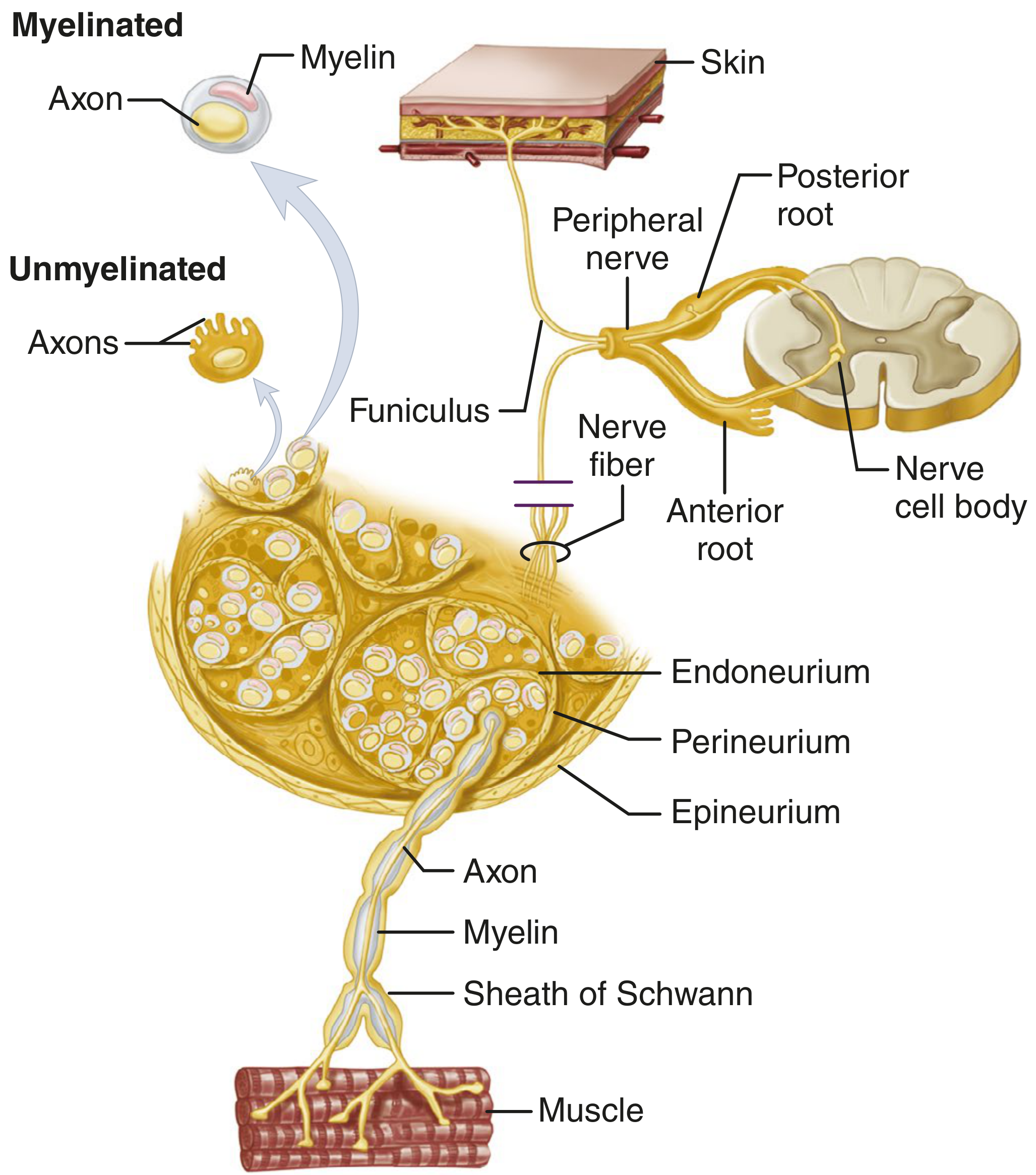
A peripheral nerve has a hierarchical organisation with three concentric connective tissue layers:
| Layer | What it surrounds | Composition |
|---|---|---|
| Endoneurium | Each individual nerve fibre (axon + Schwann cell) | Loose connective tissue; collagen fibrils run parallel and around nerve fibres; mainly secreted by Schwann cells; macrophages and mast cells present |
| Perineurium | Each fascicle (bundle of nerve fibres) | Specialised epithelium-like connective tissue; metabolically active diffusion barrier contributing to the blood-nerve barrier; maintains ionic milieu of enclosed fibres |
| Epineurium | The entire nerve (all fascicles together) + fills spaces between fascicles | Dense irregular connective tissue; richly vascularised; contains longitudinal nutrient arteries; blends with dura mater at the nerve root |
Each fascicle contains several hundred axons. A typical peripheral nerve contains multiple fascicles. - Adams & Victor's Principles of Neurology, 12th Ed.
Key Differences at a Glance
| Feature | Neuron | Nerve |
|---|---|---|
| Nature | A single cell | An organ-level bundle of many axons |
| Location | CNS and PNS | PNS only (CNS equivalent = tracts) |
| Components | Cell body + dendrites + axon + terminals | Axons from many neurons + Schwann cells + connective tissue sheaths + blood vessels |
| Visibility | Microscopic | Macroscopic (visible to naked eye) |
| Connective tissue | None | Endoneurium, perineurium, epineurium |
| Function | Individual unit of signal generation and transmission | Highway carrying combined sensory and/or motor information to/from targets |
| Example | A single motor neuron in the anterior horn of the spinal cord | The sciatic nerve (contains axons from many motor and sensory neurons) |
| Cell bodies | Part of the neuron | Cell bodies are NOT inside the nerve; they reside in the CNS or in peripheral ganglia |
An Analogy
Think of a nerve like an electrical cable:
- Each individual wire inside the cable = one axon (part of one neuron)
- The rubber insulation around each wire = myelin sheath + endoneurium
- Each sub-bundle of wires = a fascicle (surrounded by perineurium)
- The outer plastic jacket of the whole cable = epineurium
- The neuron itself = the wire + the power source (the cell body back at the wall) combined
One Important Note
The cell bodies of neurons are never found inside the nerve trunk itself. They reside either:
- Inside the CNS (e.g., anterior horn motor neurons, brainstem nuclei), or
- In peripheral ganglia (e.g., dorsal root ganglia for sensory neurons, autonomic ganglia for postganglionic autonomic neurons)
Only the axons (and in the case of sensory neurons, what functionally serve as axons) travel within the nerve.
Sources: Histology: A Text and Atlas, Ross & Pawlina, 8th Ed. | Adams & Victor's Principles of Neurology, 12th Ed. | Campbell's Operative Orthopaedics, 15th Ed.
This is a shared conversation. Sign in to Orris to start your own chat.