
rashes (petechie and all) according to their size
petechiae purpura ecchymosis skin rash classification by size
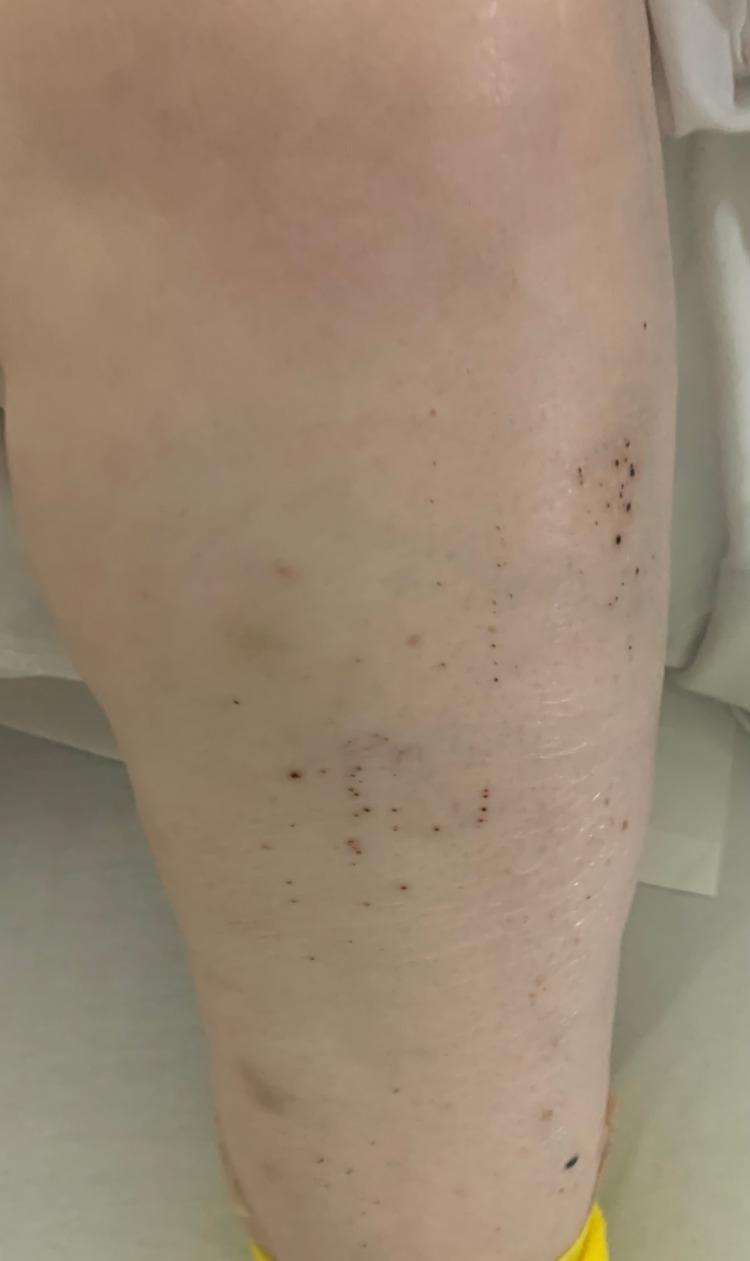
Clinical photograph of the left lower extremity demonstrating a localized rash characterized by numerous small, non-blanching, dark red to purplish-black spots consistent with petechiae. The lesions vary from pinpoint size to approximately 2-3 mm in diameter and are distributed in a scattered, patchy pattern across the anterior and lateral leg surface. Centrally, a larger, faint purplish area suggests underlying ecchymosis. The surrounding skin appears otherwise intact without evidence of ulceration or significant scaling. This presentation is highly characteristic of profound thrombocytopenia, such as Immune Thrombocytopenic Purpura (ITP), where low platelet counts lead to spontaneous microvascular hemorrhages within the dermis.
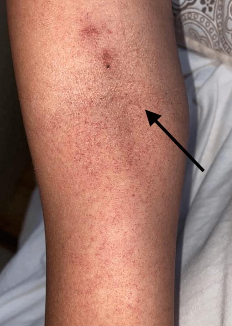
This clinical photograph displays a human extremity featuring a widespread, non-blanching petechial rash. The lesions are small, approximately 1-2 mm in diameter, appearing as reddish-purple punctate spots. They are distributed with varying density across the skin surface, creating a mottled, purpuric appearance. A black arrow points to a representative cluster of these petechiae. In the proximal portion of the image, there is a slightly larger, darker, and more confluent erythematous lesion, which may represent ecchymosis or evolving purpura. This image demonstrates characteristic dermatological manifestations often associated with systemic conditions such as thrombocytopenia, coagulopathies, or viral hemorrhagic fevers like Dengue. It is a valuable resource for medical education in dermatology, hematology, and infectious diseases, illustrating the morphology and distribution of microvascular hemorrhages in the skin.
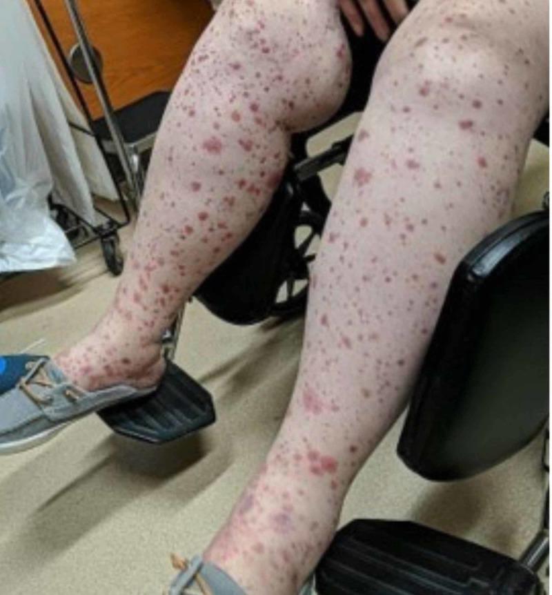
This clinical photograph shows the bilateral lower extremities of a patient demonstrating a widespread, palpable purpuric rash and associated soft tissue edema. The skin lesions are characterized by numerous non-blanching, erythematous to violaceous macules and papules of varying sizes, ranging from small petechiae to larger confluent ecchymotic patches. The distribution is dense and extends from the feet upwards to the thighs, consistent with a gravity-dependent pattern. Significant bilateral edema is visible, particularly pronounced around the ankles and shins, where the skin appears taut and shiny. These findings are hallmarks of small-vessel vasculitis, such as Henoch-Schönlein purpura (IgA vasculitis), reflecting systemic extravasation of red blood cells and perivascular inflammation. This image serves as a classic educational example for dermatology and internal medicine in recognizing the visual manifestations of systemic vasculitic syndromes and distinguishing between petechiae, purpura, and ecchymosis.
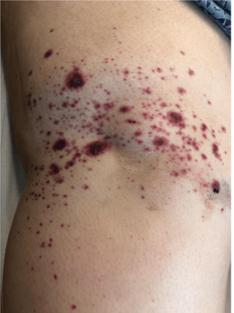
This clinical photograph shows a close-up view of a cutaneous eruption on a human extremity, specifically identified as a purpuric rash. The primary lesions consist of non-blanching, petechiae (pinpoint spots) and larger ecchymoses (purple-red blotches) that range in size from approximately 1 mm to several centimeters. The morphology includes flat macules and slightly raised, palpable papules, characteristic of palpable purpura. The lesions exhibit a range of colors from bright red to dark violaceous and purplish-black, indicating areas of acute hemorrhage and more significant dermal blood accumulation. The distribution is diffuse and irregular across the limb, with several areas showing higher density or clustering of lesions. There is no evidence of active ulceration, crusting, or diffuse erythema in the surrounding skin. This visual presentation is highly suggestive of small-vessel vasculitis, such as IgA vasculitis (Henoch-Schönlein purpura), which often presents with this type of leukocytoclastic vasculitis (LCV) pattern on the lower or upper extremities.
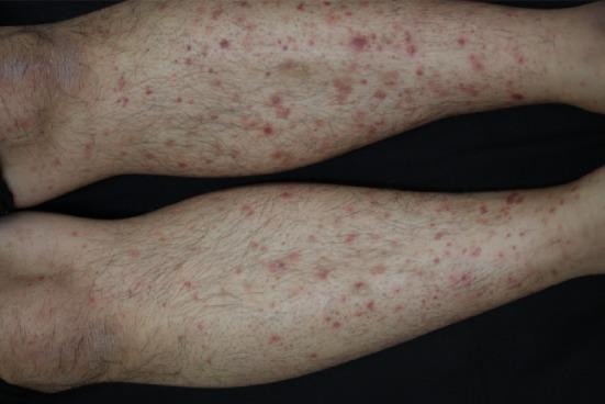
This clinical photograph shows a bilateral, symmetrical purpuric rash on the lower extremities of a patient. The lesions are non-blanching, reddish-purple macules and small papules, consistent with palpable purpura. They vary in size from pinpoint petechiae to several millimeters in diameter. The distribution is widespread across the shins and calves, with a tendency toward coalescence in certain areas. The surrounding skin appears otherwise normal in texture without obvious edema, ulceration, or secondary scaling. This presentation is characteristic of small-vessel vasculitis, specifically anaphylactoid purpura (Henoch-Schönlein purpura), which often presents with cutaneous involvement following a dependent distribution. The educational focus is on the morphological identification of vasculitic skin lesions and their clinical association with systemic conditions such as IgA nephropathy.
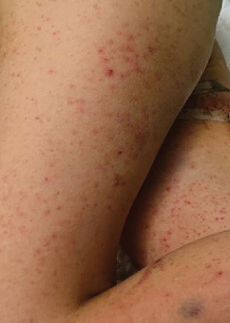
Clinical photograph of a patient's lower extremities exhibiting a nascent skin rash, characterized by a widespread distribution of erythematous macules and papules. The lesions are small, relatively uniform in size (petechial-like), and vary in color from bright red to darker purplish hues, suggesting possible early purpura. The distribution is dense and symmetric across the thighs and lower legs, with occasional clustering of papules. In addition to the primary rash, there is evidence of clinical intervention, including medical adhesive tape and a localized ecchymosis or bruise likely associated with a previous venipuncture or intravenous site. This visual presentation is educationally significant for recognizing the early dermatological manifestations of systemic conditions such as Henoch-Schönlein purpura (IgA vasculitis), sepsis-induced coagulopathy, or viral exanthems in a pediatric or adult hospital setting.
Skin Rashes/Lesions Classified by Size
Primary Lesions
Flat (Non-palpable) Lesions
| Lesion | Size | Description |
|---|---|---|
| Macule | ≤ 5-10 mm | Flat, circumscribed color change - no elevation or depression. Cannot be felt, only seen. |
| Patch | > 5-10 mm | Larger flat color change; essentially a large macule. |
Note: Robbins Basic Pathology uses ≤5 mm for macule; Robbins Pathologic Basis of Disease uses ≤10 mm. The 5 mm cutoff is more commonly tested.
Raised (Palpable) Solid Lesions
| Lesion | Size | Description |
|---|---|---|
| Papule | ≤ 5-10 mm | Elevated, dome-shaped or flat-topped solid lesion. Confined to epidermis/upper dermis. |
| Plaque | > 5-10 mm | Elevated, flat-topped broad lesion. Often formed by coalescence of papules. |
| Nodule | > 5-10 mm | Elevated solid lesion, but extends deeper into dermis/subcutis (deeper than a plaque). |
| Tumor | > 2 cm | Large solid mass, benign or malignant. |
| Wheal (Hive) | Variable | Itchy, transient, edematous elevation - pale center with erythematous rim. Caused by dermal edema (e.g., urticaria). |
Fluid-Filled Raised Lesions
| Lesion | Size | Description |
|---|---|---|
| Vesicle | ≤ 5-10 mm | Small fluid-filled blister (clear serous fluid). Examples: herpes simplex, varicella (early). |
| Bulla | > 5-10 mm | Large blister (clear fluid). "Blister" is the lay term for both vesicle and bulla. |
| Pustule | Variable | Pus-filled raised lesion (cloudy/yellow fluid). Examples: acne, folliculitis. |
Hemorrhagic (Purpuric) Lesions - The Petechiae Family
| Lesion | Size | Description |
|---|---|---|
| Petechiae | < 2-3 mm (pinpoint) | Tiny, flat, red-purple macules - the smallest hemorrhagic lesion. Caused by capillary bleeding (thrombocytopenia, Rocky Mountain spotted fever, parvovirus, scurvy, trauma). |
| Purpura | 2-10 mm | Larger red-purple macules/patches from hemorrhage into skin. May be flat ("macular purpura") or raised ("palpable purpura"). |
| Ecchymosis | > 1 cm | Large purpuric patch - what we commonly call a "bruise." May appear yellow-green as it ages (days old). |
| Hematoma | Variable (mass) | Blood collection under the skin forming a raised plaque or nodule - dermal/subdermal hemorrhage forming a palpable lump. |
"Purpuric macules are sometimes called petechiae; purpuric patches are sometimes called ecchymoses." - Fitzpatrick's Dermatology, Vol. 1
Palpable Purpura (Special Subtype)
Secondary Lesions (arising from primary lesions)
| Lesion | Description |
|---|---|
| Scale | Dry, flaky, plate-like thickening of stratum corneum |
| Crust | Dried serum, blood, or pus on skin surface (scab) |
| Erosion | Superficial loss of epidermis only - heals without scarring |
| Ulcer | Deeper loss extending into dermis/subcutis - heals with scarring |
| Fissure | Linear crack through epidermis (e.g., cracked heels) |
| Excoriation | Traumatic linear defect from scratching |
| Lichenification | Thickened, rough leathery skin from repeated rubbing |
| Scar | Fibrous replacement of dermis after injury |
| Atrophy | Loss of epidermal/dermal substance - skin appears thin, shiny |
Quick Visual Summary: Size Spectrum
← SMALLER ----------------------------------------- LARGER →

Petechiae   Purpura    Papule     Plaque      Ecchymosis   Hematoma
< 2-3 mm    2-10 mm   ≤ 5 mm     > 5 mm       > 1 cm       Mass

Macule      Patch      Vesicle    Bulla
≤ 5 mm      > 5 mm    ≤ 5 mm     > 5 mm
Clinical Pearl: Blanching Test
- Blanches with pressure = erythema (vascular dilation, blood still inside vessels) - e.g., drug rash, cellulitis, viral exanthem
- Does NOT blanch = purpura (blood outside vessels) - e.g., petechiae, ecchymosis, vasculitis, DIC, meningococcemia
- Robbins & Kumar Basic Pathology, Table 22.1 - Nomenclature of Skin Lesions
- Robbins, Cotran & Kumar Pathologic Basis of Disease, Table 25.1
- Fitzpatrick's Dermatology, Vol. 1 - Purpuric Reaction Pattern (p. 45)
- Tintinalli's Emergency Medicine - Differential Diagnosis by Primary Lesion Morphology
Clinical images showing the petechiae-purpura-ecchymosis spectrum: