
Create a details on myxoid neirofibroma focussing on histolopathology findings and it's IHC along with differential diagnosis
Reading File
Now let me run parallel searches across the library and supporting sources.
Finding Sources
Finding Sources
Searching PubMed
myxoid neurofibroma histopathology immunohistochemistry
Searching the Web
myxoid neurofibroma histopathology IHC differential diagnosis pathology
Now let me read the detailed neurofibroma sections from Fitzpatrick's and fetch the full web article on histopathological variants, plus search for images.
Reading File
Reading Web Page
https://www.mdpi.com/2296-3529/10/1/1
Reading Article
PMID: 38363063
Let me now read the full histopathology and differential diagnosis section from Fitzpatrick's, and the Dermatology 2-Volume Set section on nerve sheath myxoma differential diagnosis.
Reading File
Reading File
Now let me fetch the key histology images from Fitzpatrick's and read the IHC section.
Reading File
Loading Image
Loading Image
I now have comprehensive data from multiple authoritative sources. Let me compile the complete, detailed response.
Myxoid Neurofibroma: Histopathology, IHC, and Differential Diagnosis
Overview
Myxoid neurofibroma (myxoid cNF) is a stromal variant of cutaneous neurofibroma defined by extensive deposition of mucin/glycosaminoglycans throughout the tumor matrix, to the point that the myxoid change is the dominant feature. It belongs to the broader family of peripheral nerve sheath tumors and represents a benign entity. It may occur sporadically or in the setting of NF1, and is classically a component of NAME syndrome (Nevi, Atrial myxoma, Myxoid neurofibroma, Ephelides). In the past it was described under multiple synonyms including nerve sheath myxoma, bizarre cutaneous neurofibroma, and lobular neuromyxoma - though current classification separates nerve sheath myxoma (true neurothekeoma) as a distinct entity. Malignant transformation of the myxoid subtype has not been reported.
Histopathological Findings
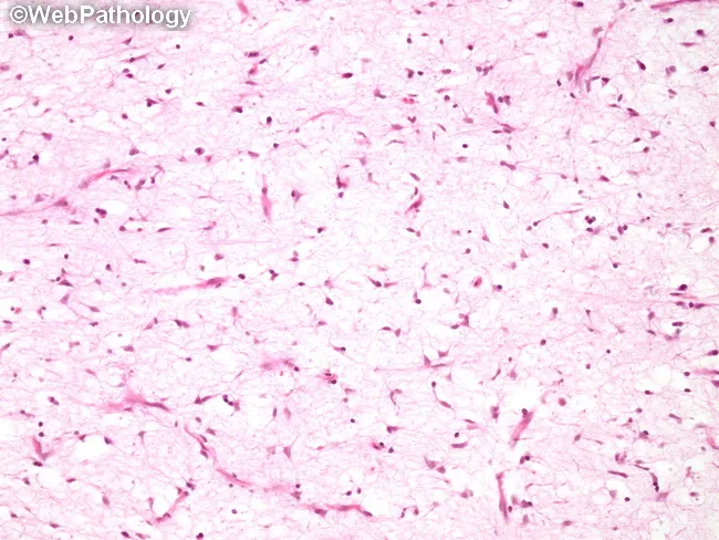
Architecture (Low Power)
- Non-encapsulated, unencapsulated dermal lesion, typically well-demarcated at low magnification
- Centered in the dermis but may extend into superficial subcutis
- The stroma is predominantly myxoid/mucinous, with the myxoid matrix being the hallmark feature - described as "extensive deposition of stromal mucin"
- Low to moderate overall cellularity (often strikingly hypocellular, which creates diagnostic difficulty on FNA/cytology)
- No true capsule; may have a peripheral condensation of fibrous tissue
Cellular Features (High Power)
- Cells have wavy, serpentine, or "comma-shaped" nuclei - a defining feature of Schwann cell lineage
- Scant, poorly-defined cytoplasm; cell borders are indistinct
- Cells are haphazardly arranged in the mucinous matrix, not forming tight fascicles
- Single-layer thin fascicles of spindle cells are present
- Nuclear pleomorphism may occasionally be present (degenerative atypia), but significant atypia raises the concern for MPNST
- Mitoses are absent or extremely rare (< 1 per 50 HPF is the accepted threshold)
- Mast cells are scattered throughout the stroma - a useful diagnostic clue favoring neurofibroma over its mimics
- Vacuolated histiocytes ("muciphages" or "pseudolipoblasts") may be present within the mucinous pools
Stromal Features
- Matrix is rich in hyaluronic acid-type mucin (stains with Alcian blue, colloidal iron)
- Ropey collagen bundles ("shredded collagen") are intermingled within the mucoid background - a characteristic finding highlighted in cytology specimens
- Thin-walled blood vessels and scattered lymphocytes may be present
- In diffuse-type myxoid neurofibroma, a branching plexiform vascular network may be prominent, mimicking myxoid liposarcoma
Axons
- Axons course through the tumor mass - a unique feature compared to most mimics
- These are difficult to see on H&E but are demonstrable with neurofilament immunostain
- The ratio of axons to Schwann cells is < 1:1 (distinguishing neurofibroma from neuroma)
Additional Notes
- Floret-like multinucleated giant cells (CD34+) may be present, as in spindle cell/pleomorphic lipoma
- Organoid pseudo-Meissnerian body-like structures may be seen, especially in diffuse type
Source: Fitzpatrick's Dermatology, Histopathology section; MDPI Diagnostics 2022 (Practical Approach to Peripheral Nerve Sheath Tumors)
Immunohistochemistry (IHC) Profile
The IHC panel of myxoid neurofibroma reflects its composite cellular origin (Schwann cells, perineurial cells, and CD34+ fibroblasts):
| Marker | Result | Cell Type Labeled |
|---|---|---|
| S100 protein | Positive (diffuse, cytoplasmic + nuclear) | Schwann cells |
| SOX10 | Positive | Schwann cells |
| CD34 | Variable/focal positive | CD34+ endoneurial fibroblasts |
| EMA (Epithelial Membrane Antigen) | Focal/variable | Perineurial cells (outlines tumor lobules) |
| Neurofilament | Positive | Axons traversing the tumor |
| Collagen IV | Positive (borders cells) | Basement membrane |
| Ki-67 / MIB-1 | Very low proliferation index | - |
| GFAP | Variable positive | - |
| p16 | Negative/retained | Loss would suggest MPNST |
| H3K27me3 | Retained | Loss suggests MPNST |
Key IHC points:
- The combination of S100 (+) + SOX10 (+) + CD34 (variable) + EMA (focal perineurial) is characteristic
- The Schwann cell population is the dominant S100/SOX10-positive component
- CD34 positivity in scattered fibroblast-like cells is helpful in supporting nerve sheath lineage
- MART-1/Melan-A, p53, cytokeratin, and desmin are negative - helping exclude melanocytic, epithelial, and smooth muscle lesions
- The pattern of S100(+) and SOX10(+) with absent or only focal EMA helps separate from perineurioma (EMA+/S100-)
Source: Fitzpatrick's Dermatology, IHC Table; MDPI Diagnostics 2022; [Shaker et al., Diagnostic Cytopathology 2024, PMID 38363063]
Differential Diagnosis
Myxoid neurofibroma sits within the broad category of myxoid soft tissue tumors, which is one of the most challenging areas in soft tissue pathology. The key differentials are:
1. Nerve Sheath Myxoma (True Neurothekeoma / NSM)
- Historically conflated with myxoid neurofibroma; now recognized as a distinct entity
- Architecture: multilobulated/multinodular with distinct fibrous septa separating myxoid lobules - more organized than myxoid NF
- Cells: epithelioid and stellate Schwann cells in syncytial-like aggregates, ring-shaped multipolar cells
- IHC: S100 strongly positive, GFAP positive, CD57 positive; EMA-positive perineurial cells at periphery of lobules
- Distinguishing feature: the lobular architecture is the most reliable separator from myxoid NF
2. Dermatofibrosarcoma Protuberans (DFSP), Myxoid Variant
- Architecture: storiform/cartwheel pattern of spindle cells infiltrating subcutis in a honeycomb pattern
- IHC: CD34 strongly and diffusely positive; S100 negative
- Genetics: COL1A1-PDGFB translocation t(17;22) - confirmatory by FISH or RT-PCR
- Key distinction: S100 negativity and strong diffuse CD34 exclude myxoid NF
3. Superficial Angiomyxoma (Cutaneous Myxoma)
- Loosely cellular myxoid lesion with prominent thin-walled blood vessels
- Cells are stellate to spindle without wavy nuclei; no Schwann cell differentiation
- IHC: S100 negative, CD34 variable - key negative S100 separates from myxoid NF
- Associated with Carney complex (NAME/LAMB syndromes) - note these syndromes also include myxoid NF
- Mucin: hyaluronic acid-rich (same as NF); erosion/ulceration of overlying epidermis is a clue
4. Myxoid Liposarcoma
- Contains a plexiform capillary network ("chicken wire" vasculature) - the most helpful architectural feature
- Cells: primitive round to oval lipoblasts in varying stages of differentiation; univacuolated lipoblasts are characteristic
- IHC: S100 variable, MDM2/CDK4 usually negative (unlike dedifferentiated variant); DDIT3 (CHOP) nuclear expression is highly sensitive/specific ([PMID 32815829])
- Genetics: FUS-DDIT3 or EWSR1-DDIT3 translocation - definitive
- Key distinction: presence of lipoblasts + chicken-wire vasculature + DDIT3 rearrangement
5. Low-Grade Myxofibrosarcoma (Myxoid MFH)
- Curvilinear blood vessels ("pseudovascular spaces"), mild nuclear pleomorphism even at low grade
- IHC: S100 negative, CD34 negative
- Usually larger, deeper, and in older patients; higher mitotic rate than NF
- No axons
6. Low-Grade Fibromyxoid Sarcoma (Evans Tumor)
- Alternating fibrous and myxoid zones ("swirling pattern")
- Cells: bland spindle cells with little atypia; can be confused with NF in myxoid zones
- IHC: MUC4 strongly positive (highly sensitive/specific marker); S100 negative
- Genetics: FUS-CREB3L2 translocation
7. Schwannoma (Antoni B Areas)
- Antoni B areas in schwannoma are hypocellular and myxoid, closely resembling myxoid NF
- Distinguished by: encapsulation, alternating Antoni A (cellular, Verocay bodies) and Antoni B areas; the absence of axons within the lesion (unlike NF)
- IHC: S100 strongly and diffusely positive (in NF, only Schwann cells are S100+, not all cells); NF2/merlin often lost in schwannoma
- CD34: often negative in schwannoma vs. variable in NF
8. Perineurioma (Intraneural or Soft Tissue)
- Composed of perineurial cells with elongated bipolar processes
- IHC: EMA strongly positive; S100 negative; GLUT-1 positive; claudin-1 positive
- This is the clearest IHC separator: EMA+/S100- is perineurioma; S100+/EMA-or-focal is NF
9. Myxoid Neurofibroma vs. MPNST with Myxoid Change
- MPNST may arise from NF1-associated neurofibroma (especially plexiform type)
- Features of malignancy: high cellularity, marked nuclear atypia, mitoses > 4/10 HPF, necrosis, loss of H3K27me3 (trimethylation of H3 at lysine 27), loss of p16
- IHC: H3K27me3 loss is highly specific for MPNST (retained in benign NF); S100 becomes patchy/focal
Summary Table
| Entity | S100 | CD34 | EMA | SOX10 | Key Distinguishing Feature |
|---|---|---|---|---|---|
| Myxoid Neurofibroma | + (Schwann cells) | Variable | Focal (perineurial) | + | Wavy nuclei, mast cells, axons (NF) |
| Nerve Sheath Myxoma | Strongly + | - | Focal peripheral | + | Lobular architecture, epithelioid cells |
| DFSP (myxoid) | - | Diffuse strong + | - | - | Storiform, honeycomb, COL1A1-PDGFB |
| Superficial Angiomyxoma | - | Variable | - | - | Prominent vessels, no wavy nuclei |
| Myxoid Liposarcoma | Variable | - | - | - | Chicken-wire vessels, lipoblasts, DDIT3+ |
| Perineurioma | - | - | Strongly + | - | EMA+, S100-, GLUT-1+ |
| Low-Grade Fibromyxoid Sarcoma | - | - | - | - | MUC4+, FUS-CREB3L2 |
| Schwannoma | Diffuse strong + | - | - | + | Encapsulated, Antoni A+B, no axons |
| MPNST | Focal/patchy | - | - | Focal | H3K27me3 loss, atypia, mitoses |
Clinical Context
- Presents as a solitary, asymptomatic, flesh-colored to pink-blue dermal nodule, most commonly on the face, shoulders, and arms
- Higher incidence in teenagers and young adults
- May occur in periungual/subungual locations
- Association with NF1 (may or may not be present)
- Component of NAME syndrome (Nevi, Atrial myxoma, Myxoid neurofibroma, Ephelides)
- Benign clinical course; malignant transformation has not been reported for the myxoid variant
- Treatment: simple excision
References:
- Fitzpatrick's Dermatology, 9th ed., Chapter 121 (Nerve Sheath Tumors) - histopathology and IHC tables
- Dermatology 2-Volume Set, 5th ed. (Bolognia), Chapter 115 - differential diagnosis of nerve sheath myxoma vs. neurofibroma
- [Shaker N et al., "Myxoid neurofibroma masquerading as lymphatic-venous malformation," Diagnostic Cytopathology 2024, PMID 38363063] - cytological features and IHC on excisional biopsy
- Practical Approach to Histological Diagnosis of Peripheral Nerve Sheath Tumors, Diagnostics 2022 - S100/SOX10/CD34/EMA IHC profile; diffuse type with myxoid stroma
- Histopathological Variants of Cutaneous Neurofibroma, Dermatopathology 2023 - myxoid cNF definition, NAME syndrome association, pseudolipoblasts
This is a shared conversation. Sign in to Orris to start your own chat.