
Whats cephalohemetoma
Reading File
A cephalohematoma is a collection of blood between the periosteum (the fibrous covering of the skull) and the skull bone in a newborn. It is caused by rupture of periosteal blood vessels, usually from birth trauma. Because it is subperiosteal, it is always limited by suture lines and does not cross them.
Searching sources...
Finding Sources
Finding Sources
Reading File
Reading File
Reading File
Loading Image
Loading Image
Cephalohematoma
A cephalohematoma is a subperiosteal collection of blood in a newborn - blood accumulates between the periosteum and the outer surface of the skull bone, caused by tearing of the diploic and/or emissary veins during mechanical birth trauma.
Anatomy - Why it doesn't cross suture lines
The periosteum is firmly attached at every suture line, so the blood collection is physically confined to a single bone. This is the single most important distinguishing feature.
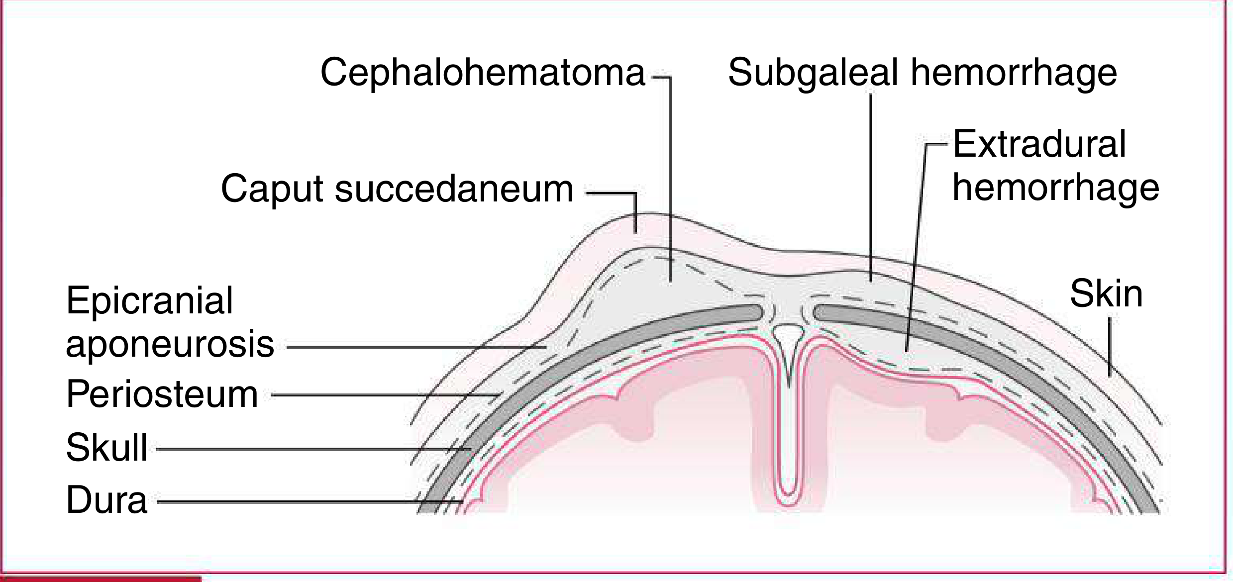
(Harriet Lane Handbook, 23rd ed.)
Key Features
| Feature | Detail |
|---|---|
| Location | Between periosteum and skull bone; usually over the right parietal bone |
| Crosses sutures? | No - strictly confined by suture lines |
| Cause | Rupture of diploic/emissary veins from birth trauma or vacuum extraction |
| Size | 1-5 cm; swelling may increase for the first 12-24 hours after birth |
| Feel on exam | Initially firm; becomes more fluctuant after 48 hours; distinct, well-defined margins |
| Timing | Not apparent at birth, grows over hours; resolves over weeks |
| Incidence | Less than 1% of newborns |
Causes / Risk Factors
- Instrumental delivery (vacuum extraction, forceps)
- Higher birth weight
- Prolonged or difficult labor
Complications
- Neonatal jaundice - the breaking down of the accumulated blood increases the bilirubin load
- Skull fracture - an underlying linear fracture may be present (reported in up to 25% of cases)
- Calcification - if the hematoma does not fully reabsorb, the periphery can calcify
- Rarely: superinfection (if the overlying skin is breached, e.g., after fetal scalp electrode use)
- Association with hemophilia - spontaneous cephalohematoma can be the presenting sign of an underlying coagulopathy
Comparison with Similar Conditions
| Caput Succedaneum | Cephalohematoma | Subgaleal Hemorrhage | |
|---|---|---|---|
| Tissue layer | Subcutaneous (above aponeurosis) | Subperiosteal | Below epicranial aponeurosis |
| Crosses sutures | Yes | No | Yes (can be massive) |
| Present at birth | Yes, maximal at birth | Grows after birth | Progressive after birth |
| Resolution | 48-72 hours | Weeks | Weeks |
| Severity | Minimal | Rarely severe | Can be life-threatening |
(Harriet Lane Handbook, 23rd ed. - Table 18.3)
Management
-
Most cases resolve spontaneously - no intervention needed
-
Do not aspirate (increases infection risk)
-
Monitor bilirubin levels for jaundice
-
X-ray or skull US if fracture is suspected
-
If calcification occurs, it usually remodels over months to years; surgical removal is rarely needed
-
The Essentials of Forensic Medicine and Toxicology, 36th ed., p. 429
-
Fitzpatrick's Dermatology, Vol. 1
-
Harriet Lane Handbook, 23rd ed.
-
Rosen's Emergency Medicine
This is a shared conversation. Sign in to Orris to start your own chat.