


Path physiology hematology
- Transcribe them into clean typed notes
- Correct mistakes and explain concepts
- Make short exam revision notes
- Create practice questions from them
Transcribe them into clean notes and give summary
Merck Manual red blood cell indices MCV MCH MCHC RDW iron deficiency anemia hepcidin ferroportin
iron deficiency anemia AND hepcidin
Hematology: RBC Indices and Iron Deficiency Anemia
1. Red Blood Cell (RBC) Indices
- Hemoglobin (Hb)
- Hematocrit / packed cell volume (Hct/PCV)
- RBC count
A. Mean Corpuscular Volume (MCV)
| MCV | Type of anemia | Common causes |
|---|---|---|
| <80 fL | Microcytic anemia | Iron-deficiency anemia, thalassemia, sideroblastic anemia, anemia of chronic inflammation/disease |
| 80-100 fL | Normocytic anemia | Acute blood loss, hemolytic anemia, aplastic anemia, early anemia of chronic disease |
| >100 fL | Macrocytic anemia | Vitamin B12 deficiency, folate deficiency, liver disease, alcohol use, hypothyroidism |
B. Mean Corpuscular Hemoglobin (MCH)
C. Mean Corpuscular Hemoglobin Concentration (MCHC)
D. Red Cell Distribution Width (RDW)
- Increased variation in RBC size is called anisocytosis.
- Normal RDW: approximately 11.5-14.5%
2. Packed Cell Volume (PCV) / Hematocrit (Hct)
- Male: 40-54%
- Female: 36-46%
3. Common Reference Values
| Test | Typical adult reference range |
|---|---|
| Hemoglobin, male | 13-17 g/dL |
| Hemoglobin, female | 12-15 g/dL |
| RBC count, male | 4.5-6.0 million/µL |
| RBC count, female | 4.0-5.5 million/µL |
| WBC count | 4,000-11,000/µL |
| Platelet count | 150,000-450,000/µL, or 1.5-4.5 lakh/µL |
4. Anemia
Anemia is not defined simply as “deficiency of Hb and RBC count.” Hb is the primary diagnostic measure. RBC count can be low, normal, or even high, such as in thalassemia trait.
5. Iron-Deficiency Anemia
Causes / Etiology
- Inadequate dietary iron intake
- Increased requirement
- Pregnancy
- Infancy, childhood, and adolescence
- Chronic blood loss
- Heavy menstrual bleeding
- Gastrointestinal blood loss, such as peptic ulcer disease, malignancy, or hemorrhoids
- Parasitic infestation
- Especially hookworm in endemic areas
- Malabsorption
- Celiac disease
- Reduced gastric acid or post-gastrectomy states
- Other small-intestinal disorders
- Reduced intake or impaired absorption associated with alcohol use, usually in the context of poor nutrition or gastrointestinal/liver disease
6. Intestinal Iron Absorption and Regulation
Iron absorption pathway
- Dietary non-heme iron is mainly present as ferric iron (Fe³⁺).
- At the brush border of the duodenum, duodenal cytochrome b (Dcytb) reduces Fe³⁺ to ferrous iron (Fe²⁺).
- DMT1 transports Fe²⁺ into the enterocyte.
- Iron can be stored within enterocytes as ferritin or exported into blood through ferroportin.
- During export, Fe²⁺ is oxidized to Fe³⁺, chiefly by hephaestin.
- Fe³⁺ binds transferrin for transport in plasma to tissues, especially bone marrow, liver, and other cells.
Hepcidin-ferroportin regulation
- Hepcidin is made by the liver.
- It binds to ferroportin and causes ferroportin internalization and degradation.
- Therefore, increased hepcidin reduces:
- Intestinal iron absorption
- Release of iron from macrophages
- Release of stored iron from hepatocytes
- High hepcidin does not cause iron deficiency directly.
- In uncomplicated iron deficiency, hepcidin decreases. This allows ferroportin to remain active and increases iron absorption and release into plasma.
- Hepcidin is increased by high iron stores and inflammation, contributing to iron-restricted erythropoiesis in anemia of chronic inflammation.
7. Signs and Symptoms of Anemia
- Fatigue
- Weakness
- Reduced exercise tolerance
- Dizziness or light-headedness
- Pallor
- Dyspnea on exertion
- Palpitations or tachycardia in more severe anemia
- Pica, especially craving ice
- Brittle nails or koilonychia
- Glossitis
- Angular cheilitis
- Hair loss in some people
8. Blood Film Terminology
- Anisocytosis: Variation in RBC size
- Poikilocytosis: Variation in RBC shape
Quick Summary
- MCV measures cell size: low in microcytic anemia, normal in normocytic anemia, and high in macrocytic anemia.
- MCH measures hemoglobin amount per RBC.
- MCHC measures hemoglobin concentration within packed RBCs.
- RDW measures RBC size variation and rises with anisocytosis.
- Iron-deficiency anemia is usually microcytic and hypochromic, often with increased RDW.
- Major causes of iron deficiency are poor intake, increased physiological demand, chronic blood loss, hookworm, and malabsorption.
- Iron is absorbed mainly in the duodenum as Fe²⁺ through DMT1, exported by ferroportin, and transported by transferrin.
- Hepcidin inhibits ferroportin. Hepcidin is generally low in pure iron deficiency and high during inflammation.
Megaloblastic anemia absorption cycle normal and deficiency both detailed with picture
diagram of vitamin B12 absorption stomach intrinsic factor terminal ileum cubilin receptor
megaloblastic anemia peripheral blood smear macro ovalocytes hypersegmented neutrophils image
vitamin B12 deficiency AND megaloblastic anemia
Merck Manual megaloblastic macrocytic anemia vitamin B12 folate absorption deficiency
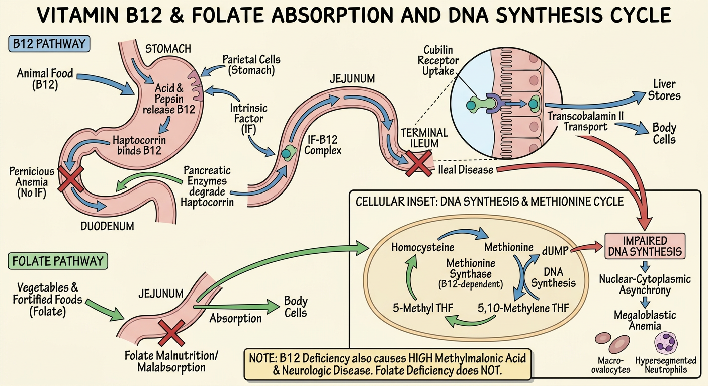
An accurate labeled medical teaching diagram of vitamin B12 and folate absorption and the folate-methionine DNA synthesis cycle. Show B12 path: animal food, stomach acid and pepsin release B12, haptocorrin, pancreatic enzymes in duodenum, intrinsic factor from parietal cells, terminal ileum cubilin receptor uptake, transcobalamin II transport to cells and liver stores. Show folate path: vegetables and fortified foods, jejunum absorption. Then a cellular inset: B12-dependent methionine synthase converts homocysteine to methionine and converts 5-methyl THF to THF; folate provides 5,10-methylene THF for dUMP to dTMP DNA synthesis. Include deficiency blocks for pernicious anemia or ileal disease and folate malnutrition/malabsorption, leading to impaired DNA synthesis, nuclear-cytoplasmic asynchrony, megaloblastic anemia, macro-ovalocytes, hypersegmented neutrophils. Include a clear note that B12 deficiency also causes high methylmalonic acid and neurologic disease, while folate deficiency does not. Use correct terminology, simple textbook style, no patient-specific details.
Megaloblastic Anemia: Vitamin B12 and Folate
Definition
- Vitamin B12 (cobalamin)
- Folate (vitamin B9)
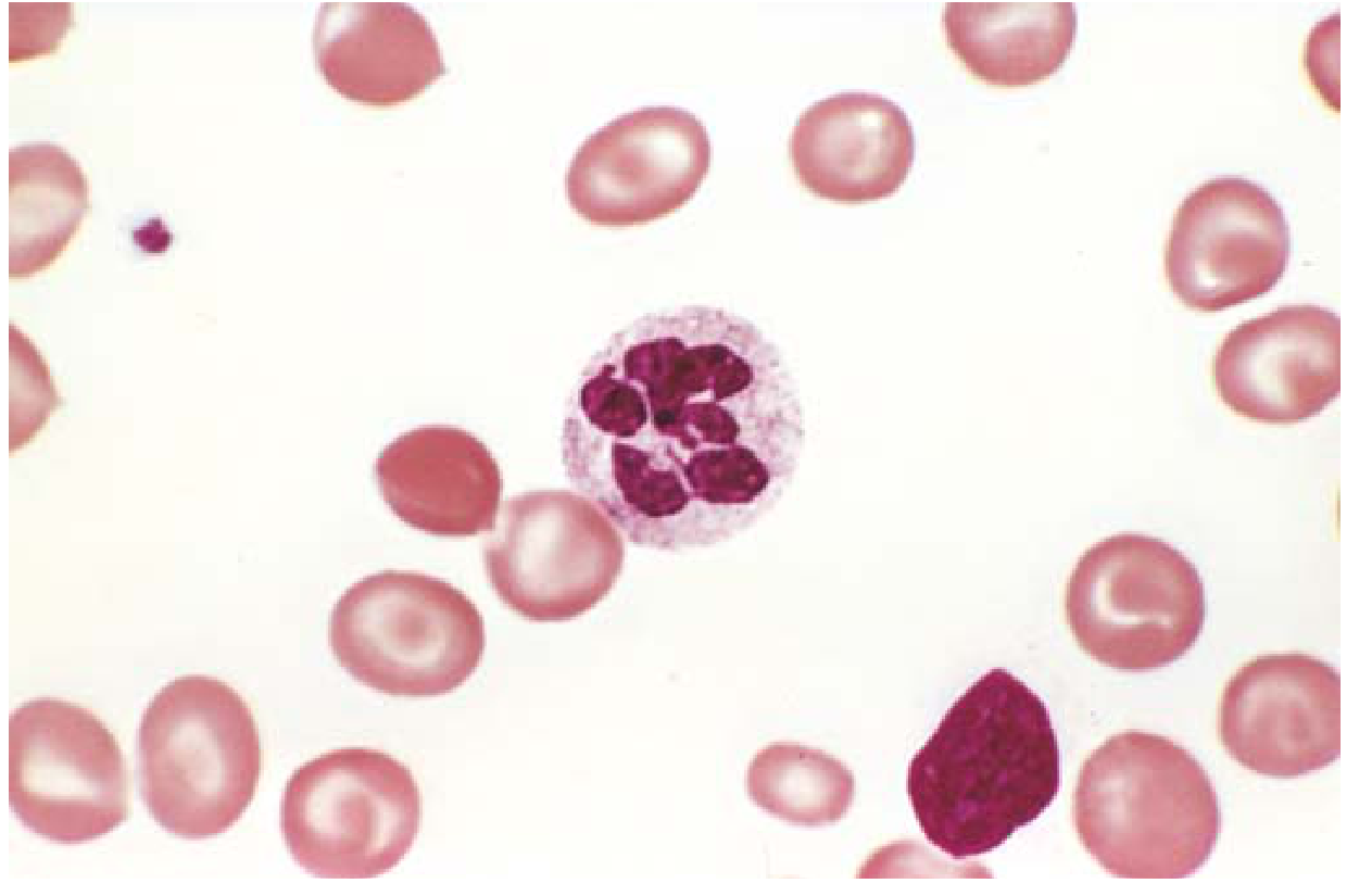
- Macro-ovalocytes
- Hypersegmented neutrophils, usually 5 or more lobes
- Anisocytosis and poikilocytosis
- Sometimes leukopenia and thrombocytopenia
1. Normal Vitamin B12 Absorption Cycle
Dietary source
- Meat
- Fish
- Eggs
- Milk and dairy products
Steps of absorption
Step 1: Stomach
- Dietary B12 is bound to food proteins.
- Gastric acid and pepsin release B12 from these proteins.
- Free B12 binds to haptocorrin, also called R-binder or R-protein, which is produced in saliva and stomach.
Step 2: Duodenum
- Pancreatic proteases digest haptocorrin.
- B12 is released and binds to intrinsic factor (IF).
- Intrinsic factor is a glycoprotein made by gastric parietal cells.
Step 3: Terminal ileum
- The B12-IF complex reaches the terminal ileum.
- It binds to the cubilin receptor complex on ileal enterocytes.
- The complex is taken into enterocytes by receptor-mediated endocytosis.
Step 4: Blood and tissues
- B12 is released from the enterocyte and enters plasma attached to transcobalamin II.
- It travels to:
- Bone marrow for erythropoiesis
- Liver for storage
- Nervous tissue and other cells
2. Normal Folate Absorption Cycle
Dietary source
- Green leafy vegetables
- Legumes
- Citrus fruits
- Liver
- Fortified grains
Absorption
- Dietary polyglutamate folate is converted at the intestinal brush border into absorbable monoglutamate forms.
- Folate is absorbed mainly in the duodenum and proximal jejunum.
- In blood it is transported to tissues.
- In cells, folate is converted to its active form, tetrahydrofolate (THF).
3. Folate-B12 Metabolic Cycle and DNA Synthesis
A. Folate cycle
- Dihydrofolate reductase (DHFR) converts dihydrofolate, DHF, to THF.
- THF accepts and transfers one-carbon units.
- 5,10-methylene THF donates a methyl group in the reaction:
- dTMP is required to make DNA.
B. Role of vitamin B12: the methyl-folate trap
Why B12 deficiency causes megaloblastic anemia
- 5-methyl THF cannot be converted back to THF.
- Folate becomes trapped as 5-methyl THF.
- The cell has less usable THF for DNA synthesis.
- dTMP synthesis declines.
- Nuclear maturation fails.
- Megaloblasts develop in the bone marrow.
4. B12's Additional Pathway
- Increased methylmalonic acid (MMA)
- Increased homocysteine
- Increased homocysteine
- Normal MMA
5. Vitamin B12 Deficiency
Causes
Reduced intake
- Strict vegan diet without B12 supplementation
- Severe malnutrition
Defective gastric phase
- Pernicious anemia: autoimmune destruction of parietal cells and/or antibodies against intrinsic factor
- Atrophic gastritis
- Total or partial gastrectomy
- Gastric bypass surgery
- Long-term acid suppression may contribute in susceptible people
Defective intestinal absorption
- Terminal ileal disease, especially Crohn disease
- Ileal resection
- Bacterial overgrowth
- Fish tapeworm infection
- Pancreatic insufficiency, which impairs haptocorrin digestion
Clinical features
General anemia features
- Fatigue
- Weakness
- Pallor
- Dyspnea on exertion
- Palpitations
Gastrointestinal features
- Glossitis: sore, smooth, red tongue
- Reduced appetite
- Weight loss
- Diarrhea
Neurologic features: unique to B12 deficiency
- Peripheral neuropathy
- Paresthesia
- Loss of vibration and position sense
- Ataxia
- Cognitive or psychiatric changes
- Subacute combined degeneration of the spinal cord in severe deficiency
Laboratory pattern
| Investigation | B12 deficiency |
|---|---|
| MCV | High, often >100 fL |
| Peripheral smear | Macro-ovalocytes, hypersegmented neutrophils |
| Serum B12 | Low |
| Homocysteine | High |
| Methylmalonic acid | High |
| LDH | High due to ineffective erythropoiesis |
| Indirect bilirubin | May be high |
| Reticulocyte count | Usually low |
| WBC and platelets | May be reduced |
6. Folate Deficiency
Causes
Reduced dietary intake
- Poor nutrition
- Alcohol use disorder
- Elderly or socially vulnerable patients
- Prolonged overcooking of food
Increased requirement
- Pregnancy
- Lactation
- Infancy and adolescence
- Chronic hemolytic anemia
- Malignancy
Malabsorption
- Celiac disease
- Proximal small-bowel disease
- Extensive small-bowel surgery
- Bariatric surgery
Drugs affecting folate
- Methotrexate
- Trimethoprim
- Pyrimethamine
- Phenytoin
- Sulfasalazine
Features
- Macro-ovalocytes
- Hypersegmented neutrophils
- Glossitis
- Weakness and fatigue
- Sometimes diarrhea
Laboratory pattern
| Investigation | Folate deficiency |
|---|---|
| MCV | High |
| Serum folate | Low |
| Homocysteine | High |
| Methylmalonic acid | Normal |
| Neurologic signs | Absent |
7. B12 versus Folate Deficiency
| Feature | Vitamin B12 deficiency | Folate deficiency |
|---|---|---|
| Main absorption site | Terminal ileum | Duodenum and proximal jejunum |
| Need for intrinsic factor | Yes | No |
| Body stores | Large, lasts years | Limited, depleted in months |
| Homocysteine | Increased | Increased |
| Methylmalonic acid | Increased | Normal |
| Neurologic manifestations | Present or possible | Absent |
| Common causes | Pernicious anemia, ileal disease/resection, vegan diet | Poor diet, alcohol, pregnancy, malabsorption, drugs |
| Blood film | Macro-ovalocytes, hypersegmented neutrophils | Same |
| Anemia treatment | B12 replacement | Folate replacement after ruling out B12 deficiency |
8. Important Clinical Warning
One-line exam summary
Vitamin B12 and folate deficiency impair thymidine synthesis and DNA replication, causing nuclear-cytoplasmic asynchrony, megaloblastic bone marrow, macro-ovalocytes, and hypersegmented neutrophils. B12 deficiency additionally raises methylmalonic acid and causes neurologic manifestations.
Permicius anemia
Merck Manual pernicious anemia autoimmune metaplastic atrophic gastritis diagnosis intrinsic factor antibodies treatment
pernicious anemia AND diagnosis
pernicious anemia histology autoimmune atrophic gastritis intrinsic factor diagram
Pernicious Anemia
Basic mechanism
Normal B12 absorption
- Dietary vitamin B12 is released from food in the stomach.
- It initially binds haptocorrin (R-protein).
- In the duodenum, pancreatic enzymes degrade haptocorrin.
- B12 binds intrinsic factor (IF), which is secreted by gastric parietal cells.
- The B12-IF complex travels to the terminal ileum.
- It binds ileal receptors and B12 is absorbed.
- B12 is transported in blood by transcobalamin II and stored mainly in the liver.
What goes wrong in pernicious anemia?
- Gastric parietal cells, especially the H+/K+-ATPase
- Intrinsic factor
- Atrophy of gastric body and fundus mucosa
- Low gastric acid or achlorhydria
- Reduced or absent intrinsic factor
- Failure of B12 absorption in the terminal ileum
Causes and associations
Cause
- Autoimmune metaplastic atrophic gastritis
Common autoimmune associations
- Autoimmune thyroid disease, especially Hashimoto thyroiditis
- Type 1 diabetes mellitus
- Vitiligo
- Addison disease
Clinical features
Symptoms of anemia
- Fatigue and weakness
- Pallor
- Dyspnea on exertion
- Palpitations
- Dizziness
Gastrointestinal signs
- Glossitis: smooth, red, painful tongue
- Reduced appetite
- Weight loss
- Diarrhea may occur
Neurologic features of B12 deficiency
- Tingling or numbness in hands and feet
- Loss of vibration and joint-position sense
- Gait ataxia
- Weakness
- Memory, mood, or cognitive changes
- Subacute combined degeneration of the spinal cord in advanced disease
Peripheral blood and bone marrow findings
CBC
- Low hemoglobin
- Increased MCV, often >100 fL
- Reticulocyte count low
- Leukopenia and thrombocytopenia may occur in severe disease
Peripheral smear
- Macro-ovalocytes
- Hypersegmented neutrophils with 5 or more nuclear lobes
- Anisocytosis and poikilocytosis
Macro-ovalocytes and hypersegmented neutrophils are characteristic. Robbins & Kumar Basic Pathology, p. 396.
Laboratory findings
| Test | Typical finding |
|---|---|
| Serum vitamin B12 | Low |
| MCV | High |
| Serum methylmalonic acid | High |
| Homocysteine | High |
| LDH | High |
| Indirect bilirubin | May be high |
| Anti-intrinsic factor antibody | Positive, highly specific |
| Anti-parietal cell antibody | Sensitive but less specific |
| Serum gastrin | Often high due to achlorhydria |
| Pepsinogen I | Low in advanced gastric atrophy |
Antibody interpretation
- Anti-intrinsic factor antibody: More specific, but may be absent in some cases.
- Anti-parietal cell antibody: More sensitive but not specific, since it may be positive in other autoimmune or gastric disorders.
Differential diagnosis
| Disorder | B12 | MMA | Homocysteine | Neurologic signs |
|---|---|---|---|---|
| Pernicious anemia / B12 deficiency | Low | High | High | May be present |
| Folate deficiency | Normal | Normal | High | Absent |
| Non-megaloblastic macrocytosis, such as alcohol or liver disease | Usually normal | Normal | Usually normal | Not due to B12 deficiency |
Treatment
- Intramuscular hydroxocobalamin or cyanocobalamin initially, then maintenance injections at regular intervals
- High-dose oral B12 may be used in selected patients under medical supervision, because passive absorption can still occur
Important caution
One-line exam answer
Pernicious anemia is an autoimmune atrophic gastritis causing loss of gastric parietal cells and intrinsic factor, leading to impaired terminal-ileal absorption of vitamin B12 and resulting in megaloblastic anemia with possible neurological manifestations.