
Hi l need everything about ecg and the most common thing and thing make my prof happy of me
Reading File
Before I dive in, let me clarify what level you need so I can give you the most useful answer:
Asking for Preferences
What is your level / course? · What aspect of ECG matters most for your exam / professor?
Finding Sources
Finding Sources
Finding Sources
Finding Sources
Harrison's has a dedicated "clinical interpretation of the ECG" section. Let me read that, along with Braunwald's ECG content and search for arrhythmias/blocks content simultaneously.
Reading File
Finding Sources
Finding Sources
Let me read the full Harrison's ECG section and Goldman-Cecil conduction sections, plus fetch some key ECG images.
Reading File
Reading File
Reading File
Excellent content found. Let me now read the Harrison's ECG section from the beginning to get the fundamentals (waves, intervals, leads, axis), and fetch key ECG images.
Reading File
Loading Image
Now let me read the Harrison's actual ECG basics section (earlier in the block) and get the Braunwald's ECG MI section plus fetch a key normal ECG / STEMI image.
Reading File
Loading Image
I now have rich content from Harrison's, Goldman-Cecil, and Washington Manual. Let me get the Harrison's section on the basics of the P wave, ST changes, MI patterns, plus fetch one more key ECG image (the MI/STEMI one).
Reading File
Loading Image
Here is your complete, professor-pleasing ECG guide - sourced from Harrison's 22e, Goldman-Cecil Medicine, Braunwald's Heart Disease, and The Washington Manual.
📋 The Complete ECG Guide for Clinical Medical Students
PART 1 - ECG Basics: The Paper, Leads & Waves
The ECG Paper
- Standard speed: 25 mm/sec
- Each small square = 1 mm = 0.04 sec (40 ms)
- Each large square = 5 mm = 0.20 sec (200 ms)
- Amplitude standard: 10 mm = 1 mV
The 12 Leads - What They "See"
| Lead Group | Leads | Territory |
|---|---|---|
| Inferior | II, III, aVF | Inferior wall (RCA) |
| Lateral | I, aVL, V5, V6 | Lateral wall (LCx) |
| Anterior (septal) | V1, V2 | Septum (LAD) |
| Anterior | V3, V4 | Anterior wall (LAD) |
| Right ventricular | V1 (+ V3R, V4R) | RV |
- aVR is the "mirror" lead - normally negative
- Precordial lead placement: V1 right sternal border 4th ICS; V2 left sternal border 4th ICS; V3-V6 progressing laterally (see diagram below)
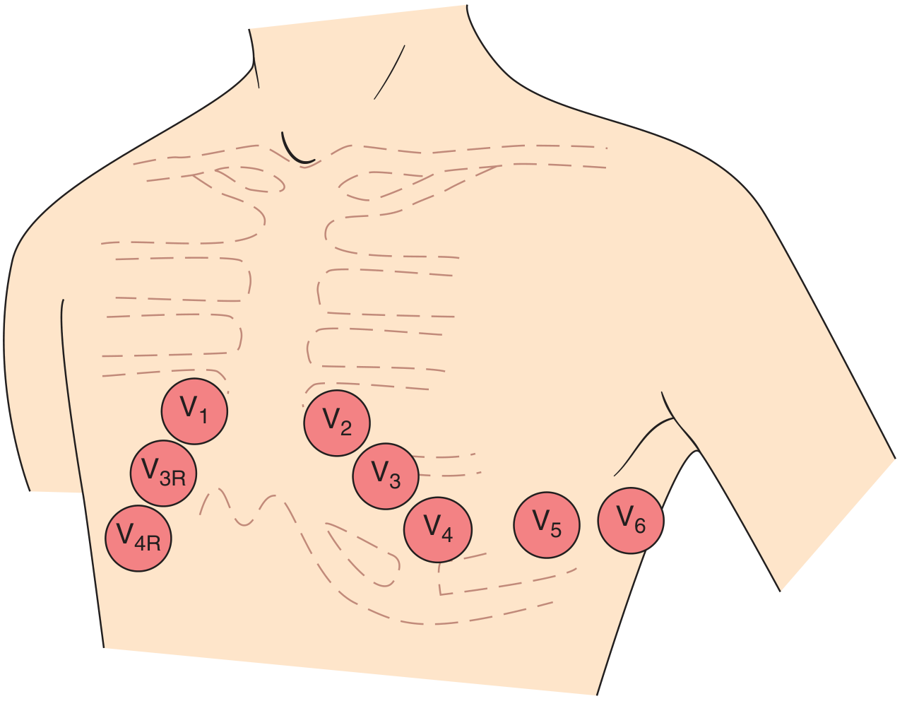
(Harrison's 22e, Fig. 247-5 - Horizontal plane (chest/precordial) lead placement)
The Normal ECG Waveform - A to Z
          R
          |
    P     |          T          U
   /\     |         /\         /\
--/  \----|--------/  \-------/  \--  (baseline)
          |\      /
          | \    /
          |  \  /
          |   \/
          S
   |--PR--|--QRS--|--ST--|----QT----|
| Component | What it represents | Normal values |
|---|---|---|
| P wave | Atrial depolarization | Duration < 120 ms; amplitude < 2.5 mm; positive in I, II, aVF |
| PR interval | AV conduction time (atria → AV node → His-Purkinje) | 120-200 ms (3-5 small squares) |
| QRS complex | Ventricular depolarization | < 120 ms (< 3 small squares) |
| ST segment | Early ventricular repolarization (isoelectric normally) | Flat at baseline |
| T wave | Ventricular repolarization | Upright in I, II, V3-V6; inverted normally in aVR, V1 |
| QT interval | Total ventricular repolarization | < 440 ms (men), < 460 ms (women); correct with Bazett: QTc = QT/√RR |
| U wave | After-depolarization of Purkinje fibers | Upright, small; prominent in hypokalemia |
Key professor tip: The T wave is normally opposite in polarity to the QRS only in bundle branch blocks (secondary change). If the T wave goes the "wrong way" in a normal QRS, it is a PRIMARY repolarization abnormality (ischemia, electrolytes, drugs). - Harrison's 22e
Heart Rate Calculation
- Regular rhythm: 300 ÷ number of large squares between R-R peaks
- 1 large square = 300 bpm, 2 = 150, 3 = 100, 4 = 75, 5 = 60, 6 = 50
- Irregular rhythm: Count QRS complexes in a 10-second strip × 6
QRS Axis (Frontal Plane)
- Normal: -30° to +90°
- Left axis deviation (LAD): more negative than -30° → most common cause is left anterior fascicular block
- Right axis deviation (RAD): more positive than +90° → RVH, PE, left posterior fascicular block
- Quick trick:
- Lead I positive + aVF positive = normal axis
- Lead I positive + aVF negative = LAD
- Lead I negative + aVF positive = RAD
- Both negative = "Northwest" / extreme axis
R-Wave Progression (Precordial Leads)
- R wave normally grows from V1 to V5 (transition at V3-V4 where R = S)
- Poor R-wave progression (small r waves persisting through V4) = anterior MI or LVH
- Diagram of ventricular depolarization vectors:
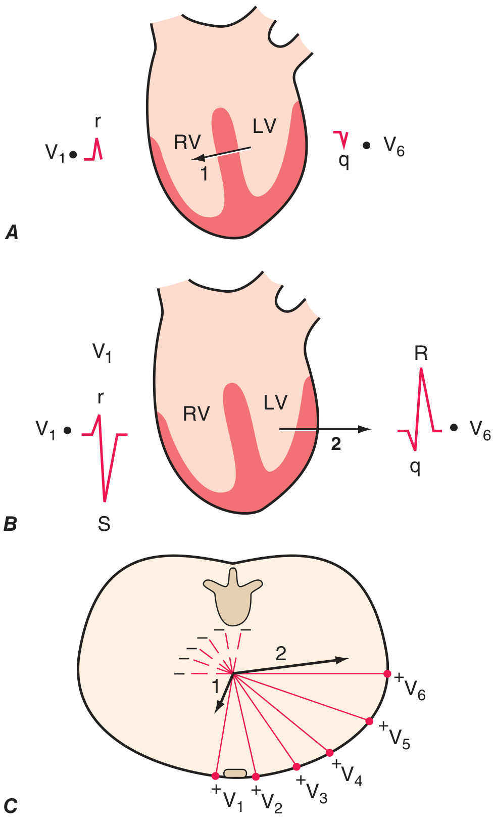
(Harrison's 22e, Fig. 247-6 - Two phases of ventricular depolarization. Phase 1: septal, right to left → small r in V1, small q in V6. Phase 2: LV-dominant, leftward/posterior → deep S in V1, tall R in V6)
PART 2 - The Systematic Approach (14 Steps - Harrison's)
Every ECG should be read in this order - professors love this:
- Standardization/calibration (10mm = 1mV, 25mm/sec)
- Rhythm (sinus vs. non-sinus)
- Heart rate
- PR interval / AV conduction
- QRS duration
- QT/QTc interval
- Mean QRS electrical axis
- P wave morphology
- QRS voltages
- R-wave progression (precordial)
- Abnormal Q waves
- ST segments
- T waves
- U waves
Always compare with prior ECGs when available. - Harrison's 22e, "Clinical Interpretation of the ECG"
PART 3 - Arrhythmias
Bradyarrhythmias
Sinus Bradycardia: Rate < 60 bpm, normal P-QRS-T morphology
AV Blocks
| Block | ECG Feature | Location | Danger |
|---|---|---|---|
| 1st Degree | PR > 200 ms, no dropped beats | AV node | Benign |
| 2nd Degree Mobitz I (Wenckebach) | Progressively lengthening PR → dropped QRS; "group beating"; RR shortens before dropped beat | AV node (proximal) | Usually benign, rarely progresses |
| 2nd Degree Mobitz II | Fixed PR interval, then sudden dropped QRS with no warning | His-Purkinje (distal) | DANGEROUS - may progress to complete block |
| 3rd Degree (Complete) | P waves and QRS completely dissociated; atrial rate > ventricular rate | AV node or infranodal | EMERGENCY - needs pacing |
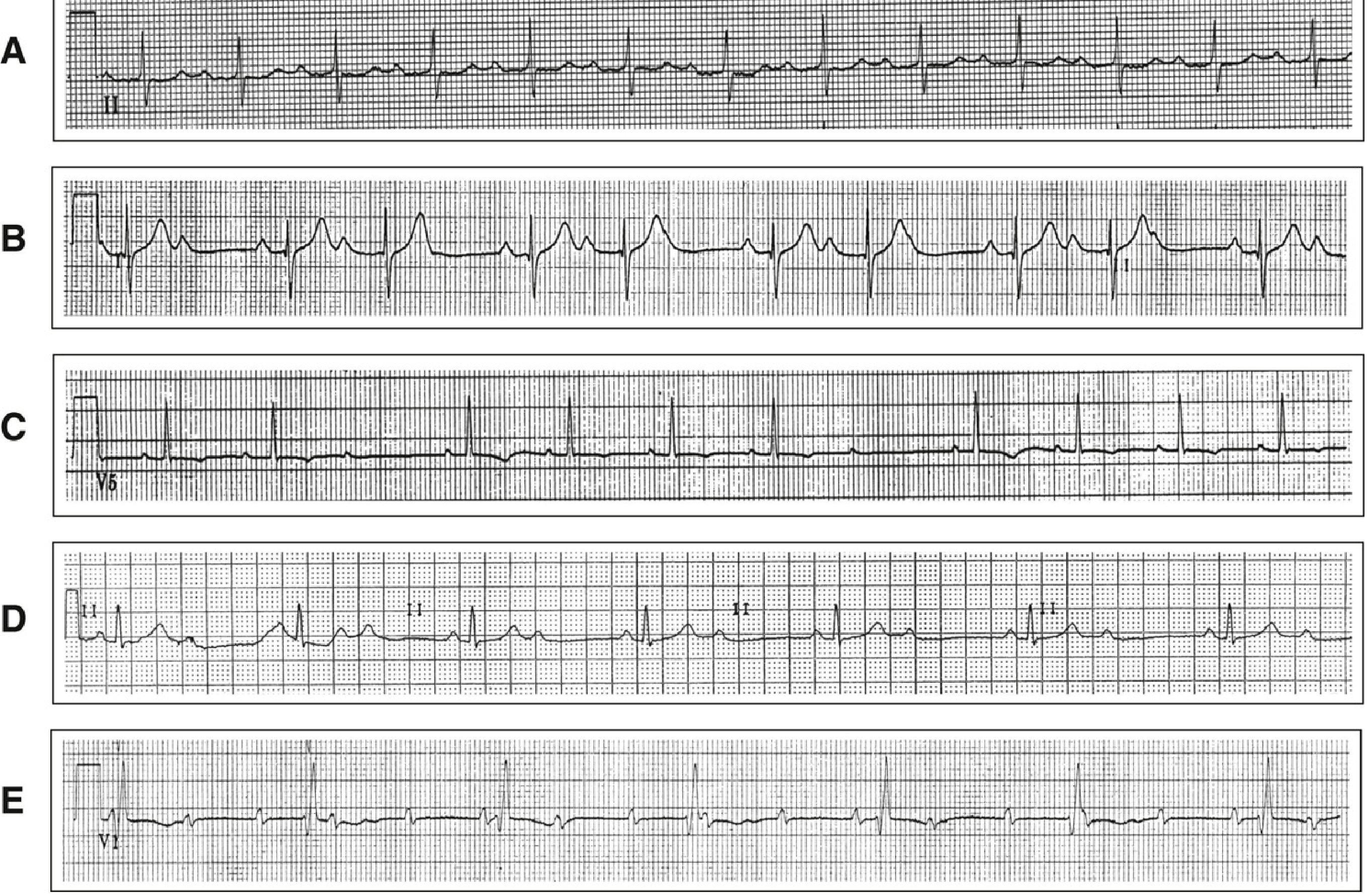
(Washington Manual, Fig. 7-5: A=1st degree AVB, B=Mobitz I, C=Mobitz II, D=2:1 block, E=Complete heart block with junctional escape)
Key teaching point: 2:1 block is difficult to classify as Mobitz I vs II. Narrow QRS + concomitant first-degree block = likely Mobitz I (proximal). Wide QRS + bundle branch block = likely Mobitz II (distal, dangerous). - Washington Manual
Tachyarrhythmias
Narrow Complex (QRS < 120 ms) = Supraventricular
| Rhythm | Key ECG Features |
|---|---|
| Sinus Tachycardia | Rate 100-160, normal P before every QRS, gradual onset/offset |
| Atrial Fibrillation (AF) | Irregularly irregular; NO distinct P waves; fibrillatory baseline; ventricular rate variable |
| Atrial Flutter | "Sawtooth" flutter waves at ~300/min; regular ventricular rate (usually 150 = 2:1 block); most common ratio 2:1 |
| SVT (AVNRT) | Sudden onset/termination; rate 150-250; P waves hidden in or just after QRS |
Professor pearl: AF is the most common sustained arrhythmia after sinus tachycardia. It is an "irregularly irregular" rhythm - no two R-R intervals are the same. - Tintinalli's EM
ECG strip showing sinus tachycardia, rapid AF, 2:1 atrial flutter, AF, and return to sinus rhythm during exercise stress test:
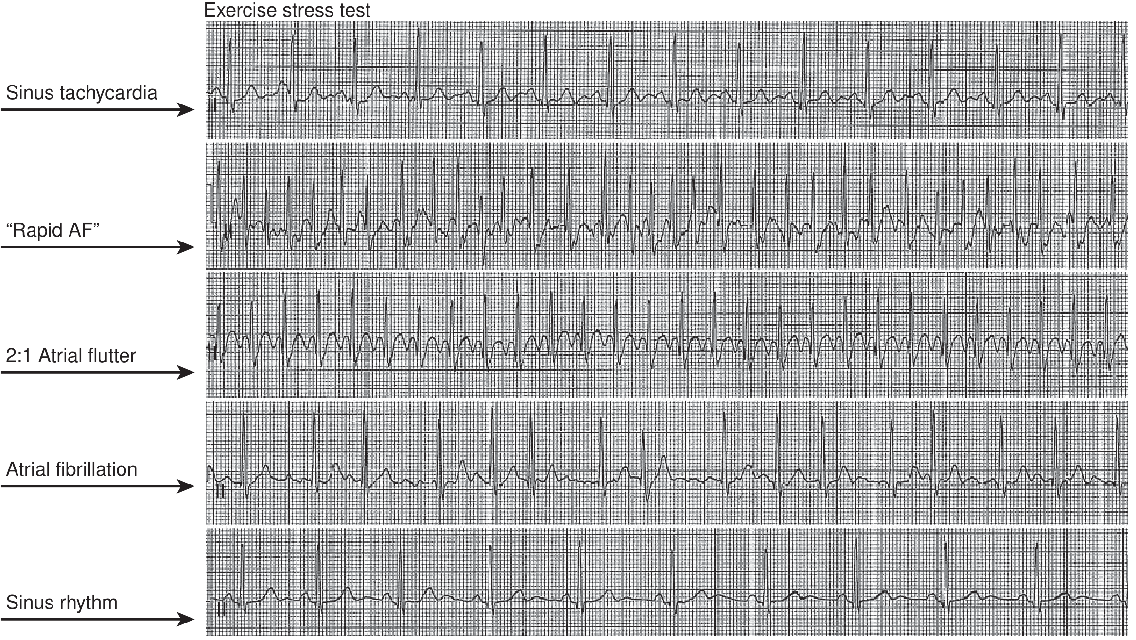
(Braunwald's Heart Disease, Fig. 65.9)
Wide Complex (QRS ≥ 120 ms)
| Rhythm | Key Features |
|---|---|
| Ventricular Tachycardia (VT) | Rate >100, wide QRS, AV dissociation (P waves march through independently), fusion beats, capture beats |
| VF | Totally chaotic, no identifiable QRS - EMERGENCY |
| SVT with aberrancy | Wide complex SVT (BBB pattern), but history/maneuvers help distinguish from VT |
Rule of thumb: Wide complex tachycardia = VT until proven otherwise.
PART 4 - Bundle Branch Blocks
Diagnosis requires QRS ≥ 120 ms (3 small squares)
Right Bundle Branch Block (RBBB)
- rSR' ("rabbit ears" or M-pattern) in V1
- Wide S wave in I, V5, V6
- T-wave inversion in V1-V3 (secondary change, expected)
- Can be normal variant OR indicate RV strain, PE, congenital heart disease
Left Bundle Branch Block (LBBB)
- Broad, notched R wave ("W"-shaped in V1, "M"-shaped in V5/V6)
- No septal q waves in lateral leads (I, V5, V6)
- T-wave inversion in V5, V6 (secondary, expected)
- LBBB in a chest pain patient = treat as STEMI equivalent (it hides ischemia and is an independent risk marker)
- Isolated LBBB in an otherwise healthy person carries 2× increased cardiovascular risk
Mnemonic:
- WiLLiaM MaRRoW: W in V1 = LBBB (Left); M in V1 = RBBB (Right)
- Or: LBBB = "Leave the patient" (more dangerous); RBBB = "Reassure" (usually benign)
(Goldman-Cecil Medicine, "Intraventricular Blocks")
PART 5 - Myocardial Ischemia and Infarction
Mechanism (Why ECG Changes Occur)
Acute ischemia lowers resting membrane potential and shortens action potential duration → voltage gradient between normal and ischemic zones → currents of injury → ST deviation. - Harrison's 22e
Evolutionary Changes in STEMI
| Stage | ECG Finding |
|---|---|
| Hyperacute (minutes) | Tall, broad, peaked "hyperacute" T waves |
| Early (hours) | ST elevation at J point |
| Established | ST elevation + Q wave formation |
| Old (hours-days) | T-wave inversion; Q waves persist |
| Chronic | Persistent Q waves (scar), T waves may normalize |
Localization of MI by Lead Groups
| Leads with ST Elevation | Territory | Culprit Artery |
|---|---|---|
| II, III, aVF | Inferior | RCA (80%) |
| V1-V4 | Anterior/Septal | LAD |
| I, aVL, V5-V6 | Lateral | LCx |
| V1-V2 (tall R + ST depression) | Posterior | RCA / LCx |
| V1 + inferior leads (ST elevation in V3R, V4R) | Right Ventricle | Proximal RCA |
NSTEMI / Unstable Angina
- ST depression (subendocardial ischemia) or T-wave inversion
- No ST elevation, but troponin distinguishes NSTEMI (elevated) from UA (normal)
Wellens Syndrome (professor favourite!)
- Deeply inverted or biphasic T waves in V2-V3 in a pain-FREE patient
- Indicates critical LAD stenosis - do NOT stress test!
De Winter T Waves
- ST depression + tall peaked T waves in V1-V6 = proximal LAD occlusion equivalent (STEMI variant - needs cath lab)
PART 6 - Other High-Yield ECG Patterns
LVH (Left Ventricular Hypertrophy)
- Sokolow-Lyon criteria: S in V1 + R in V5 or V6 ≥ 35 mm
- R in aVL ≥ 11 mm (Cornell criterion)
- Often associated with "strain pattern" (ST depression + T inversion in lateral leads)
RVH (Right Ventricular Hypertrophy)
- Tall R in V1 (R > S in V1)
- Right axis deviation
- ST depression + T inversion in V1-V3
Pericarditis
- Diffuse ST elevation (saddle-shaped, concave upward) in multiple lead groups (not fitting one territory)
- PR depression (reciprocal PR elevation in aVR) - pathognomonic early sign
- No reciprocal ST depression (vs. STEMI)
Pulmonary Embolism (PE)
- Sinus tachycardia (most common, ~44%)
- S1Q3T3 pattern: deep S in lead I, Q wave + T inversion in lead III
- New RBBB, right axis deviation
- T inversions in V1-V4 (RV strain)
Hyperkalemia
- Peaked/tented T waves (early)
- Wide QRS (later)
- Flat/absent P waves
- Sine-wave pattern → VF (severe)
Hypokalemia
- Prominent U waves (larger than T in same lead)
- T-wave flattening
- Prolonged QT
- Risk of torsades de pointes
Hypercalcemia / Hypocalcemia
- Hypercalcemia: Short QT (short ST segment)
- Hypocalcemia: Long QT (prolonged ST segment)
Long QT Syndrome
- QTc > 440 ms (men), > 460 ms (women)
- Risk of torsades de pointes (polymorphic VT) → VF → sudden death
- Causes: congenital channelopathies, drugs (amiodarone, sotalol, quinolones, antipsychotics), electrolyte disturbance (↓K, ↓Mg, ↓Ca)
Wolff-Parkinson-White (WPW)
- Short PR interval (< 120 ms)
- Delta wave (slurred upstroke of QRS)
- Wide QRS
- Due to accessory pathway (Bundle of Kent) bypassing AV node
Brugada Syndrome
- Coved-type ST elevation in V1-V2 (right chest leads)
- RBBB pattern
- Risk of sudden cardiac death - important channelopathy
PART 7 - The "Things That Impress a Professor" Checklist
When presenting an ECG to your professor, say ALL of these:
- "I will use a systematic approach..." (then use the 14-step Harrison's method)
- Always state rate, rhythm, axis, intervals first
- Describe P waves: "present/absent, morphology, relation to QRS"
- Know the difference between primary vs. secondary ST-T changes
- In any wide-complex tachycardia: "This is VT until proven otherwise"
- In any new LBBB with chest pain: "This is a STEMI equivalent"
- Know reciprocal changes (ST depression in leads opposite to ST elevation) - confirms STEMI
- State the "territory and likely culprit artery" for any ischemic pattern
- Mention QTc (corrected QT), not just QT
- With AV blocks: always classify (1st/2nd Mobitz I or II/3rd) and state the clinical risk
Quick-Reference Summary Card
| Normal Value | Number to Know |
|---|---|
| PR interval | 120-200 ms |
| QRS duration | < 120 ms |
| QTc (men) | < 440 ms |
| QTc (women) | < 460 ms |
| Normal axis | -30° to +90° |
| LVH (Sokolow) | S-V1 + R-V5/6 ≥ 35 mm |
| STEMI threshold | ≥ 1 mm ST elevation in 2 contiguous leads (≥ 2 mm in V1-V3) |
| Complete BBB | QRS ≥ 120 ms |
| AF flutter rate | ~300/min atrial, ~150/min ventricular (2:1) |
Sources: Harrison's Principles of Internal Medicine 22e (2025) - "Clinical Interpretation of the ECG"; Goldman-Cecil Medicine International Edition, 2 Vol Set - "Conduction Abnormalities" & "Intraventricular Blocks"; Braunwald's Heart Disease, 2 Vol Set - "Resting Electrocardiogram"; The Washington Manual of Medical Therapeutics - "AV Conduction Disturbances"; Tintinalli's Emergency Medicine - "Atrial Fibrillation/Flutter"
Now a need tips for me and practice this thing
Searching the Web
best free ECG practice websites tools for medical students 2024 2025
Here are your practical tips + the best free resources to actually get good at ECGs fast.
🧠 How to Actually Learn ECGs (Not Just Memorize Them)
The #1 Mindset Shift
Most students try to memorize patterns. That fails. Instead, ask "why" for every finding:
- Why does LBBB hide ischemia? (altered depolarization changes ST baseline)
- Why is Mobitz II dangerous? (infranodal block - escape rhythm is unreliable)
- Why does PE cause S1Q3T3? (acute RV dilation rotates the heart electrically)
When you understand the mechanism, the pattern sticks forever.
Study Tips by Stage
Stage 1 - Build the Foundation (Week 1)
- Read ECGs in one fixed order every single time (use the 14-step Harrison's system from the guide above) - never "spot read"
- Draw the normal ECG waveform from memory every day until it's automatic
- Learn the five boxes trick for axis (I + aVF quadrant method) - 30 seconds per ECG
- Memorize normal values as numbers, not ranges: "PR is 3-5 small boxes, QRS is under 3 boxes, QT is under half the RR interval"
Stage 2 - Pattern Recognition (Week 2)
- Learn the "Big 5" patterns your prof will definitely ask:
- STEMI (ST elevation, territory, reciprocal changes)
- AF (irregularly irregular, no P waves)
- Complete heart block (complete P-QRS dissociation)
- LBBB vs RBBB (WiLLiaM MaRRoW mnemonic)
- VT vs SVT with aberrancy (AV dissociation = VT)
- For each pattern: describe it out loud as if presenting to your professor
Stage 3 - Volume Practice (Week 3 onward)
- Do at least 5 ECGs per day - this is the only thing that works
- Time yourself: aim to read a full 12-lead in under 3 minutes
- Read one "difficult" or "tricky" case weekly (Wellens, De Winter, posterior MI, Brugada)
Memory Tricks That Actually Work
| Pattern | Trick |
|---|---|
| RBBB vs LBBB in V1 | WiLLiaM MaRRoW - W in V1 = LBBB, M in V1 = RBBB |
| Rate from R-R | 300, 150, 100, 75, 60, 50 (one large box each) |
| Axis quadrant | I + aVF both positive = normal. I positive, aVF negative = LAD |
| Mobitz I vs II | "Wenckebach WARMS up then DROPS" (lengthens then drops) |
| PE on ECG | S1Q3T3 = "1, 2, 3 PE" |
| Hyperkalaemia | Peaks early (peaked T), then P waves disappear, then wide QRS, then sine wave = "Tall tents, flat prairie, wide roads, no more" |
| Long QT risk | "Drugs that kill: Class IA, III antiarrhythmics, macrolides, fluoroquinolones, antipsychotics, antiemetics (metoclopramide)" |
| Pericarditis PR | PR depression - the "D" in periocarDitis drops below baseline |
Common Mistakes to Avoid
- Calling rate without rhythm first - always state rhythm before rate
- Missing reciprocal changes - for every STEMI, always look at the opposite leads for ST depression (confirms the diagnosis)
- Ignoring the P wave - most arrhythmia diagnoses live or die on P-wave analysis
- Not correcting QT - always report QTc not raw QT
- Diagnosing ischemia in LBBB - LBBB makes ST interpretation unreliable (use Sgarbossa criteria if needed)
- Forgetting to compare - always ask "is there a prior ECG?" - this is the most clinically useful step
Free Practice Resources
Websites (Free, No Login Needed)
- ECG Wave-Maven (Harvard): ecg.bidmc.harvard.edu - 400+ real clinical cases with answers. The gold standard for self-study.
- Practical Clinical Skills: practicalclinicalskills.com/ecg - short courses, drills, and quizzes
- ECG Made Simple: ecgmadesimple.ca - free videos + interactive materials
- Dr. Smith's ECG Blog: hqmeded-ecg.blogspot.com - real cases daily, great for advanced learning
Apps (Free or Freemium)
- ECG Quiz (iOS/Android) - rapid-fire pattern recognition
- Cardioserv - good for systematic approach drills
- Geeky Medics ECG - medical student focused
YouTube Channels
The best channel for visual learners - clear, fast, and clinical:
(Dr. Burns / LifeInTheFastLane - ECG Interpretation Masterclass)
Your Weekly Practice Schedule
| Day | What to Do | Time |
|---|---|---|
| Monday | 5 ECGs on Wave-Maven (systematic read each one) | 20 min |
| Tuesday | 5 ECGs - focus on arrhythmias only | 20 min |
| Wednesday | 5 ECGs - focus on ischemia/MI patterns | 20 min |
| Thursday | 5 ECGs - focus on conduction/blocks | 20 min |
| Friday | 5 mixed ECGs timed (3 min each), then review mistakes | 20 min |
| Weekend | Read 1 interesting case from Dr. Smith's blog | 10 min |
Total: ~1.5 hours/week - enough to be significantly ahead of your classmates in 4 weeks.
The "Professor Presentation" Formula
Every time you read an ECG out loud, use this exact structure:
"This is a 12-lead ECG recorded at 25 mm/sec and 10 mm/mV. The rhythm is [sinus/AF/etc], rate is [X] bpm, the PR interval is [X] ms, QRS duration is [X] ms, and QTc is [X] ms. The axis is [normal/LAD/RAD]. P waves are [present/absent/abnormal]. There are [no/these] ST changes: [describe with leads]. My conclusion is [diagnosis]. The most important clinical concern is [action]."
This structure covers all 14 Harrison's parameters and will consistently impress.
Bottom line: Volume beats perfection. Read 5 ECGs a day for 4 weeks and you will be in the top 10% of your class. Use Wave-Maven as your daily practice tool - it has real cases with teaching points written by Harvard cardiologists.
This is a shared conversation. Sign in to Orris to start your own chat.