Here is a comprehensive reference covering all 18 topics in your syllabus.
Thyroid Diseases: Complete Clinical Reference
1. Clinical Classification of Thyroid Diseases
Nikolaev Classification (O.V. Nikolaev, 1949 - Soviet/Russian standard)
Used widely in post-Soviet medical practice. Classifies thyroid diseases by:
A. By functional state:
- Euthyroid conditions (normal function)
- Hypothyroid conditions (reduced function)
- Hyperthyroid/thyrotoxic conditions (excess function)
B. By morphology:
- Diffuse goiter
- Nodular goiter
- Diffuse-nodular (mixed) goiter
C. By degree of enlargement (Nikolaev's goiter grades, 0-V - see Topic 4)
WHO Classification (2001, updated)
Goiter grades (WHO 2001):
| Grade | Description |
|---|
| 0 | No palpable or visible goiter |
| 1 | Palpable goiter, not visible with neck in normal position |
| 2 | Visible goiter with neck in normal position |
Broader disease categories (ICD-based):
- Simple and unspecified goiter
- Thyrotoxicosis (hyperthyroidism)
- Hypothyroidism
- Thyroiditis
- Other thyroid disorders (nodules, cancer, congenital anomalies)
Bailey & Love Surgical Classification of Thyroid Swellings:
| Category | Type | Examples |
|---|
| Simple goiter (euthyroid) | Diffuse hyperplastic | Physiological, pubertal, pregnancy |
| Multinodular goiter | |
| Toxic | Diffuse (Graves' disease) | |
| Multinodular toxic | |
| Toxic adenoma | |
| Neoplastic | Benign / Malignant | |
| Inflammatory | Autoimmune | Hashimoto's, chronic lymphocytic |
| Granulomatous | de Quervain's |
| Fibrosing | Riedel's |
| Infective | Acute bacterial, viral thyroiditis |
(Bailey and Love's Short Practice of Surgery, 28th Edition)
2. Congenital Anomalies of the Thyroid Gland
The thyroid develops from an endodermal outgrowth at the foramen cecum of the tongue, descending via the thyroglossal duct to its final position. Anomalies arise from errors in this process:
Agenesis / Aplasia
- Complete absence of thyroid tissue
- Leads to severe congenital hypothyroidism (cretinism if untreated)
- Accounts for ~30-35% of congenital hypothyroidism
Hypoplasia
- Inadequately developed but present gland
- Causes congenital hypothyroidism of variable severity
Ectopic thyroid
- Thyroid tissue located anywhere along the descent path
- Most common: lingual thyroid (at foramen cecum) - may be the only thyroid tissue
- Others: sublingual, mediastinal, intrathoracic
- Lingual thyroid presents as a mass at base of tongue; may cause dysphagia/dysphonia
Thyroglossal duct cyst
- Persistence of the thyroglossal duct
- Most common congenital neck mass in children
- Presents as a midline cystic swelling that moves on swallowing AND on tongue protrusion
- Treatment: Sistrunk operation (excision of cyst + central portion of hyoid bone)
Hemiagenesis / Thyroid lobe agenesis
- Absence of one lobe (usually left)
- Often discovered incidentally; may be euthyroid
Pyramidal lobe persistence
- Remnant of the thyroglossal duct as a superior thyroid lobe extension
- Present in ~50% of people; clinically relevant in goiter/hyperthyroidism
Accessory thyroid tissue
- Small nodules of ectopic thyroid alongside the normal gland
3. Concept of Endemic and Sporadic Goiter: Prevalence and Etiology
Definitions
Endemic goiter: Goiter affecting >10% of the population in a geographic region. By WHO definition, a community is iodine-deficient if the goiter prevalence exceeds 5% in school-age children.
Sporadic goiter: Goiter occurring in individuals within a non-endemic (iodine-sufficient) region; accounts for isolated cases.
Prevalence
- Approximately 200 million people worldwide have goiter
- Endemic areas: inland/mountainous regions far from the sea (Alps, Himalayas, Andes, Central Africa, parts of Central Asia)
- Sporadic: occurs at ~5% prevalence even in iodine-sufficient populations
Etiological Factors
Endemic goiter:
- Iodine deficiency - primary cause; dietary iodine <50 mcg/day leads to inadequate thyroid hormone synthesis → compensatory TSH rise → gland enlargement
- Goitrogens in food: cassava (thiocyanate), millet, soya, brassicas, water pollutants (fluoride, calcium excess)
- Selenium deficiency (selenium is required for iodothyronine deiodinases)
- Genetic susceptibility modifying TSH receptor signaling
Sporadic goiter:
- Dyshormonogenesis: Inherited enzyme defects in thyroid hormone synthesis (organification defects, iodotyrosine dehalogenase deficiency, thyroglobulin synthesis defects)
- Goitrogen exposure (drugs: lithium, amiodarone, aminoglutethimide; foods as above)
- Autoimmune stimulation (non-toxic phase of Hashimoto's)
- Physiological: puberty, pregnancy (hCG has weak TSH-like activity; increased renal iodine clearance)
- Microadenoma of the pituitary secreting excess TSH (rare)
(Bailey and Love's Short Practice of Surgery, 28th Ed; Costanzo Physiology 7th Ed)
4. Pathogenesis and Classification of Thyroid Enlargement Grades
Pathogenesis
The fundamental mechanism is inadequate thyroid hormone production relative to demand:
- Reduced T3/T4 (from iodine deficiency, dyshormonogenesis, or increased peripheral demand)
- Compensatory TSH rise from the anterior pituitary
- TSH-driven hypertrophy and hyperplasia of follicular cells
- Diffuse hyperplastic goiter - early, potentially reversible stage
- Involution and re-stimulation cycles → heterogeneous nodularity
- Multinodular goiter - irreversible stage; some nodules become autonomous
Additional growth factors including immunoglobulins and local cytokines contribute. Nodularity reflects clonal variation in sensitivity to growth stimuli.
Grades of Thyroid Enlargement
Nikolaev (Russian) Classification - 5 grades:
| Grade | Criteria |
|---|
| 0 | No enlargement |
| I | Enlargement of isthmus palpable |
| II | Lobes and isthmus palpable, neck contour normal |
| III | Goiter visible - "thick neck" |
| IV | Clearly visible goiter, changes neck shape |
| V | Very large goiter causing compression/displacement |
WHO Classification (2001) - 3 grades:
| Grade | Criteria |
|---|
| 0 | No goiter (not palpable or visible) |
| 1 | Palpable but not visible with neck in normal position |
| 2 | Visible with neck in normal position |
(The WHO classification simplified from the earlier Perez/Stanbury 3-grade system)
5. Clinical Presentation of Endemic and Sporadic Goiter
Both types are typically euthyroid at presentation (simple goiter).
Symptoms from gland enlargement:
- Neck swelling (cosmetic concern)
- Dysphagia - compression of esophagus by large/retrosternal goiter
- Dyspnea and stridor - tracheal compression (especially retrosternal extension)
- Hoarseness - rarely from RLN compression (raises concern for malignancy)
- Superior vena cava syndrome - with large retrosternal goiter (rare)
- Pemberton's sign: facial plethora, JVP elevation when arms raised above head (retrosternal goiter)
Systemic symptoms (if euthyroid): absent
Physical signs:
- Swelling moves on swallowing (key diagnostic feature)
- May be diffuse (hyperplastic) or nodular
- Soft/firm consistency; cystic areas from hemorrhage into nodules
- Tracheal deviation may be visible/palpable
Retrosternal goiter:
- May have no visible neck mass
- Presents with obstructive symptoms
- Diagnosed by CT/MRI
Transition to toxic (hyperthyroid) state can occur with long-standing nodular goiter as nodules become autonomous.
6. Diagnostic Methods for Endemic and Sporadic Goiter
History and Physical Examination
- Area of origin (endemic vs. sporadic), duration, rate of growth
- Family history, drug history (lithium, amiodarone)
- Symptoms of compression, hypo/hyperthyroidism
Laboratory Tests
- TSH (first-line): suppressed in hyperthyroidism, elevated in hypothyroidism; normal in simple goiter
- Free T3, Free T4: to assess functional status
- Anti-TPO, anti-Tg antibodies: elevated in autoimmune thyroiditis; may be mildly elevated in endemic goiter
- Serum thyroglobulin: marker for iodine deficiency; elevated in goiter
- Urine iodine concentration (UIC): population-level and individual assessment; <100 mcg/L indicates deficiency
- Calcitonin: if medullary thyroid cancer suspected
Imaging
- Ultrasound (US): first-line imaging; defines goiter size, echotexture, nodules, vascularity; identifies features suspicious for malignancy (microcalcifications, irregular margins, hypoechogenicity, tall-wide ratio >1)
- CT/MRI: evaluation of retrosternal extension, tracheal compression, mediastinal involvement
- Chest X-ray: tracheal deviation, retrosternal mass, calcifications
Radionuclide Studies
- 99mTc scintigraphy or 123I uptake scan: distinguishes hot (functioning) vs. cold (non-functioning) nodules; cold nodules carry higher malignancy risk (~5-15%)
- Radioiodine uptake (RAIU): elevated in iodine deficiency, normal or low in thyroiditis
Fine-Needle Aspiration Cytology (FNAC/FNA)
- Indicated for nodules >1 cm with suspicious features on US, or any nodule growing
- Bethesda reporting system:
- Thy1: Non-diagnostic
- Thy2: Non-neoplastic (benign)
- Thy3: Follicular (indeterminate)
- Thy4: Suspicious for malignancy
- Thy5: Malignant
(Bailey and Love's Short Practice of Surgery, 28th Ed)
7. Conservative Treatment and Surgical Management of Endemic/Sporadic Goiter
Conservative Treatment
Iodine supplementation (endemic goiter):
- Iodized salt programs (most effective public health measure)
- Potassium iodide or iodate supplementation
- In early hyperplastic stages, iodine can achieve partial regression
Levothyroxine (T4) suppression therapy:
- Dose: 0.15-0.2 mg/day to suppress TSH
- Early hyperplastic goiters may regress over several months
- Nodular goiters: limited effectiveness; >50% of benign nodules regress spontaneously over 10 years
- Risks: atrial fibrillation, osteoporosis (especially in postmenopausal women)
- Current evidence does not strongly support routine T4 suppression for nodular goiters
Observation:
- Most patients with multinodular euthyroid goiter are asymptomatic and require monitoring only
Indications for Surgery
- Suspected or confirmed malignancy (FNA Thy4-5, suspicious US features)
- Pressure symptoms (dysphagia, stridor, dyspnea) not explained otherwise
- Retrosternal goiter causing compression
- Cosmetic deformity unacceptable to patient
- Progressive enlargement
- Toxic transformation (secondary thyrotoxicosis)
- Failure of conservative treatment
Types of Surgical Intervention
| Operation | Description | Indication |
|---|
| Hemithyroidectomy (lobectomy) | Removal of one lobe + isthmus | Unilateral disease, solitary nodule |
| Subtotal thyroidectomy | Leaves 2-8 g of normal tissue bilaterally | Bilateral benign goiter (historical); higher recurrence risk |
| Dunhill procedure | Total lobectomy one side + subtotal on other | Asymmetric bilateral disease |
| Near-total thyroidectomy | Leaves <1 g of tissue near RLN/parathyroids | Cancer, toxic goiter |
| Total thyroidectomy | Complete removal | Cancer, large bilateral goiter, Graves' disease |
Current preference: Total thyroidectomy is increasingly favored for bilateral disease (eliminates recurrence risk, allows RAI if cancer found incidentally).
(Bailey and Love's Short Practice of Surgery, 28th Ed)
8. Concept of Toxic Goiter: Prevalence and Etiology
Definitions
Toxic goiter: Thyroid enlargement accompanied by hyperthyroidism (thyrotoxicosis).
Diffuse Toxic Goiter (Graves' disease): An autoimmune disorder characterized by diffuse thyroid enlargement, hyperthyroidism, and extrathyroidal manifestations (ophthalmopathy, dermopathy).
Toxic (Thyrotoxic) Adenoma (Plummer's disease): A solitary autonomous hyperfunctioning thyroid nodule, typically ≥2 cm, that produces thyroid hormones independently of TSH.
Prevalence
- Graves' disease: most common cause of hyperthyroidism (~70-80% of cases); prevalence ~0.5% in the general population; F:M ratio ~7-10:1; peak age 20-50 years
- Toxic adenoma: ~5-10% of hyperthyroidism causes; toxic multinodular goiter ~15-20%
- Overall hyperthyroidism prevalence: ~1% in women, 0.1% in men
Etiological Factors
Graves' disease:
- Autoimmune: TSH receptor stimulating antibodies (TSH-RAb / TRAb / TSI) bind TSH receptor → continuous unregulated stimulation of thyroid follicular cells
- Genetic predisposition: HLA-DR3, HLA-B8 association; CTLA-4 gene polymorphisms; family clustering
- Environmental triggers: iodine excess, infections (Yersinia enterocolitica has TSH receptor-like antigen), stress, smoking (strong risk factor for ophthalmopathy), postpartum period, interferon-alpha therapy
Toxic adenoma:
- Somatic activating mutations in the TSH receptor gene (most common: codon 619, 623, 632) or Gs-alpha subunit → constitutive cAMP signaling → autonomous growth and function
- Not autoimmune; no TSH-RAb
- Surrounding normal thyroid is suppressed (visible on scan as "hot nodule" with absent uptake elsewhere)
(Mulholland and Greenfield's Surgery 7e; Bailey and Love 28th Ed)
9. Thyrotoxicosis: Pathogenesis, Classification by Severity and Stage, Treatment
Pathogenesis
Excess thyroid hormones (T3 > T4 in potency) act on virtually every organ:
- Metabolic: Increased oxygen consumption, heat production, BMR elevation; weight loss despite hyperphagia; gluconeogenesis and glycogenolysis
- Cardiovascular: Increased heart rate, cardiac output; decreased peripheral vascular resistance; atrial fibrillation (especially elderly)
- Neuromuscular: Tremor, hyperreflexia, proximal myopathy
- Bone: Accelerated remodeling → osteoporosis
- Hematopoietic: Mild anemia, elevated SHBG
- Adrenal: Increased cortisol clearance → relative adrenal insufficiency during crisis
In Graves' disease: TRAb stimulates TSH receptors without feedback inhibition → persistent excess T3/T4 production and gland hypertrophy.
Classification by Severity (Graves'/Thyrotoxicosis)
Nikolaev / Classic Russian Classification (by degree of thyrotoxicosis):
| Grade | Features |
|---|
| Mild (Grade I) | Slight nervousness, resting HR 80-100 bpm, weight loss <10%, no AF, mild goiter, working capacity maintained |
| Moderate (Grade II) | HR 100-120 bpm, significant weight loss 10-20%, tremor, exophthalmos, goiter enlarged, working capacity reduced |
| Severe (Grade III) | HR >120 bpm, severe weight loss >20%, fibrillation, myopathy, marked exophthalmos, signs of visceral involvement (heart, liver), disabled |
Thyroid Storm (Thyrotoxic Crisis): Medical emergency; precipitated by surgery, infection, trauma, or RAI in undertreated patients. Features: hyperpyrexia (>38.5°C), severe tachycardia, CNS disturbance (agitation, psychosis, coma), GI symptoms, cardiovascular collapse.
Treatment Methods
1. Antithyroid Drugs (ATDs) - Thionamides:
- Methimazole (carbimazole in Europe) - first-line for most patients; blocks thyroid peroxidase (TPO), inhibiting organification of iodide; carbimazole is converted to methimazole in vivo
- Propylthiouracil (PTU) - also inhibits peripheral T4→T3 conversion; preferred in pregnancy (1st trimester) and thyroid storm
- Side effects: rash, agranulocytosis (1:500, usually within 90 days - fever + pharyngitis → stop immediately, check WBC); hepatotoxicity (PTU > methimazole)
- Titration block-and-replace or dose titration regimens; usual duration 12-18 months
- Remission rate: ~45% for Graves'; recurrence common
2. Beta-blockers:
- Propranolol, atenolol - symptomatic control of tachycardia, tremor, anxiety, heat intolerance
- Propranolol also inhibits peripheral T4→T3 conversion at high doses
- Used adjunctively; not curative
3. Radioactive Iodine (RAI / 131I):
- Destroys thyroid tissue by beta radiation
- Advantages: non-surgical, definitive, outpatient
- Leads to hypothyroidism in majority → lifelong levothyroxine
- Contraindicated in pregnancy, breastfeeding, active ophthalmopathy (can worsen)
4. Surgery (Thyroidectomy):
Thyroid Storm Treatment:
- PTU 200-250 mg q4h (blocks synthesis and conversion)
- Lugol's iodine 1 hour after PTU (blocks hormone release)
- IV glucocorticoids (hydrocortisone 100 mg q8h)
- Beta-blockade (propranolol IV)
- Supportive: cooling, fluids, treatment of precipitating cause
(Mulholland and Greenfield's Surgery 7e; Bailey and Love 28th Ed)
10. Clinical Presentation of Diffuse Toxic Goiter and Thyrotoxic Adenoma; Ocular Signs
Diffuse Toxic Goiter (Graves' Disease)
Classic triad: Hyperthyroidism + Diffuse goiter + Exophthalmos (only all three present together in ~50%)
General/metabolic:
- Weight loss despite increased appetite
- Heat intolerance, excessive sweating
- Fatigue, weakness (proximal myopathy)
- Diarrhea / frequent stools
Cardiovascular:
- Palpitations, sinus tachycardia (HR often 90-130 at rest)
- Atrial fibrillation (especially in elderly)
- Wide pulse pressure
- High-output cardiac failure (severe/prolonged disease)
Neuromuscular:
- Fine tremor of outstretched hands
- Hyperreflexia
- Anxiety, irritability, emotional lability, psychosis (rare)
- Insomnia
Thyroid:
- Diffuse, smooth, soft/firm goiter; bilateral enlargement
- Bruit over gland (from increased vascularity) - pathognomonic of Graves'
- Thrill may be palpable
Dermatological:
- Warm, moist, fine/silky skin
- Pretibial myxedema (Graves' dermopathy): non-pitting, waxy thickening of the skin over anterior shins - specific to Graves'
- Thyroid acropachy (clubbing + periosteal reaction - rare)
- Onycholysis
Reproductive:
- Oligomenorrhea/amenorrhea in women
- Gynecomastia, decreased libido in men
- Impaired fertility
Thyrotoxic Adenoma (Plummer's Disease)
- Symptoms of hyperthyroidism as above, but typically milder onset
- Palpable solitary nodule (>2 cm) in one lobe
- No ophthalmopathy (no autoimmune component)
- No pretibial myxedema
- Surrounding thyroid tissue suppressed → no bruit
- Cardiovascular manifestations (AF) prominent in elderly patients
Ocular Signs of Hyperthyroidism
Sympathomimetic signs (any cause of thyrotoxicosis - from excess catecholamine sensitivity):
| Sign | Description |
|---|
| Von Graefe's sign | Upper lid lag - lid fails to follow eyeball on downward gaze |
| Dalrymple's sign | Lid retraction - white sclera visible above cornea (staring appearance) |
| Stellwag's sign | Infrequent blinking |
| Möbius' sign | Impaired convergence |
| Joffroy's sign | Absence of forehead wrinkling on upward gaze |
| Kocher's sign | Jerky upward movement of upper lid on upward gaze |
| Rosenbach's sign | Fine tremor of closed eyelids |
True Graves' Ophthalmopathy (infiltrative - specific to Graves'):
- Proptosis/exophthalmos (bilateral, though often asymmetric): caused by glycosaminoglycan deposition + lymphocyte infiltration of orbital fat and extraocular muscles (especially inferior and medial recti)
- Periorbital edema
- Chemosis (conjunctival edema)
- Corneal exposure → keratitis, ulceration
- Diplopia (from restrictive myopathy of extraocular muscles)
- Compressive optic neuropathy (severe: visual field defect, reduced acuity) - emergency
Clinical Activity Score (CAS): Used to assess activity of Graves' ophthalmopathy (scores pain, redness, swelling, impaired function); CAS ≥3 = active disease
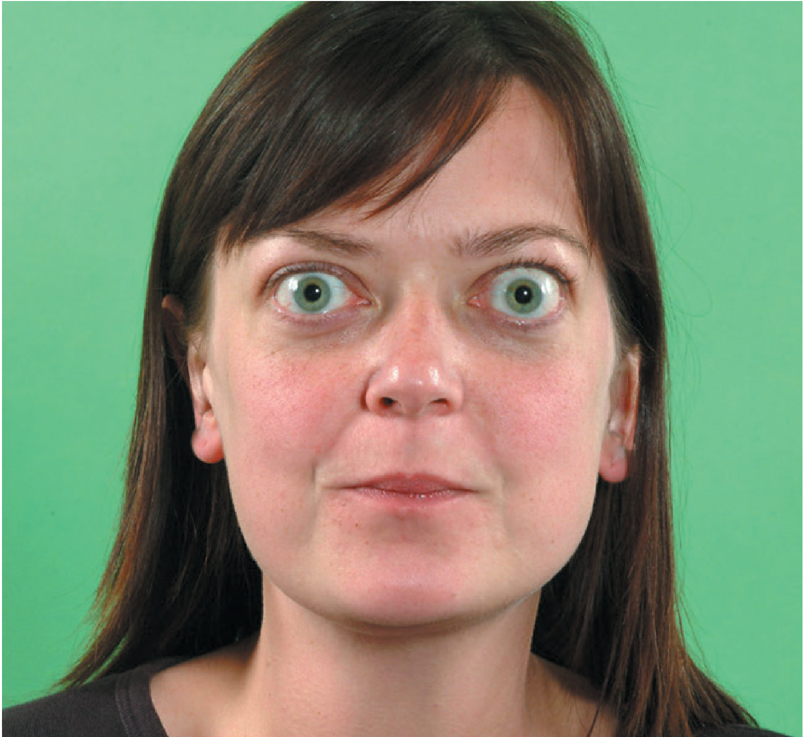
Graves' disease: characteristic exophthalmos and stare
(Bailey and Love's Short Practice of Surgery, 28th Ed)
11. Conservative Treatment of Diffuse Toxic Goiter
Goal: Restore euthyroidism; ideally achieve long-term remission.
Antithyroid Drugs (Thionamides) - Primary Medical Treatment
Agents:
- Carbimazole (converted to methimazole): 10-40 mg/day initially in divided doses; maintenance 5-10 mg/day
- Methimazole (preferred globally): equivalent dosing
- Propylthiouracil (PTU): 100-200 mg q6-8h initially; used when methimazole not tolerated, in pregnancy (1st trimester), or thyroid storm
Mechanism: Block TPO-catalyzed organification of iodide; carbimazole/methimazole also have modest immunomodulatory effects (reducing TRAb titers)
Titration approaches:
- Dose titration: Start high, reduce as TSH/T4 normalize
- Block-and-replace: Higher dose ATD + levothyroxine to prevent hypothyroidism (less dose adjustment needed)
Duration: 12-18 months typical; up to 24 months in severe cases
Remission rate: ~40-50% after one course; predictors of remission: small goiter, mild disease, TRAb normalization
Monitoring:
- TFTs every 4-6 weeks initially
- FBC before starting and if fever/sore throat develops (agranulocytosis)
- LFTs if PTU used
Adjuncts:
- Beta-blockers (propranolol 40 mg TDS or atenolol 50 mg OD): rapid symptomatic control
- Iodine (Lugol's solution): pre-operatively to reduce vascularity; reduces hormone release acutely
- Potassium perchlorate: rarely used; blocks iodine uptake
Failure / indications to escalate: Recurrence after ATDs → consider RAI or surgery
(Bailey and Love's Short Practice of Surgery, 28th Ed; Mulholland & Greenfield's Surgery 7e)
12. Indications for Surgery and Types of Surgical Interventions for Diffuse Toxic Goiter
Indications for Surgery in Graves' Disease
- Large goiter (>75 g) causing compressive symptoms
- Failure or intolerance of ATDs (agranulocytosis, hepatotoxicity)
- Patient preference for definitive cure without RAI
- Active or severe Graves' ophthalmopathy (RAI may worsen; surgery preferred)
- Concurrent thyroid malignancy (suspected or proven)
- Pregnancy with poor ATD control or side effects (surgery in 2nd trimester)
- Young patients (avoid lifelong RAI follow-up)
- Planned pregnancy soon (RAI requires 6-12 month delay)
- Non-compliance with medication
(Cummings Otolaryngology)
Types of Operations
| Operation | Notes |
|---|
| Total thyroidectomy | Preferred; eliminates recurrence risk; requires lifelong T4 replacement |
| Near-total thyroidectomy | Leaves <1 g near RLN; very low recurrence |
| Subtotal thyroidectomy (Nikolaev/Kocher) | Historically standard; leaves ~2-4 g (enough for normal function); ~10-15% recurrence; still used in some centers |
Current consensus: Total or near-total thyroidectomy is preferred over subtotal because it eliminates recurrence and simplifies long-term management.
For toxic adenoma:
- Hemithyroidectomy (lobectomy) is the operation of choice
- Low rate of postoperative hypothyroidism (2-3%) vs. total thyroidectomy (20%)
- Euthyroid state returns within 2-3 days
- RAI is an alternative but may leave palpable nodule and has higher hypothyroidism rate
(Mulholland and Greenfield's Surgery 7e)
13. Indications and Contraindications for Surgical Treatment of Thyrotoxic Goiter; Surgical Techniques
Indications (as above, Topic 12, plus)
Absolute indications:
- Proven thyroid cancer in toxic goiter
- Severe compression (respiratory failure, dysphagia)
- Failure of all medical therapy
Relative indications:
- Young patients, large goiters, ophthalmopathy, patient preference, planned pregnancy
Contraindications to Surgery
Absolute:
- Uncorrected thyrotoxicosis (risk of thyroid storm) - requires preoperative preparation
- Severely compromised cardiorespiratory function precluding general anesthesia
Relative:
- Mild/moderate Graves' disease in older patients (RAI preferred)
- Previous thyroid surgery (increased complication risk)
- Active, uncontrolled systemic disease
Surgical Technique (Thyroidectomy)
Steps:
- Positioning: Supine with neck extended (sandbag under shoulders)
- Incision: Kocher's collar incision (2 cm above clavicle, in skin crease)
- Subplatysmal flaps raised superiorly and inferiorly
- Midline division of strap muscles (sternohyoid and sternothyroid) if needed
- Identification and preservation of parathyroid glands: superior (near junction of upper and middle third of posterior thyroid) and inferior (near lower pole)
- Inferior thyroid artery ligated distal to parathyroid blood supply branches
- Identification of the recurrent laryngeal nerve (RLN): traced from tracheoesophageal groove; most common locations for injury: posterior to inferior pole, at intersection with inferior thyroid artery, and at Berry's ligament
- Berry's ligament division: RLN must be in direct vision; pre-emptive bipolar to small vessels within ligament; layer-by-layer dissection
- Superior pole vessels divided close to gland
- Tubercle of Zuckerkandl mobilized to expose RLN at its terminus
- Delivery of gland and division of isthmus
- Hemostasis, closure of strap muscles, subcutaneous and skin layers
- Drain placement: most surgeons omit routine drains
Intraoperative Neuromonitoring (IONM):
- Electrode on endotracheal tube senses vocal cord signals; grounding on skin
- Negative predictive value ~100% for ruling out nerve injury
- Standard of care in many centers; does not replace visual identification
(Bailey and Love 28th Ed; Current Surgical Therapy 14e; Mulholland & Greenfield 7e)
14. Preoperative Preparation and Postoperative Management
Preoperative Preparation
Essential: Patient must be euthyroid before surgery - failure to achieve this risks thyroid storm.
Steps:
- ATDs (carbimazole/methimazole or PTU) for 6-8 weeks until TFTs normalize (TSH recovers)
- Beta-blockers (propranolol) for cardiovascular stability; continued perioperatively
- Lugol's iodine (potassium iodide solution): given 10-14 days preoperatively - reduces thyroid vascularity by 30-40% and decreases intraoperative blood loss; 0.3 mL TID (Lugol's 5%); Wolff-Chaikoff effect blocks hormone release acutely
- Cardiovascular optimization: Control AF, heart failure if present
- Preoperative laryngoscopy: Baseline vocal cord assessment; detect any pre-existing RLN palsy
- Calcium/PTH: Baseline levels
- Informed consent: Risks of RLN injury, hypoparathyroidism, bleeding, hypothyroidism
Postoperative Management
Immediate (first 24-48 hours):
- Monitor for bleeding - risk of airway compression from hematoma; keep suture removal kit at bedside; re-explore and evacuate hematoma if airway compromise
- Calcium monitoring: Check serum calcium at 6, 12, 24 hours; check PTH at 4-6 hours
- Hypocalcemia (most common complication, from parathyroid ischemia): tingling perioral/fingertips (Chvostek, Trousseau signs) → oral/IV calcium replacement
- RLN injury: Assess voice quality postoperatively; hoarseness warrants laryngoscopy
Medications:
- Levothyroxine: Started immediately post-total thyroidectomy
- Calcium ± calcitriol: For temporary or permanent hypoparathyroidism
- Beta-blockers: Continued 2-4 weeks postoperatively (TSH may remain suppressed; risk of thyrotoxic rebound)
- Analgesia: Regular paracetamol ± NSAIDs
Long-term:
- TFTs at 6-8 weeks then annually
- Optimize levothyroxine dose (aim for TSH 0.5-2.5 mIU/L; lower TSH target if cancer)
- Bone density (if prolonged TSH suppression)
15. Hypothyroidism: Definition, Classification, Clinical Manifestations, Treatment
Definition
Hypothyroidism is a syndrome resulting from insufficient thyroid hormone action at the tissue level, due to deficient secretion of thyroid hormones or (rarely) tissue resistance to their action.
Classification
Primary hypothyroidism (most common, ~95% of cases):
- Thyroid gland itself is deficient
- TSH elevated; T3/T4 low
- Causes:
- Autoimmune (Hashimoto's thyroiditis) - most common in iodine-sufficient areas
- Post-radioiodine or post-surgical hypothyroidism
- Iodine deficiency (most common worldwide)
- Drugs: amiodarone, lithium, interferon-alpha
- Congenital: athyreosis, dyshormonogenesis
- External radiation to neck
Secondary (central) hypothyroidism:
- Pituitary insufficiency → reduced TSH secretion
- TSH low/normal; T4 low
- Causes: pituitary adenoma/tumor, Sheehan's syndrome, hypophysitis, cranial irradiation
Tertiary hypothyroidism:
- Hypothalamic insufficiency → reduced TRH → reduced TSH
- TRH, TSH, T4 all reduced
- Causes: craniopharyngioma, hypothalamic tumors, irradiation
Subclinical hypothyroidism: Elevated TSH with normal free T4; asymptomatic or mild symptoms; treat if TSH >10 mIU/L or if symptomatic
Congenital hypothyroidism: Thyroid agenesis/dysgenesis or dyshormonogenesis; screened at birth; levothyroxine must be started within first 2 weeks to prevent irreversible intellectual disability (cretinism)
Clinical Manifestations
| System | Symptoms | Signs |
|---|
| General | Fatigue, cold intolerance, weight gain, constipation | Bradycardia, hypothermia |
| Skin | Dry, coarse skin; hair loss; brittle nails | Non-pitting edema (myxedema), yellow tinge (carotenemia), loss of outer eyebrow (Hertoghe's sign) |
| Cardiovascular | Dyspnea | Pericardial effusion, cardiac enlargement, bradycardia |
| Neurological | Cognitive slowing, depression, forgetfulness | Delayed relaxation of tendon reflexes (classic), carpal tunnel syndrome, cerebellar ataxia |
| Reproductive | Menorrhagia, infertility | Galactorrhea |
| Muscular | Myalgia, cramps | Myopathy, pseudohypertrophy |
| Other | Deafness, macroglossia, hoarseness | Goiter (Hashimoto's) or absent thyroid |
Myxedema coma: Extreme hypothyroidism; precipitated by cold, infection, sedatives; features - hypothermia, severe bradycardia, hypotension, hypoventilation, coma; high mortality; treat with IV levothyroxine (500 mcg loading dose) ± T3 and IV hydrocortisone.
Laboratory: TSH elevated (primary); T4 low; hypercholesterolemia; raised CPK; anemia; hyponatremia.
Treatment
Levothyroxine (T4) - treatment of choice:
- Oral synthetic T4, fully absorbed
- Dose: 1.6 mcg/kg/day (adults); start low (25-50 mcg/day) in elderly and cardiac patients, titrate up every 4-6 weeks
- Full replacement in young, otherwise healthy adults: 1.6-2.0 mcg/kg/day
- Target: TSH 0.5-2.5 mIU/L (or 0.1-0.5 mIU/L if post-thyroid cancer)
- Take on empty stomach, 30-60 min before food; avoid co-administration with calcium, iron, PPI
T3/T4 combination therapy: Occasionally used for persistent symptoms despite optimized T4; not routine
(ROSEN's Emergency Medicine; Frameworks for Internal Medicine; Goldman-Cecil Medicine)
16. Thyroiditis and Strumitis: Etiology, Pathogenesis, Clinical Presentation, Diagnosis, Treatment
Hashimoto's Thyroiditis (Chronic Autoimmune / Lymphocytic Thyroiditis)
Etiology: Autoimmune; HLA-DR3, HLA-DR5 association; female predominance (F:M 7:1)
Pathogenesis: T-cell mediated cytotoxicity and autoantibodies (anti-TPO [most sensitive], anti-thyroglobulin) destroy thyroid follicular cells → progressive follicular destruction → lymphocytic infiltration with germinal center formation → hypothyroidism
Clinical presentation:
- Initial goiter (diffuse, firm, rubbery, lobulated "pebble-stone" feel)
- Transient thyrotoxicosis (Hashitoxicosis) early in some
- Progressive hypothyroidism
- May be euthyroid for years
- Associated with other autoimmune conditions (T1DM, Addison's, vitiligo, pernicious anemia)
Diagnosis: Elevated anti-TPO (>90%), anti-Tg; TSH elevated/normal; US: diffuse heterogeneous hypoechoic gland with increased vascularity; FNA rarely needed
Treatment: Levothyroxine when hypothyroid or TSH >10; monitoring when euthyroid; goiter rarely requires surgery
De Quervain's Thyroiditis (Subacute Granulomatous / Painful Thyroiditis)
Etiology: Likely viral (Coxsackie, adenovirus, mumps, ECHO, EBV); often follows URTI; seasonal clustering
Pathogenesis: Viral injury → follicular destruction → release of preformed thyroid hormone → thyrotoxicosis → recovery phase → hypothyroidism (transient) → return to euthyroid
Clinical presentation:
- Prodrome: URTI, malaise, fever
- Thyroid pain - anterior neck pain, tender, hard enlarged gland; pain radiates to jaw/ear
- Thyrotoxic symptoms (mild-moderate, from hormone release not overproduction)
- Sequence: thyrotoxicosis (4-8 weeks) → hypothyroidism (4-12 weeks) → euthyroid (~90% recover)
Diagnosis: Elevated ESR (often >50, sometimes >100 - dramatic); normal/low WBC; low RAI uptake (distinguishes from Graves' - in de Quervain's, gland cannot trap iodine due to damage); normal or low anti-TPO (may transiently rise); IL-6 elevated
Treatment:
- NSAIDs (aspirin, ibuprofen) for mild-moderate pain
- Corticosteroids (prednisolone 40-60 mg/day then taper) for severe pain/systemic symptoms - rapid resolution
- Beta-blockers for symptomatic thyrotoxicosis
- Levothyroxine if hypothyroid phase is prolonged/severe
- Self-limiting; recurrence ~5%
Riedel's Thyroiditis (Fibrous / Invasive Thyroiditis)
Etiology: Unknown; possibly part of IgG4-related systemic disease
Pathogenesis: Replacement of thyroid parenchyma by dense fibrous tissue extending beyond the thyroid capsule into surrounding structures
Clinical presentation:
- Hard, "stony," non-tender goiter - literally "woody" consistency, fixed to surrounding structures
- Mimics thyroid cancer (must distinguish)
- Compressive symptoms: dysphagia, stridor, dyspnea
- Extension to surrounding structures - can fibrosis retroperitoneum, orbit, mediastinum (multifocal fibrosclerosis)
- Usually euthyroid, rarely hypothyroid
Diagnosis: FNA/core biopsy (distinguishes from cancer); CT for extent; IgG4 levels may be elevated
Treatment: Corticosteroids (may slow progression); tamoxifen (anti-fibrotic); surgery (isthmusectomy) for airway decompression - not curative; levothyroxine if hypothyroid
Acute (Suppurative) Thyroiditis
Etiology: Bacterial (Staphylococcus, Streptococcus, Haemophilus, E. coli); rare; usually via persistent thyroglossal duct or piriform sinus fistula (especially in children)
Pathogenesis: Hematogenous seeding or direct extension → abscess formation
Clinical presentation:
- Acute anterior neck pain, fever, dysphagia
- Tender, warm, erythematous, fluctuant thyroid swelling
- Leukocytosis
Treatment: IV antibiotics; surgical drainage if abscess; excision of fistulous tract to prevent recurrence
Strumitis
Strumitis refers to acute inflammation of a pre-existing goiter (most often nodular), usually from bacterial infection or hemorrhage into a nodule. Clinical presentation is similar to acute thyroiditis but within an already enlarged gland. Treatment: antibiotics, drainage if suppurated; aspiration of hemorrhagic cysts.
17. Complications During and After Thyroid Surgery: Clinical Presentation, Treatment; Prevention of RLN Injury
Intraoperative Complications
Hemorrhage:
- Injury to superior/inferior thyroid arteries or thyroid veins
- Control with pressure, ligation, bipolar cautery
RLN injury (intraoperative):
- Transection, stretch, crush, burn, ischemia
- Presentation: immediate hoarseness/voice change if anesthesia reversed
- If transected: immediate re-anastomosis or immediate referral to laryngologist
Early Postoperative Complications
1. Hemorrhage/Hematoma (1-2%):
- Occurs within first 6-12 hours
- Rapidly expanding hematoma can compress trachea → airway emergency
- Clinical features: swelling, stridor, neck distension, respiratory distress
- Treatment: Immediate wound opening at bedside to decompress (suture removal kit should be at bedside of ALL post-thyroidectomy patients) → return to OR for formal re-exploration and hemostasis
2. Recurrent Laryngeal Nerve (RLN) Palsy:
- Unilateral: hoarseness, breathy voice, aspiration (incomplete glottic closure)
- Bilateral: stridor, severe respiratory compromise → may require tracheostomy
- Rate: unilateral permanent palsy <0.5-1% in experienced hands; temporary palsy 5-10%
- Transient palsy from edema/ischemia: usually recovers within 6-12 weeks
- Assessment: laryngoscopy
- Treatment of permanent palsy: medialization laryngoplasty (injection or thyroplasty)
3. Hypoparathyroidism / Hypocalcemia (most common complication):
- From parathyroid devascularization, accidental removal, or ischemia
- Temporary: 15-30% after total thyroidectomy; permanent: 1-3%
- Symptoms: perioral tingling, carpopedal spasm, Chvostek's sign, Trousseau's sign, tetany, seizures
- Treatment:
- Mild: oral calcium carbonate 1.5-3 g/day + calcitriol 0.25-0.5 mcg/day
- Severe/symptomatic: IV calcium gluconate (10 mL of 10% solution slowly)
- Monitoring: serum calcium q4-6h for 24h; PTH at 4-6h postop (PTH <10 pg/mL predicts hypocalcemia)
4. Thyroid Storm / Thyrotoxic Crisis:
- In inadequately prepared Graves' patients
- Treatment: PTU + Lugol's + beta-blockade + glucocorticoids + supportive care (see Topic 9)
Late Complications
5. Hypothyroidism:
- Expected after total thyroidectomy → lifelong levothyroxine
- May develop after subtotal thyroidectomy (15-40%)
- Treatment: levothyroxine replacement
6. Recurrence of hyperthyroidism (after subtotal thyroidectomy for Graves'):
- Rate ~10-15%; treatment: ATDs, RAI, or completion thyroidectomy
7. Wound complications: Keloid scar, infection (rare), seroma
8. Superior laryngeal nerve (SLN) injury:
- External branch injury → loss of cricothyroid muscle function → lowered voice pitch, reduced projection, voice fatigue
- Important for singers, professionals
Prevention of RLN Injury
The key principles (from Bailey and Love, Current Surgical Therapy):
- Visual identification of the RLN - mandatory; "No cut, burn, or tie unless the RLN is identified"
- Systematic approach: Dissect from inferior pole upward in tracheoesophageal groove; spread perpendicular to nerve axis with blunt dissection
- Gentle retraction of the thyroid medially; avoid excessive traction
- Careful dissection at Berry's ligament - most dangerous zone; pre-emptive bipolar, layer-by-layer dissection
- Awareness of anatomic variations:
- RLN may be anterior to inferior thyroid artery (higher risk)
- Branching may occur proximal to cricothyroid muscle
- Non-recurrent laryngeal nerve (0.5-1% on right) - associated with anomalous subclavian artery
- Tubercle of Zuckerkandl - mobilize anteriorly to expose RLN terminus
- Intraoperative neuromonitoring (IONM): Negative predictive value ~100%; does not replace visualization
- Preoperative laryngoscopy - documents baseline function; identifies unsuspected pre-existing palsy
- Minimize use of energy devices near nerve - safe distance ≥3 mm from nerve with ultrasonic devices
(Bailey and Love 28th Ed; Current Surgical Therapy 14e; Mulholland & Greenfield 7e)
18. Thyroid Cancer: Classification, Clinical Presentation, Diagnosis, Differential Diagnosis, Treatment, Outcomes
Classification
Primary thyroid malignancies:
| Type | % of All Thyroid Ca | Cell of Origin | Key Features |
|---|
| Papillary thyroid cancer (PTC) | 80% | Follicular cells | Best prognosis; lymph node mets common but not prognostic; BRAF mutation (45%), RET/PTC rearrangement |
| Follicular thyroid cancer (FTC) | 10% | Follicular cells | Hematogenous spread (bone, lung); no reliable FNA diagnosis (requires capsular/vascular invasion on histology); RAS mutation, PAX8-PPARγ |
| Hürthle cell carcinoma | 3-5% | Follicular (oxyphilic) | Poorer prognosis than FTC; lower RAI uptake; higher lymph node mets |
| Medullary thyroid cancer (MTC) | 5-7% | Parafollicular C-cells | Calcitonin secreting; 25% hereditary (MEN2A, MEN2B, FMTC); RET proto-oncogene mutations |
| Anaplastic thyroid cancer (ATC) | 1-2% | Follicular (dedifferentiated) | Most lethal cancer; rapid growth; local invasion; median survival 3-6 months; 1-year survival ~20% |
| Thyroid lymphoma | <1% | Lymphoid | Usually B-cell NHL; arises on background of Hashimoto's; treated with chemotherapy ± radiation, not surgery |
Staging (ATA/AJCC TNM - simplified):
- T1: ≤2 cm confined to thyroid
- T2: 2-4 cm
- T3: >4 cm or minimal extrathyroidal extension
- T4: Significant extrathyroidal extension / invasion of surrounding structures
- N1: Regional lymph node mets
- M1: Distant mets (lung, bone most common)
Clinical Presentation
- Usually presents as a thyroid nodule - palpable or incidentally detected on imaging
- Features raising concern for malignancy:
- Rapid growth
- Hard, fixed, irregular nodule
- Hoarseness (RLN invasion - ominous sign)
- Dysphagia, stridor (local invasion)
- Cervical lymphadenopathy (especially in PTC)
- Male sex, extremes of age (<20 or >65)
- History of radiation to neck (childhood irradiation → PTC)
- Family history (MTC in MEN2, familial PTC)
- Calcitonin elevation (MTC)
ATC: Rapidly enlarging anterior neck mass; often presents with stridor, dysphagia, dysphonia; neck fullness; most patients have extrathyroidal extension at diagnosis
Diagnosis
Evaluation pathway:
- History and physical examination (risk factors, nodule features, cervical LN)
- TSH: If suppressed → RAI scan (rule out hot nodule = virtually always benign); if normal/elevated → US
- Thyroid ultrasound (US): Characterize nodule (size, echogenicity, margins, microcalcifications, vascularity, shape); TIRADS classification; examine for lymphadenopathy
- FNA biopsy (FNAC): For nodules >1 cm with suspicious US features, or any growing nodule; Bethesda classification (Thy1-5); core biopsy for FTC (capsular invasion not assessable by FNA)
- Molecular testing (Bethesda III-IV): ThyroseqTM, AfirmaTM GEC - improve diagnostic accuracy for indeterminate nodules
- CT neck/chest: Large or locally advanced tumors, lymph node mapping, substernal disease
- Calcitonin/CEA: For suspected MTC
- RET mutation testing: Hereditary MTC (index case + all 1st-degree relatives if positive)
(FNA Bethesda classification: Thy1 non-diagnostic, Thy2 benign, Thy3 follicular/indeterminate, Thy4 suspicious, Thy5 malignant - Bailey and Love 28th Ed)
Differential Diagnosis of Thyroid Nodule / Mass
- Benign: Simple cyst, follicular adenoma, Hashimoto's, colloid nodule, hemorrhagic cyst
- Malignant: PTC, FTC, MTC, ATC, lymphoma
- Other: Parathyroid adenoma/cyst, branchial cleft cyst, lymph node, lipoma, cervical abscess
Treatment
Differentiated thyroid cancer (PTC and FTC):
- Surgery: Total thyroidectomy for lesions >4 cm, bilateral disease, high-risk features, any FTC; thyroid lobectomy acceptable for low-risk PTC ≤1-4 cm without extrathyroidal extension or lymphadenopathy
- Central neck dissection (Level VI): Therapeutic if clinical/radiologic N1a; prophylactic in high-risk PTC (controversial but commonly performed)
- Lateral neck dissection (Levels II-V): For confirmed lateral LN mets
- Radioactive iodine (RAI, 131I): After total thyroidectomy for intermediate/high-risk disease; ablates remnant thyroid tissue and micrometastases; not effective in Hürthle cell, MTC, ATC
- TSH suppression therapy: Levothyroxine post-operatively to TSH <0.5 (intermediate risk) or <0.1 mIU/L (high risk) for 1-5 years, then relax to low-normal
- External beam radiation (EBRT): Residual/unresectable PTC/FTC; ATC (combined modality)
- Targeted therapy for ATC and progressive DTC: BRAF/MEK inhibitors (dabrafenib + trametinib for BRAF V600E-mutated ATC); lenvatinib, sorafenib for RAI-refractory DTC
Medullary thyroid cancer (MTC):
- Total thyroidectomy + central neck dissection
- Lateral dissection if calcitonin >200 pg/mL or known lateral LN mets
- RET-directed therapy: vandetanib, cabozantinib for advanced/progressive MTC
- Genetic counseling and RET testing of family members mandatory
Anaplastic thyroid cancer (ATC):
- Multimodal: surgery (rarely curative) + chemoradiation (doxorubicin + radiation)
- Targeted therapy: dabrafenib + trametinib (FDA approved for BRAF V600E-mutant ATC)
- Tracheostomy often required
- Most patients treated with palliative intent
Thyroid lymphoma:
- Chemoimmunotherapy (CHOP ± rituximab) ± radiation
- Surgery only for airway decompression
Long-Term Outcomes
| Type | 10-year Survival | Notes |
|---|
| PTC (confined to gland) | >95% | Excellent; recurrence manageable with RAI |
| PTC (with distant mets) | ~40% | |
| FTC (low risk) | ~85% | |
| FTC (with distant mets) | ~50% | |
| Hürthle cell | ~80% | Less favorable than FTC |
| MTC (Stage I-II) | ~80-90% | |
| MTC (Stage IV) | ~20-25% at 10 years | |
| ATC | ~5-10% at 1 year | Essentially uniformly fatal |
Surveillance after differentiated thyroid cancer:
- Thyroglobulin (Tg) + anti-Tg antibodies: tumor marker; should be undetectable after total thyroidectomy + RAI; rising Tg = recurrence
- Whole body RAI scan: 6-12 months post-ablation; then based on risk
- Neck US: annually
- CT/PET-CT: if Tg elevated with negative US/RAI scan
(Current Surgical Therapy 14e; Mulholland & Greenfield's Surgery 7e; Bailey and Love 28th Ed)
Quick Reference: Key Drug Doses
| Drug | Indication | Dose |
|---|
| Carbimazole | Hyperthyroidism | 10-40 mg/day (induction); 5-10 mg/day (maintenance) |
| Propylthiouracil | Hyperthyroidism (pregnancy/storm) | 100-200 mg q6-8h |
| Propranolol | Symptomatic thyrotoxicosis | 40 mg TDS or titrate |
| Levothyroxine | Hypothyroidism | 1.6 mcg/kg/day; start 25-50 mcg in elderly |
| Prednisolone | de Quervain's thyroiditis | 40-60 mg/day then taper |
| Lugol's iodine | Preoperative preparation | 0.3 mL TDS ×10-14 days |
| IV Calcium gluconate | Postop hypocalcemia | 10 mL of 10% solution slowly IV |
Sources: Bailey and Love's Short Practice of Surgery (28th Ed) - Bailey and Love's, p. 883-900; Mulholland and Greenfield's Surgery 7e; Current Surgical Therapy 14e; Frameworks for Internal Medicine; Goldman-Cecil Medicine; Costanzo Physiology 7th Ed; ROSEN's Emergency Medicine.