
Posterior relation of radial artery
radial artery forearm wrist posterior relations anatomy diagram
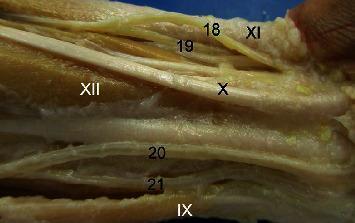
This clinical photograph displays a detailed anatomical dissection of the distal forearm and wrist region in a Macaca fuscata specimen, specifically providing a caudal (posterior) view of the right forelimb. The image highlights the complex neurovascular and musculoskeletal architecture relevant to comparative anatomy. Key arterial structures labeled include the superficial branch of the radialis artery (18), the deep branch of the radialis artery (19), the ulnaris artery profundus ramus (20), and the superficial branch of the ulnaris artery (21). Associated musculature and tendinous structures are also identified, including the Brachioradialis (IX), the tendon of the Palmaris longus (X), the Flexor carpi ulnaris tendon (XI and XIII), and the Flexor digitorum superficialis (XII). The dissection illustrates the distal branching patterns of the radial and ulnar arteries as they transition toward the palmar arches, serving as a model for understanding primate vascular variations and their similarities to human upper limb anatomy. The educational focus is on the spatial relationships between the major arteries of the forearm and the surrounding flexor tendons.
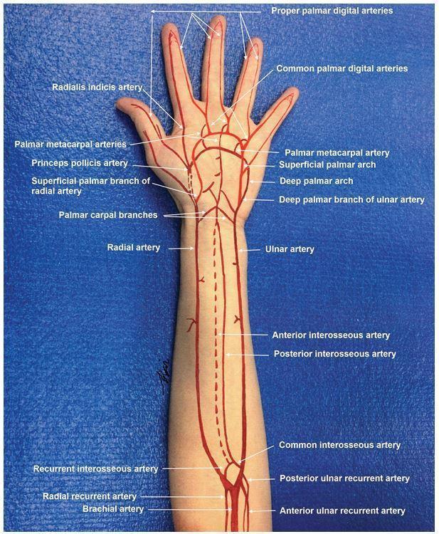
Anatomical diagram superimposed on a photograph of a human arm, illustrating the arterial circulation from the distal humerus to the fingertips. The brachial artery is shown bifurcating into the radial and ulnar arteries. Key forearm vessels are labeled, including the radial and ulnar recurrent arteries, common interosseous artery, and the anterior and posterior interosseous arteries. In the hand, the illustration details the complex vascular anastomoses: the superficial palmar arch (primarily from the ulnar artery) and the deep palmar arch (primarily from the radial artery). Specific terminal branches are identified, such as the princeps pollicis artery, radialis indicis artery, palmar metacarpal arteries, common palmar digital arteries, and proper palmar digital arteries supplying each digit. This educational graphic serves as a guide for understanding upper extremity vascular anatomy, essential for surgical planning, trauma assessment, and performing procedures like the Allen's test.
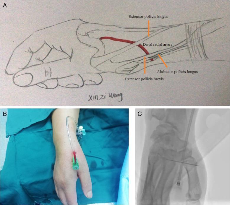
A multi-panel educational figure illustrating the anatomy and clinical application of distal radial artery (DRA) access. Panel A is an anatomical diagram of the hand and wrist highlighting the anatomical snuffbox. It depicts the distal radial artery in relation to key tendons: the extensor pollicis longus forms the medial border, while the abductor pollicis longus and extensor pollicis brevis form the lateral border. Panel B is a clinical photograph showing the procedural setup for distal transradial access (dTRA), featuring a 6Fr sheath successfully inserted into the snuffbox region with a connector tube attached. Panel C is a fluoroscopic diagnostic image (X-ray) providing a radiological view of the puncture site. It demonstrates the radiopaque sheath positioned within the DRA, localized near the radial styloid process and carpal bones. This figure is designed for medical professionals and interventionalists to understand the topographical and radiological landmarks necessary for arterial cannulation at the distal forearm/hand junction.
Posterior Relations of the Radial Artery
1. In the Forearm (Upper to Lower)
| Level | Posterior Relation |
|---|---|
| Upper forearm | Tendon of biceps brachii and supinator |
| Middle forearm | Pronator teres (its insertion on the radius) |
| Upper 2/3 (general) | Radial head of flexor digitorum superficialis |
| Lower 2/3 | Flexor pollicis longus |
| Distal forearm | Pronator quadratus and the lower end of the radius |
- At its origin (cubital fossa), the artery lies on the tendon of biceps and the supinator muscle.
- In the proximal half, its proximal portion lies between the pronator teres and brachioradialis muscles, resting on structures of the deep layer.
- In the distal forearm, it lies directly on the pronator quadratus and the distal radius - this is why the radial pulse can be felt by pressing the artery against the underlying bone here. - Gray's Anatomy for Students, p. 903
2. At the Wrist (Anatomical Snuffbox)
- Scaphoid (navicular)
- Trapezium (greater multangular)
3. In the Hand (First Interosseous Space)
- First dorsal interosseous muscle
Summary Table
| Segment | Posterior Relations |
|---|---|
| At origin (cubital fossa) | Tendon of biceps, supinator |
| Upper forearm | Pronator teres insertion, radial head of FDS |
| Middle forearm | Flexor pollicis longus |
| Lower forearm | Pronator quadratus, lower end of radius |
| Anatomical snuffbox | Scaphoid, trapezium |
| 1st interosseous space | 1st dorsal interosseous muscle |
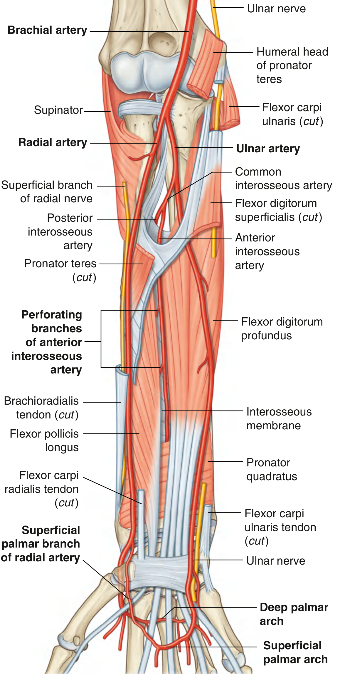
Sources: Gray's Anatomy for Students (9780323934237); Color Atlas of Human Anatomy Vol. 2 (9783132424487); Imaging Anatomy: Bones, Joints, Vessels and Nerves Vol. 3 (9781626239845)