
Describe imaging of cavitory lesions of lung
Reading File
Now let me search the medical library for cavitary lung lesions imaging content in parallel:
Finding Sources
Finding Sources
Finding Sources
Good results. Now let me read the key sections in parallel:
Reading File
Finding Sources
Finding Sources
Reading File
Reading File
Reading File
Excellent content. Let me now read the remaining key sections - the cavitary lung diseases imaging and differential diagnosis sections, and get more content from Murray & Nadel:
Reading File
Reading File
Finding Sources
Reading File
Finding Sources
Now let me fetch key images and get a few more imaging-specific details:
Loading Image
Loading Image
Finding Sources
I now have comprehensive content. Let me also fetch one more key image - the Nocardia abscess CXR/CT composite:
Loading Image
Finding Sources
Reading File
I now have comprehensive material from multiple authoritative textbooks. Let me compose the full answer.
Imaging of Cavitary Lesions of the Lung
Definition
A lung cavity is a gas-filled space with a well-defined wall (greater than 4 mm thick) within a nodule, mass, or area of parenchymal consolidation. When a cavity exceeds 2 cm, results from liquefactive necrosis, and contains necrotic debris or fluid due to microbial infection, it is termed a lung abscess. Cavities may be solitary or multiple, acute or subacute. - Goldman-Cecil Medicine, p. 919
Plain Chest Radiograph (CXR)
The CXR is the initial and most widely used modality. Key findings include:
- Thick-walled radiolucency within an area of consolidation or mass
- Air-fluid level - the hallmark of a communicating cavity; the fluid level is horizontal and may change position with posture
- On frontal view, the air-fluid level appears as a horizontal line; the level should be equal or similar in length when compared with the lateral view (a parenchymal lesion). If the air-fluid level is significantly longer on one view, this favors an empyema (pleural collection) rather than a true abscess
- The surrounding infiltrate or consolidation is common with infectious cavities
- Location: aspiration-related abscesses typically occur in the posterior segment of the upper lobes or superior segment of the lower lobes (dependent zones in a supine patient). Due to bronchial anatomy, the right lung is more commonly affected
Limitations of CXR
- Cannot reliably determine wall thickness, inner wall character, or relationship to pleura
- May miss small cavities or those obscured by consolidation
- Cannot differentiate abscess from empyema reliably
Primary tuberculosis (TB) on CXR - Figure 53.1 from Murray & Nadel:
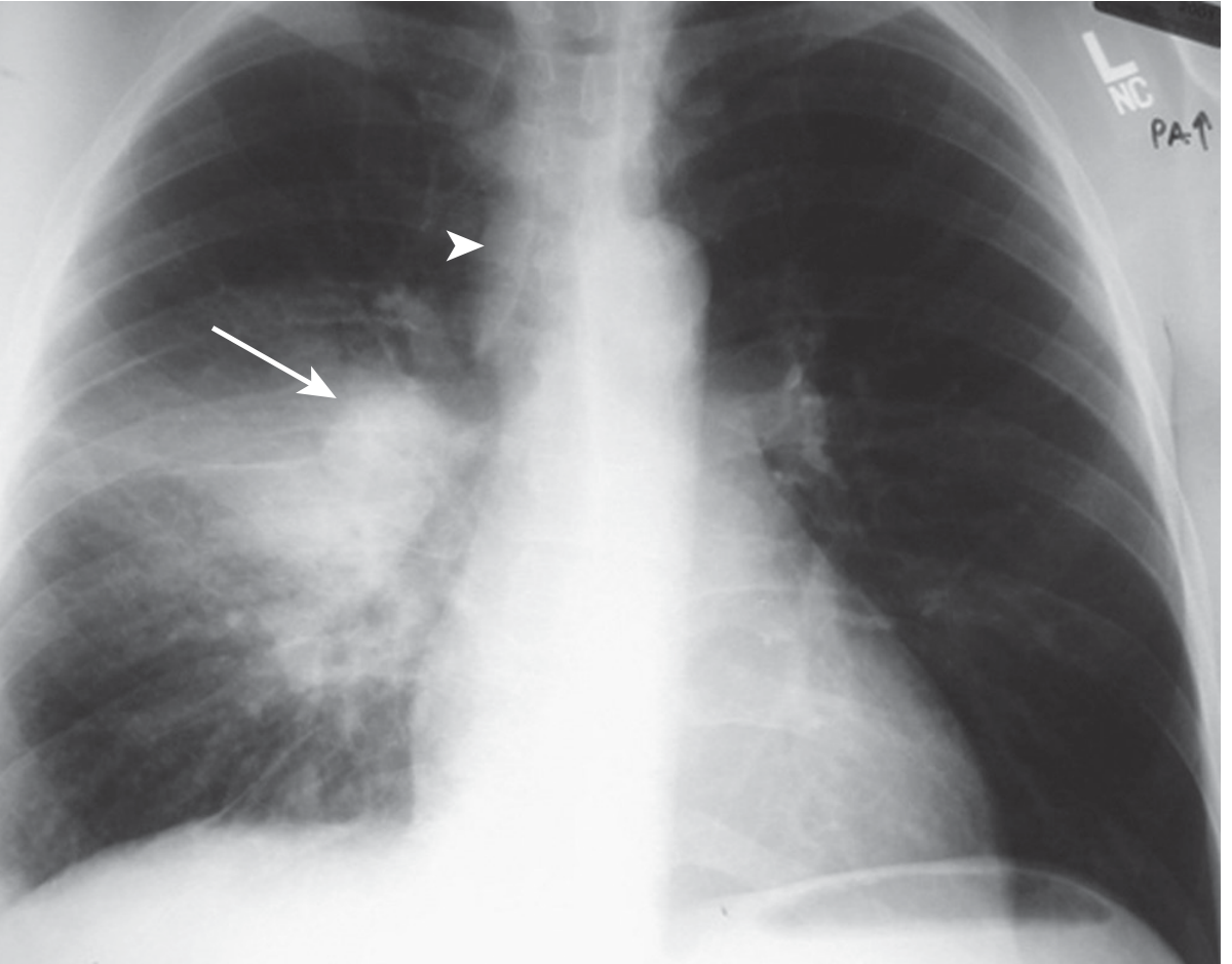
Primary TB: consolidation (arrow) with ipsilateral hilar lymphadenopathy (arrowhead). - Murray & Nadel's Textbook of Respiratory Medicine
Computed Tomography (CT)
CT is the modality of choice for characterizing cavitary lesions. It provides:
- Wall thickness and regularity
- Inner wall character
- Relationship to pleura and adjacent structures
- Associated lymphadenopathy, pleural effusion, satellite nodules
CT Features by Diagnosis
1. Lung Abscess (Infectious/Bacterial)
- Spherical or polygonal shape - similar dimensions on frontal and lateral views
- Thick, shaggy ("ragged") inner wall - highly characteristic
- Air-fluid level within the cavity
- Surrounding consolidation or ground-glass opacity (perilesional inflammation)
- Destroys rather than displaces adjacent structures and pulmonary vessels
- Does not compress adjacent lung
- Lymphadenopathy and pleural effusion may accompany severe infection
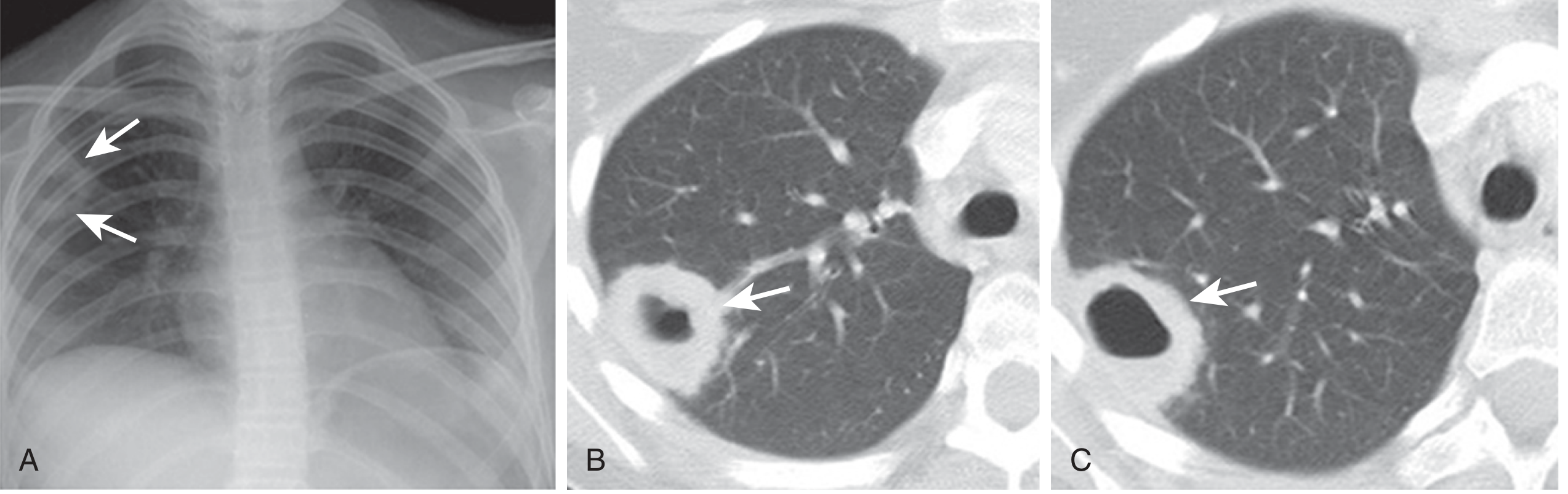
Nocardia asteroides lung abscess. (A) CXR: poorly defined opacity in subpleural right upper lobe. (B-C) CT lung windows: subpleural right upper lobe cavity (arrows) containing an air-fluid level. The cavity appearance is nonspecific - culture/biopsy is required. - Murray & Nadel's Textbook of Respiratory Medicine
2. Malignant Cavitary Lesion (e.g. Squamous Cell Carcinoma)
- Wall thickness ≥ 16 mm strongly favors malignancy (though variable by tumour type)
- Nodular, irregular inner wall (eccentric nodularity)
- Associated lymphadenopathy and pleural effusion favor malignancy
- Squamous cell carcinoma (both primary and metastatic) cavitates more often than adenocarcinoma or small cell carcinoma
- PET scan helps assess extrathoracic disease
3. Empyema (vs. Abscess - critical distinction for management)
| Feature | Lung Abscess | Empyema |
|---|---|---|
| Shape | Round/spherical | Lenticular/elliptical |
| Margins | Thick, shaggy/ragged inner wall | Smooth inner AND outer margins |
| Chest wall angle | Acute angles (parenchymal) | Obtuse angles (pleural/extraparenchymal) |
| Adjacent lung | Destroyed/necrosed | Compressed and displaced |
| Pulmonary vessels | Destroyed | Displaced around lesion |
| Air-fluid level | Similar length on frontal and lateral | Markedly different lengths on frontal vs. lateral |
| Enhancement | Absent or patchy rim | Prominent rim - "split pleura sign" after IV contrast |
Murray & Nadel's Textbook of Respiratory Medicine, p. 488
4. Tuberculosis
- Reactivation (post-primary) TB: cavitation is common; upper lobes (apical and posterior segments of RUL, apicoposterior segment of LUL) are the classic sites
- Cavities may be thin or thick-walled; satellite nodules ("tree-in-bud" pattern) represent bronchogenic spread
- HIV-related TB: with low CD4 counts, cavitation is less common; lower zone infiltrates and lymphadenopathy predominate
- A cavity on imaging cannot reliably indicate whether TB is active or a sterile residual of old disease from a single study - serial imaging or microbiological data is needed
- Healing results in fibrotic scarring with calcification
5. Septic Emboli
- Multiple distinct nodules, 1-3 cm in diameter, in different stages of cavitation
- Subpleural wedge-shaped infiltrates (representing infarcts)
- Bilateral and peripheral distribution
- Echocardiography is indicated to rule out valvular vegetations (endocarditis)
6. Fungal Infections
- Invasive Pulmonary Aspergillosis: early CT shows the halo sign (ground-glass opacity surrounding a nodule or mass - representing hemorrhagic infarction). As necrosis progresses, the air crescent sign appears - a crescent of air between the necrotic core and surrounding tissue, highly suggestive of invasive fungal infection. Present in 5-12.5% of cases; cannot be distinguished from mucormycosis on imaging alone
- Cryptococcus: solitary or multiple nodules, sometimes cavitating, more common in immunocompromised patients (especially HIV)
- Cavity wall in fungal disease can be thin or thick; the air-crescent sign specifically suggests liquefying fungal ball
7. Granulomatosis with Polyangiitis (GPA / Wegener's)
- Multiple bilateral cavitary nodules; autoimmune; noninfectious
- Associated with upper airway and renal disease
- No air-fluid level typically; more nodular, may wax and wane
8. Rheumatoid Nodules
- Peripheral, subpleural cavitary nodules
- May be thin-walled; may arise in the context of known RA
Additional Imaging Modalities
PET-CT
- Helpful when malignancy is suspected to assess metabolic activity and detect extrathoracic/nodal metastatic disease
- Active infections (TB, fungal) can also show FDG-avidity, limiting specificity
Ultrasound
- Limited role for parenchymal cavities; useful for guiding percutaneous drainage if abscess abuts the chest wall
Image-guided aspiration / CT-guided biopsy
- Required when non-invasive imaging cannot establish the diagnosis - provides material for culture and histology
Key Imaging Approach Summary
| Feature | Favors Infection | Favors Malignancy |
|---|---|---|
| Wall | Thick, shaggy/irregular inner wall | Nodular inner wall, wall ≥16 mm |
| Air-fluid level | Present | Less common |
| Surrounding opacity | Ground-glass, consolidation | Usually absent |
| Number | Solitary (or multiple if septic emboli) | Solitary; multiple if metastatic |
| Location | Posterior upper lobe / superior lower lobe (aspiration) | Any, often central |
| Lymphadenopathy | May be present (TB, fungal) | Common (malignancy) |
| Pleural effusion | Suggests empyema complication | More common in malignancy |
Sources:
- Goldman-Cecil Medicine International Edition (2-Volume Set), Chapter 78: Cavitary Lung Diseases, p. 919
- Murray & Nadel's Textbook of Respiratory Medicine (2-Volume), Chapter 50: Lung Abscess; Chapter 20: Radiographic Features; Chapter 53: Tuberculosis Radiology
- Fishman's Pulmonary Diseases and Disorders (2-Volume Set): Invasive Pulmonary Aspergillosis
Radiology standard textbooks only
Finding Sources
The primary radiology textbook in the library is Grainger & Allison's Diagnostic Radiology. Let me search it thoroughly now.
Finding Sources
Finding Sources
Reading File
Reading File
Finding Sources
Finding Sources
Finding Sources
Finding Sources
Finding Sources
Good - now let me read the key sections in Grainger & Allison in depth:
Reading File
Reading File
Reading File
Reading File
Reading File
Reading File
Now let me get the TB section, metastases cavitation, and vasculitis/GPA sections from Grainger & Allison:
Reading File
Reading File
Reading File
Loading Image
Loading Image
Reading File
Loading Image
Now let me get the metastases / solitary pulmonary mass section and the vasculitis / GPA section:
Reading File
Finding Sources
Finding Sources
Loading Image
Reading File
Loading Image
I now have comprehensive and detailed content exclusively from Grainger & Allison's Diagnostic Radiology. Let me compile the full answer.
Imaging of Cavitary Lesions of the Lung
Source: Grainger & Allison's Diagnostic Radiology
Definition and Imaging Approach
The most useful imaging techniques for evaluating cavitary lung lesions are plain chest radiography and CT. Radiographic findings of pulmonary infections (including cavitary forms) do not provide a specific aetiological diagnosis on their own - they must be integrated with clinical findings. CT is the problem-solving tool that characterises the cavity further.
General Radiological Features of Cavitation
A cavity appears as a lucency (radiolucency on CXR, low attenuation on CT) within a mass or area of consolidation, bounded by a wall. Key imaging parameters that guide the differential diagnosis are:
| Parameter | Significance |
|---|---|
| Wall thickness | Thin (<4 mm) = benign/cystic; Thick (>8 mm), irregular = malignancy or abscess |
| Inner wall character | Smooth = benign; Nodular/irregular = malignant; Shaggy = abscess |
| Number | Solitary vs. multiple |
| Air-fluid level | Communicates with bronchus; common in abscess |
| Associated findings | Consolidation, halo, satellite nodules, lymphadenopathy, pleural effusion |
| Location | Upper lobe = TB/post-primary infection; dependent zones = aspiration |
1. Lung Abscess
CXR findings:
- Rounded opacity with thick, often poorly defined walls
- Air-fluid level when the cavity communicates with a bronchus (see CXR: cavity with air-fluid level in right upper zone below)
- Surrounding consolidation/infiltrate
CT findings:
- Large cavity with a thick, irregular ("shaggy") inner wall with intracavitary thick septa
- Air-fluid level within the cavity
- Surrounding parenchymal consolidation, ground-glass opacity
- Primarily round/spherical shape - unlike empyema
- Does not compress adjacent lung; instead destroys it
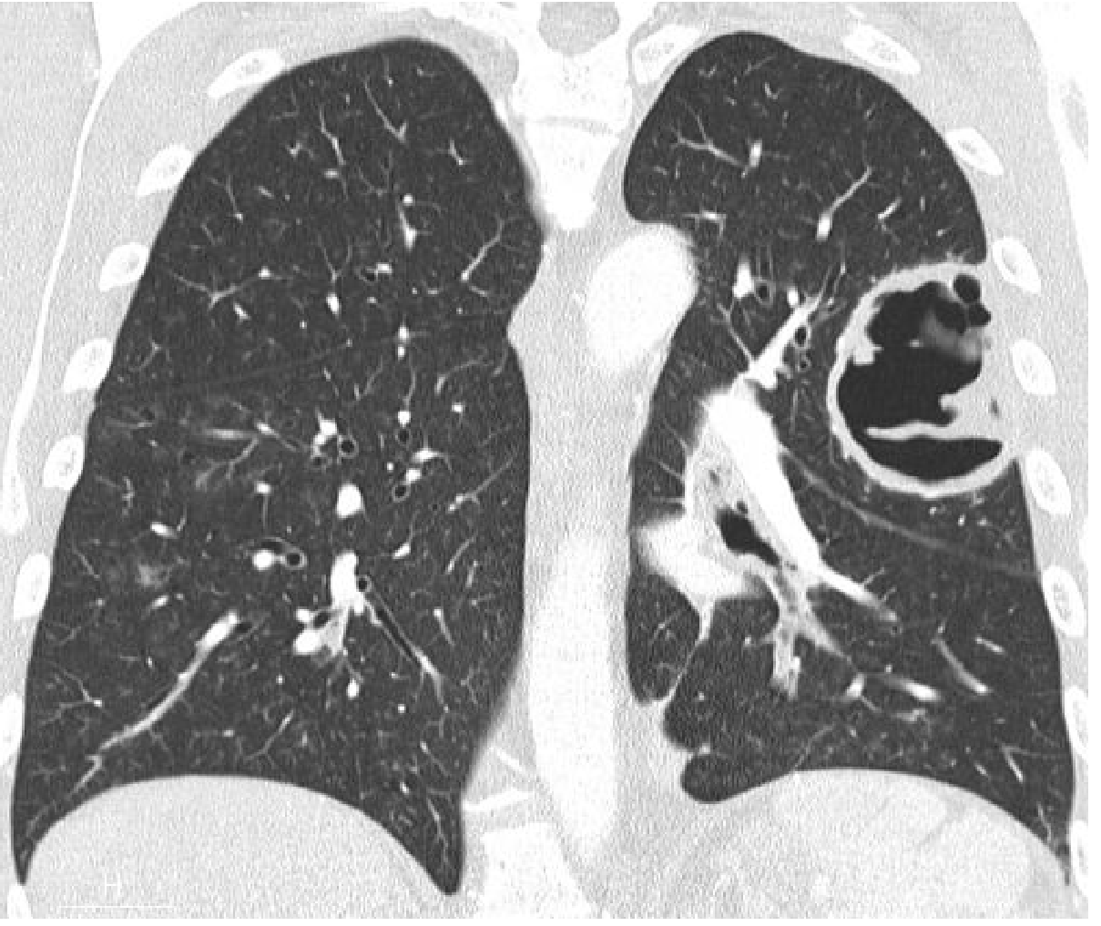
Fig. 5.4 - Lung Abscess. Coronal reformatted CT showing a large cavity in the left upper lobe with intracavitary thick septa. Pseudomonas aeruginosa on culture. - Grainger & Allison's Diagnostic Radiology
Septic emboli (haematogenous abscess):
- Multiple bilateral nodules, typically 1-3 cm, in different stages of cavitation
- A characteristic CT sign is the "feeding vessel" sign - a vessel coursing directly into a cavitating nodule
- Associated subpleural wedge infarcts
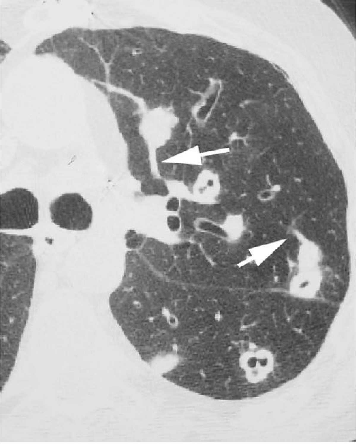
Fig. 5.6 - Septic Embolism. CT showing multiple cavitated nodules in the left upper lobe. Different vessels (arrows) course into the nodules. Blood cultures positive for S. aureus. - Grainger & Allison's Diagnostic Radiology
2. Necrotising / Cavitary Pneumonia
- Staphylococcus aureus is the classic organism: necrotising cavitary pneumonia, bilateral in ~40%, with abscess formation (15-30% of cases), pneumatoceles (thin-walled gas-filled cysts, classic sequelae), and pleural effusions (30-50%)
- Klebsiella pneumoniae: lobar consolidation with expansion of the lobe ("bulging fissure" sign), rapidly progressing to cavitation and necrosis
- Pseudomonas aeruginosa (most common nosocomial): multifocal upper lobe consolidation, large random nodules, tree-in-bud, ground-glass, necrosis
- Pulmonary gangrene: rare complication - initially small lucencies within consolidated lung; development of fragments of necrotic lung (pulmonary sequestrum) within an abscess cavity
- On contrast-enhanced CT: areas of decreased or absent enhancement indicate ischaemia/infarction within the consolidation, signifying necrotising pneumonia
Pneumatoceles vs. abscess:
- Pneumatoceles: thin-walled, no wall enhancement, anatomically located within the previous pneumonia zone
- Abscess: thick wall with enhancement
3. Empyema (vs. Lung Abscess - Critical Distinction)
CT is superior to CXR for this differentiation:
| Feature | Lung Abscess | Empyema |
|---|---|---|
| Shape | Round/spherical | Lenticular / elliptical |
| Chest wall angle | Acute angles (parenchymal) | Obtuse angles (pleural/extraparenchymal) |
| Inner/outer margins | Thick, shaggy/irregular | Smooth inner and outer margins |
| Adjacent lung | Destroyed; not displaced | Compressed and displaced |
| Pleural thickening | Absent | Present (parietal pleura) |
| Split pleura sign | Absent | Present - rim enhancement of parietal pleura after IV contrast |
| Extrapleural fat | Normal | Increased density, thickening |
| Air-fluid level length | Similar on PA and lateral | Markedly different on frontal vs. lateral views |
4. Tuberculosis
Primary TB
- Homogeneous lobar consolidation (any lobe) with ipsilateral hilar/mediastinal lymphadenopathy (most common manifestation)
- Ghon focus + ipsilateral calcified node = Ranke complex
- Cavitation is rare in uncomplicated primary TB but may occur (progressive primary TB) in one-third of untreated cases
Post-Primary (Reactivation) TB
- Predilection for upper lobes (apical and posterior segments) - absence of lymphadenopathy + upper lobe cavitation distinguishes this from primary TB
- Cavitation indicates active disease: present in 40%-80% of cases of post-primary TB
- Air-fluid levels are unusual but seen in up to 20% of cases
- Rasmussen aneurysm: contrast-enhanced CT shows a contrast-filling aneurysm within parenchymal consolidation adjacent to a cavity - rare but life-threatening (granulomatous weakening of a pulmonary arterial wall)
- "Tree-in-bud" pattern: reflects endobronchial spread; centrilobular branching opacities in the same or contralateral lung
- Healing results in scar formation, severe volume loss, pleural thickening; residual thin-walled cavities may persist in both active and inactive disease
- A tuberculoma may be present with satellite nodules in up to 80% of cases
5. Malignant Cavitary Lesions
Primary Bronchogenic Carcinoma
- Cavitation occurs in tumours of any size and is best shown on CT
- Squamous cell carcinoma is the most likely cell type to cavitate
- Wall is typically ≥ 8 mm thick with irregular thickness; may contain tumour nodules projecting into the lumen
- Sometimes walls can have smooth inner and outer margins
- Fluid levels are common
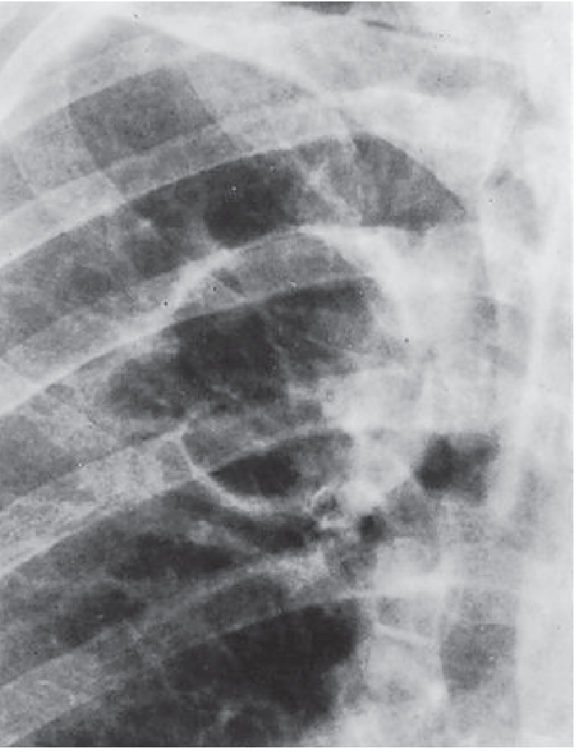
Fig. 8.13 - Cavitating Squamous Cell Carcinoma. Cavity in the right upper zone with an irregular wall in a heavy smoker. - Grainger & Allison's Diagnostic Radiology
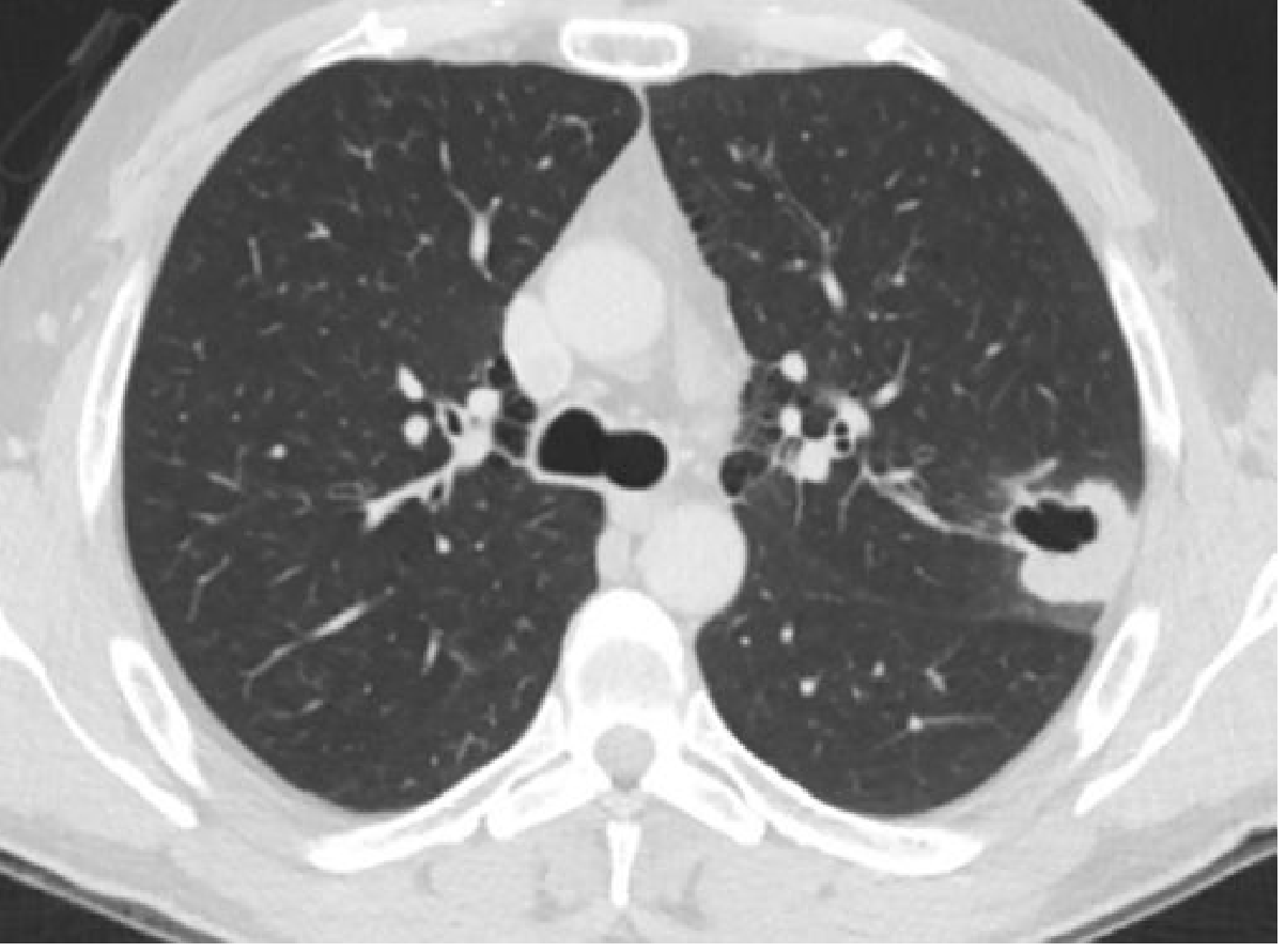
Fig. 8.14 - CT showing a cavitating squamous cell carcinoma in the left lung with variable wall thickness. - Grainger & Allison's Diagnostic Radiology
Cavitating Pulmonary Metastases
- Cavitation is seen more commonly in squamous cell than in adenocarcinoma metastases
- Multiple nodules at various stages of cavitation; bilateral distribution
6. Fungal Infections
Invasive Pulmonary Aspergillosis / Mucormycosis (Immunocompromised Host)
- "Halo sign": nodule or mass surrounded by a halo of ground-glass attenuation (represents haemorrhagic infarction around the fungal lesion)
- "Air-crescent sign": crescent of air between the necrotic fungal core and surrounding tissue; highly suggestive of invasive fungal infection; seen in 5%-12.5% of cases; cannot distinguish aspergillosis from mucormycosis on imaging alone
- Mucormycosis: lobar/multilobar consolidation, nodules, cavitation in 26%-40% of cases
Cryptococcosis
- Masses ranging from 5 mm to very large; may show a halo sign and eventually cavitate
- Seen in AIDS patients (CD4 < 100 cells/mm³)
Histoplasmosis
- Chronic pulmonary histoplasmosis radiologically resembles post-primary tuberculosis: upper lobe contraction, calcification, and cavitation
Coccidioidomycosis
- Cavitation with hilar/mediastinal adenopathy in ~20% of primary lesions
- Characteristic CT: central soft-tissue attenuation with surrounding halo of ground-glass around nodules
- Chronic fibronodular cavitary disease resembles post-primary TB
Paracoccidioidomycosis
- HRCT: ground-glass opacities, nodules, "halo" and "reversed halo" signs, consolidation, cavitation, fibrosis
7. Bronchopleural Fistula
A complication of necrotising infection or cavitation:
- Sinus tract between bronchus and pleural space
- CT shows communication between a parenchymal cavity and pleural space
- Pneumothorax or hydropneumothorax with air in pleural space
- May result from necrotising pneumonia, abscess rupture, surgery, or malignancy
Differential Diagnosis Summary (Causes of Solitary Pulmonary Mass/Cavity)
Per Grainger & Allison Table 8.5:
- Bronchial carcinoma / carcinoid
- Granuloma (TB, fungal)
- Hamartoma (cavitation is almost unknown)
- Metastasis
- Chronic pneumonia or abscess
- Hydatid cyst
- Pulmonary haematoma
- Fungus ball (aspergilloma in pre-existing cavity)
- Bronchogenic cyst / sequestration
- Arteriovenous malformation / pulmonary infarct
Source: Grainger & Allison's Diagnostic Radiology - A Textbook of Medical Imaging (Chapters 5, 8, and 19)
Pulmonary hydrated radiology
You likely mean pulmonary hydatid (Echinococcus) radiology. Let me search Grainger & Allison's Diagnostic Radiology directly.
Searching sources...
Finding Sources
Finding Sources
The main pulmonary hydatid section is in block1 and block1 (Echinococcosis). Let me read all of it now:
Reading File
Reading File
Now I need to find the earlier part of the text that describes the unruptured cyst, intact cyst radiological signs. Let me read the section just before line 3858 more carefully:
Reading File
Loading Image
The text at line 3858 begins mid-sentence. Let me check what immediately precedes this about the intact/unruptured cyst radiological features, and also read the "Hydatid" section in block5:
Reading File
Finding Sources
The block5 hydatid is hepatic (biliary), not pulmonary. The main pulmonary section in block1 had a gap - it jumped from line 3857 to 3858 mid-sentence, meaning there was content between 3846 and 3858 that was cut off describing the intact cyst features. Let me retrieve the missing text more carefully:
Finding Sources
Finding Sources
The user asked for radiology textbook only. The best pulmonary hydatid content is in Grainger & Allison block1, and Bailey & Love has relevant radiology content too (since the user asked previously about radiology textbooks). Let me now read the full Bailey & Love pulmonary hydatid section since it is detailed, and fetch the key images:
Reading File
Loading Image
Reading File
Loading Image
Loading Image
I now have all the key content and images. I note the user asked specifically for radiology standard textbooks - Grainger & Allison is the primary radiology textbook in the library. The content from Grainger & Allison block1 (lines 3830-3879) is the core, supplemented with a concise descriptive passage from the text. Let me now compose the full answer.
Pulmonary Hydatid Disease - Radiology
Source: Grainger & Allison's Diagnostic Radiology
Aetiology & Background
Hydatid disease (echinococcosis) is caused by the larval forms of Echinococcus granulosus (most common - unilocular cystic echinococcosis), E. multilocularis, and E. vogeli. It is endemic in the Mediterranean, Eastern Europe, Africa, South America, the Middle East, Australia, and New Zealand. Humans are accidental hosts, acquiring infection by ingesting ova from fomites, contaminated water, or direct contact with dogs.
The lung is the second most commonly affected organ after the liver. The right lung and lower lobes are slightly more often involved.
Cyst Structure
A hydatid cyst has two layers:
- Pericyst (outer host-derived fibrous layer)
- Laminated (ectocyst) membrane with inner germinal (endocyst) layer
This layered structure underlies all the classical radiological signs.
Epidemiology
- Usually solitary
- Multiple and/or bilateral in 10% of cases
- At the time of presentation: two-thirds are ruptured, one-third unruptured
- Size varies from very small to very large
Radiological Features - Classified by Stage
Stage 1 - Intact / Unruptured Cyst
Plain CXR:
- Well-defined, rounded or oval homogeneous opacity - often with completely smooth margins
- The intact cyst appears as a simple mass indistinguishable from a solid tumour or benign mass on CXR
- No calcification (unlike hepatic hydatid which may calcify)
- No satellite lesions
- May cause adjacent compression of lung and diaphragm elevation with large cysts
CT:
- Well-defined, round or oval cystic lesion with a thin, smooth wall
- Water-density (0 HU) content - pure fluid inside the cyst
- The pericyst wall may be visible as a thin rim
- Internal daughter cysts may be seen with E. multilocularis (alveolar type - more complex, infiltrative)
Stage 2 - Early Communication (Pericyst Erosion / Impending Rupture)
When bronchioles erode the outer pericyst, allowing air to enter the space between the pericyst and the laminated membrane:
Plain CXR:
- "Meniscus sign" (also called the "crescent sign"): a fine radiolucent crescent of air appears at the upper pole of the cyst between the outer pericyst and the inner laminated membrane
- This is considered a sign of impending rupture
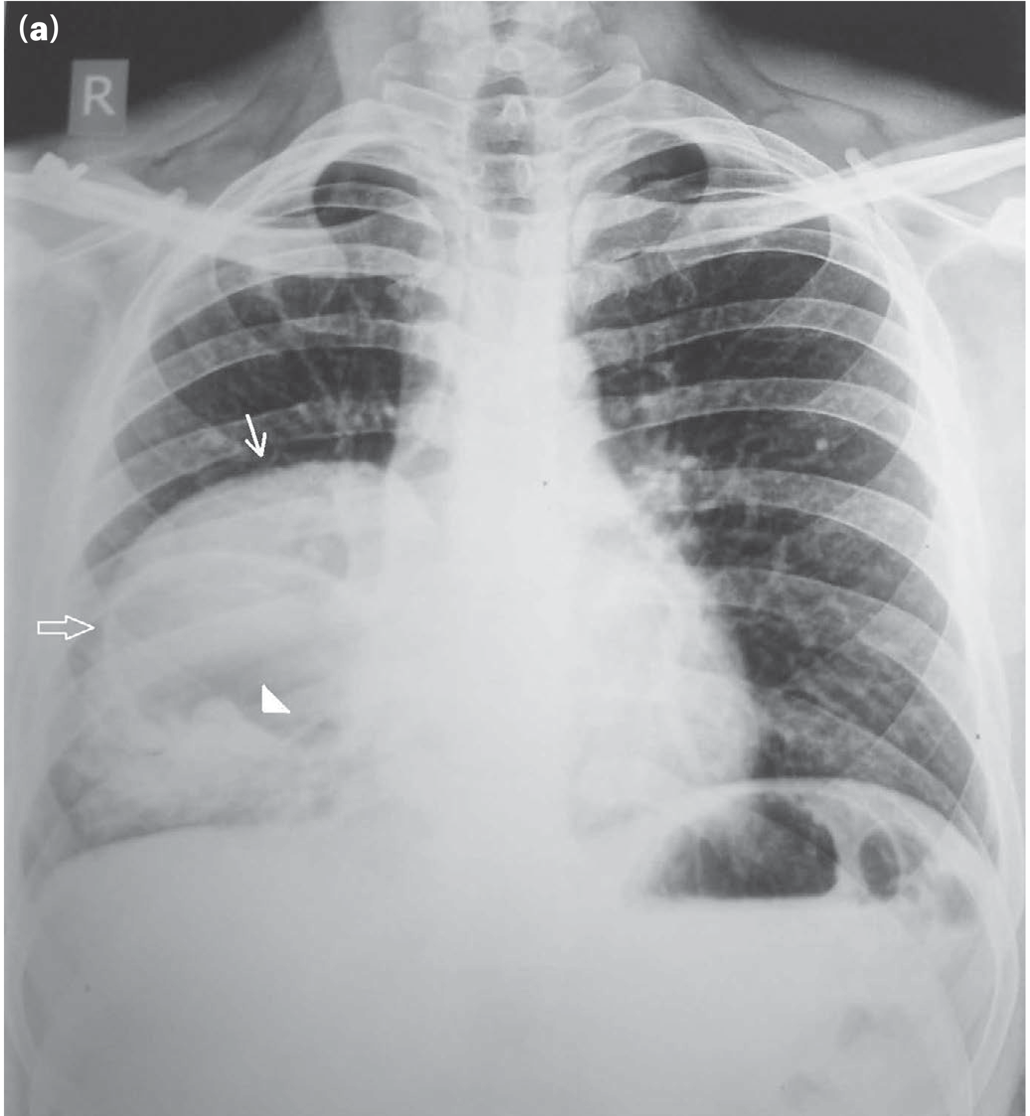
Figure 6.15 - Hydatid cysts of the lung: intact cyst (solid arrow), ruptured cyst (hollow arrow) with lamellar membrane floating like a water lily (solid arrowhead).
Stage 3 - Ruptured Cyst (Endocyst Rupture into Bronchus)
When both the pericyst and the inner endocyst (laminated membrane) rupture and communicate with the airway, multiple classical signs appear:
(A) Water-Lily Sign (Camalote Sign / Nymphaea Sign)
- The crumpled, collapsed endocyst floats on residual fluid at the bottom of the cyst cavity
- On CXR/CT it resembles a water lily (lotus leaf) or floating membrane above an air-fluid level
- Best demonstrated on CT where the floating membrane is clearly visible
- Pathognomonic of a collapsed (ruptured) pulmonary hydatid cyst
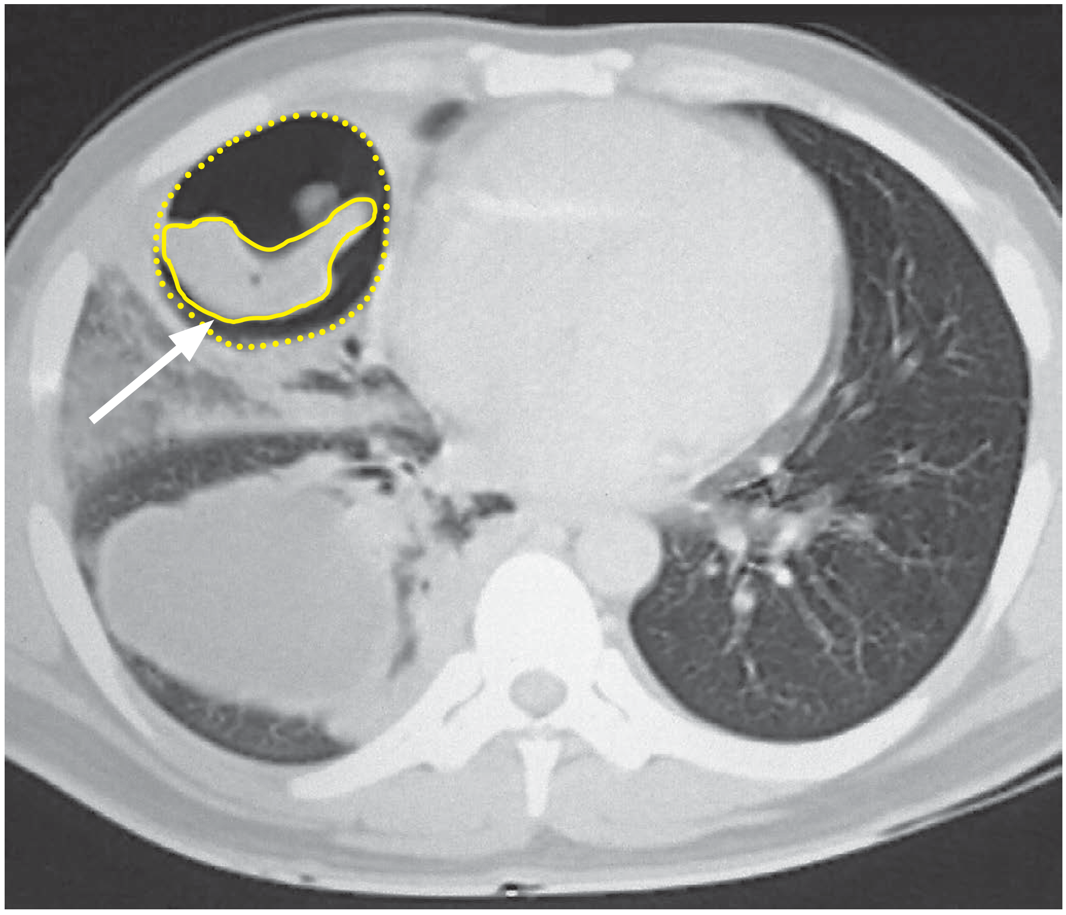
Figure 6.16 - CT showing the "water-lily sign" (arrow) in a ruptured pulmonary hydatid cyst - Bailey & Love's Short Practice of Surgery
(B) Air-Fluid Level
- A horizontal air-fluid level within the now-communicating cyst cavity
- Indicates bronchial communication has occurred and cyst contents are being partially expectorated
(C) Double Wall Sign
- Both the pericyst (outer) and the laminated membrane (inner) are visible as two distinct concentric rings on CT
- The space between them filled with air
(D) Rising Sun Sign / Serpent Sign
- When most fluid has been expectorated, the crumpled membranes lie at the bottom of the cavity
- Rising sun sign: the membrane appears along the lower arc of the cyst
- Serpent sign: the floating membrane has a wavy, snake-like configuration on CT
(E) Empty Cyst Sign
- After complete expectoration of all cyst contents, only an empty collapsed cavity remains
- This is the "empty cyst" or "ghost cyst" appearance - the wall may be visible as a thin ring
Stage 4 - Complications
1. Secondary Infection
- Secondary bacterial infection of a ruptured or partially evacuated hydatid cyst produces a lung abscess
- Appears as a thick-walled cavity with surrounding consolidation and air-fluid level, indistinguishable from a primary pyogenic abscess without clinical context
2. Pleural Rupture
- Rupture into the pleural space causes:
- Pleural effusion (hydatid fluid + daughter cysts in pleural space)
- If there is also airway communication: hydropneumothorax
- Risk of anaphylaxis from spillage of hydatid fluid
3. Massive Haemoptysis / Haemorrhage
- Aggressive invasion of bronchial or pulmonary arteries can result in massive haemoptysis
Classic Radiological Signs - Summary Table
| Sign | Mechanism | Modality | Significance |
|---|---|---|---|
| Rounded opacity | Intact cyst | CXR/CT | Unruptured; may mimic tumour |
| Meniscus / Crescent sign | Air between pericyst and laminated membrane | CXR | Impending rupture |
| Air-fluid level | Bronchial communication, partial expectoration | CXR/CT | Ruptured cyst |
| Water-lily sign (Camalote sign) | Collapsed endocyst floating on fluid | CXR/CT | Pathognomonic of ruptured cyst |
| Double wall sign | Air separating pericyst from endocyst | CT | Rupture into pericyst space |
| Serpent / Rising sun sign | Membranes lying at cyst base after fluid expectorated | CT | Largely evacuated ruptured cyst |
| Empty cyst sign | Complete expectoration of contents | CXR/CT | All contents expectorated |
Imaging Modality Comparison
| Modality | Role |
|---|---|
| CXR | Initial detection; shows rounded opacity, crescent sign, air-fluid level |
| CT | Modality of choice - best defines the floating membrane, double wall, confirms water-lily sign, demonstrates pleural involvement and secondary complications |
| MRI | Rarely used; may help characterise complex cysts or when CT is inconclusive |
| Ultrasound | Not applicable to lung parenchyma; useful for associated hepatic hydatid |
Source: Grainger & Allison's Diagnostic Radiology - A Textbook of Medical Imaging, Chapter 5 (Echinococcosis / Hydatid Disease), p. 139-140
Life cycle for radiology small 2 mark
Here is a concise 2-mark answer on the life cycle of Echinococcus granulosus (pulmonary hydatid):
Life Cycle of Echinococcus granulosus (Hydatid Disease)
Definitive host = Dog (tapeworm Taenia echinococcus lives in dog intestine)
↓ Eggs passed in dog faeces → contaminate soil, water, vegetables
Intermediate host = Sheep, cattle (and accidentally, humans)
↓ Humans ingest ova → hatch in duodenum → hexacanth embryo penetrates intestinal wall → enters portal circulation
Sites of lodgement:
- Liver (most common - 60-70%)
- Lung (second most common - 20-30%)
- Other organs (brain, kidney, bone - rare)
In the lung: embryo develops into a hydatid cyst with:
- Outer pericyst (host fibrous tissue)
- Middle ectocyst (laminated membrane)
- Inner endocyst (germinal layer - produces scolices/daughter cysts)
Cycle completes when dogs eat the infected viscera (liver/lung) of sheep or cattle containing scolices → develop into adult tapeworms in dog intestine.
Key points for 2 marks:
- Dog = definitive host; Human = accidental intermediate host
- Route: dog faeces → oral ingestion → portal blood → liver → lung
- Lung is the second most common site after liver
Imaging findingsin chest of, immunocompromised patient.
Finding Sources
Finding Sources
Finding Sources
Finding Sources
Reading File
Reading File
Good - the immunocompromised chest content is spread across the HIV section (block1 lines ~3540-3830) and the individual pathogen sections. Let me now read the dedicated HIV / changing spectrum sections and get the key fungal/Aspergillus/PCP sections:
Reading File
Finding Sources
Reading File
Reading File
Reading File
Your credits are exhausted. You can purchase a top-up or subscribe to a plan to continue using Orris. If you're already subscribed, your daily credits will reset tomorrow.
This is a shared conversation. Sign in to Orris to start your own chat.