
fuchs uveitis syndrome
I'll invoke the medical library skill first before answering.
Reading File
Now let me search the library and PubMed in parallel.
Finding Sources
Searching PubMed
"Fuchs uveitis syndrome" OR "Fuchs heterochromic iridocyclitis"
Good - found a dedicated section in Kanski's. Let me read it along with a broader paragraph search.
Reading File
Finding Sources
Let me read more of the Kanski section and fetch the clinical images.
Reading File
Let me read the remaining content and fetch the key clinical images in parallel.
Reading File
Loading Image
Loading Image
Loading Image
I now have comprehensive content. Let me compile the full answer.
Fuchs Uveitis Syndrome (FUS)
Also called Fuchs heterochromic iridocyclitis (FHC) or Fuchs heterochromic cyclitis. A classic and well-characterised chronic anterior uveitis entity.
Overview
- Type: Chronic, non-granulomatous anterior uveitis
- Age at diagnosis: Average ~40 years
- Sex/race: No predilection
- Laterality: Unilateral in 90-95% of cases
- Etiology: Uncertain. Strongest evidence implicates rubella virus. Toxoplasma gondii has also been suspected. Much of the apparent "anterior chamber (AC) activity" may be due to blood-aqueous barrier breakdown rather than true inflammation.
Clinical Features
Symptoms
- Often detected incidentally
- Gradual visual blurring (due to cataract development)
- Persistent floaters
- Patient or clinician may notice heterochromia
Signs (the "FUS constellation")
1. Heterochromia iridis
- Most effectively seen in daylight
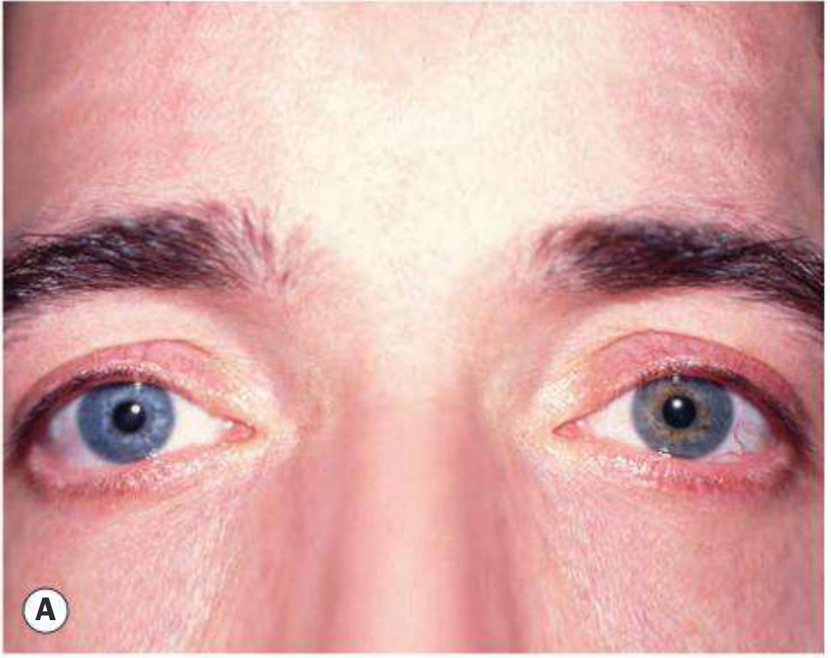
- Typically the affected eye is hypochromic (lighter) - Fig. A below
- Due to diffuse iris stromal atrophy + posterior pigment epithelial atrophy
- Exception: in blue eyes, stromal atrophy allows the pigmented posterior layer to dominate, making the eye paradoxically hyperchromic
2. Keratic precipitates (KPs)
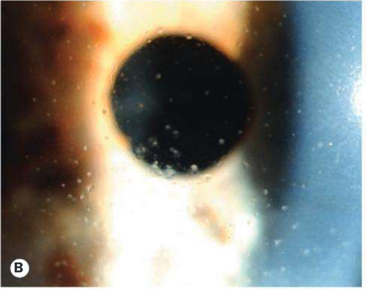
- Characteristically stellate, grey-white
- Located diffusely over the entire corneal endothelium (not just inferior as in typical uveitis)
- This diffuse stellate distribution is pathognomonic
3. Iris nodules
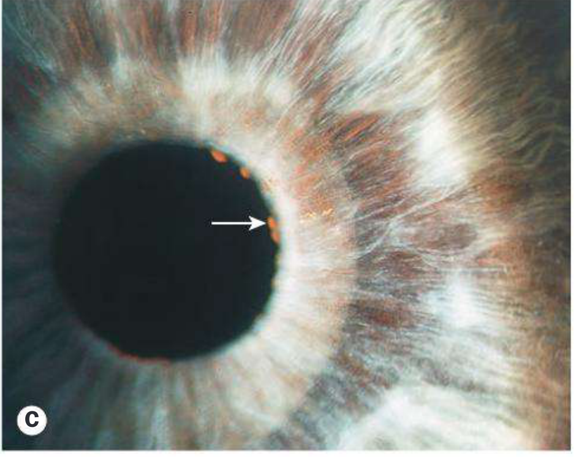
- Koeppe nodules (on pupillary border) - present in ~30% - arrow shown in Fig. C
- Russell bodies (tiny crystals) may be present on iris surface
4. Iris atrophy
- Diffuse with loss of crypts
- Iris appears smooth with a prominent sphincter pupillae
- Pigment epithelial atrophy demonstrated by retroillumination (band-like pattern)
- Fine irregular iris surface vessels commonly present
5. Anterior chamber
- Faint flare, usually only mild cellular activity
- The eye is virtually always white even during exacerbations - no ciliary injection
- No posterior synechiae (PS) - absence of PS is a distinguishing feature (may appear post-surgery)
6. Vitritis
- Anterior vitreous opacities may be dense
7. Posterior subcapsular cataract
- Extremely common - a frequent mode of presentation
8. Glaucoma
- A later manifestation, but occasionally present at diagnosis
- Develops in up to 60% of involved eyes
- Multiple mechanisms suspected
9. Gonioscopy
- Fine radial angle vessels or small irregular peripheral anterior synechiae (PAS)
- These angle vessels are the source of the Amsler sign - a small hyphema that appears on paracentesis / AC incision due to bleeding from fragile neovascular angle vessels
10. Fundus
- Peripheral choroiditis foci/scarring reported
- Possible increased incidence of retinal dialysis
- Macular edema does not occur except following surgery (a key differentiator)
Heterochromia Iridis - Differential Diagnosis
| Type | Causes |
|---|---|
| Hypochromic (lighter) | Idiopathic congenital, Horner syndrome (especially congenital), Waardenburg syndrome |
| Hyperchromic (darker) | Topical prostaglandin analogue use, oculodermal melanocytosis (naevus of Ota), ocular siderosis, diffuse iris naevus/melanoma, Sturge-Weber syndrome |
| Either | Fuchs uveitis syndrome, other chronic anterior uveitides |
Investigation
Diagnosis is clinical. Investigation is needed only to exclude alternative conditions (e.g., herpetic uveitis, sarcoidosis, toxoplasmosis).
Treatment
| Problem | Management |
|---|---|
| Chronic low-grade inflammation | Topical steroids generally NOT helpful long-term; may be used short-term for moderate/severe exacerbations |
| Monitoring | Long-term monitoring for glaucoma and cataract is mandatory |
| Cataract surgery | Carries increased risk: poor mydriasis, postoperative hyphema, increased inflammation, worsened glaucoma, zonular dehiscence. Preoperative topical/systemic steroids used by some |
| Glaucoma | Often difficult to control medically; surgical options include glaucoma drainage device or trabeculectomy + mitomycin-C |
| Vitreous opacification | Pars plana vitrectomy may be considered for visually significant cases |
Key Points for Exam / Clinic
- FUS is almost always unilateral, presents in middle age, no sex preference
- The eye is white and quiet-looking despite active inflammation
- No posterior synechiae (absence is hallmark - unlike most other anterior uveitides)
- KPs are diffuse and stellate (not inferior/mutton-fat)
- Amsler sign - bleeding from angle vessels during paracentesis
- Macular edema absent (except post-op)
- Rubella virus is the leading suspected etiology
- Glaucoma develops in up to 60% - the most significant long-term complication
- Steroids do NOT prevent progression and are not used chronically
Source: Kanski's Clinical Ophthalmology: A Systematic Approach, 10th edition, p. 459-460
This is a shared conversation. Sign in to Orris to start your own chat.