
Nerve anatomy
Reading File
Searching sources...
Finding Sources
Searching Images
nerve anatomy structure cross section myelinated fiber
Finding Sources
Finding Sources
Good results. Let me read the key sections now in parallel.
Reading File
Reading File
Reading File
Now let me fetch the key diagrams and get more content on myelinated vs unmyelinated fibers and nerve fiber types.
Loading Image
Loading Image
Finding Sources
Reading File
Reading File
Loading Image
Loading Image
Finding Sources
Reading File
Reading File
Excellent - I now have comprehensive content from authoritative textbooks. Let me compile the full answer.
Nerve Anatomy
1. Overview
A peripheral nerve is not a single wire - it is an organized bundle of many individual nerve fibers (axons), each with its own cellular sheath, wrapped together in layers of connective tissue and supplied by a dedicated blood system.
2. Connective Tissue Layers (Inside Out)
The three concentric connective tissue sheaths are the structural backbone of every peripheral nerve:
Endoneurium
- The innermost layer; loose connective tissue (reticular fibers, sparse fibroblasts, capillaries) that surrounds each individual nerve fiber (axon + Schwann cell)
- Collagen fibrils run both parallel to and circumferentially around the fiber; most are secreted by Schwann cells
- Contains mast cells and macrophages for immune surveillance and repair
- Sparse capillaries here form part of the blood-nerve barrier
- Histology A Text and Atlas With Correlated Cell and Molecular Biology, p. 981-982
Perineurium
- Groups of nerve fibers + endoneurium are bundled into fascicles by the perineurium
- 2-6 layers of flat, specialized fibrocyte-like cells with edges sealed by tight junctions - these create the main blood-nerve barrier that regulates the fascicular microenvironment
- Cells have actin microfilaments, pinocytotic vesicles, and cytoplasmic densities, resembling smooth muscle cells ultrastructurally
- Perineurial cell processes can extend inward to surround sub-groups of axons at branching points
- Junqueira's Basic Histology, 17e, p. 469-470
Epineurium
- Dense irregular fibrous coat that surrounds the entire nerve trunk and fills the spaces between fascicles
- Has a superficial dense region and a deeper, looser region containing larger blood vessels
- Extends as interfascicular epineurium between fascicle groups
- Current Surgical Therapy 14e, p. 980
Mesoneurium
- Loose connective tissue extending from the epineurium to surrounding tissues
- Allows nerve gliding during range of motion and carries the segmental (extrinsic) blood supply to the nerve
- Campbell's Operative Orthopaedics 15th Ed, p. 3840
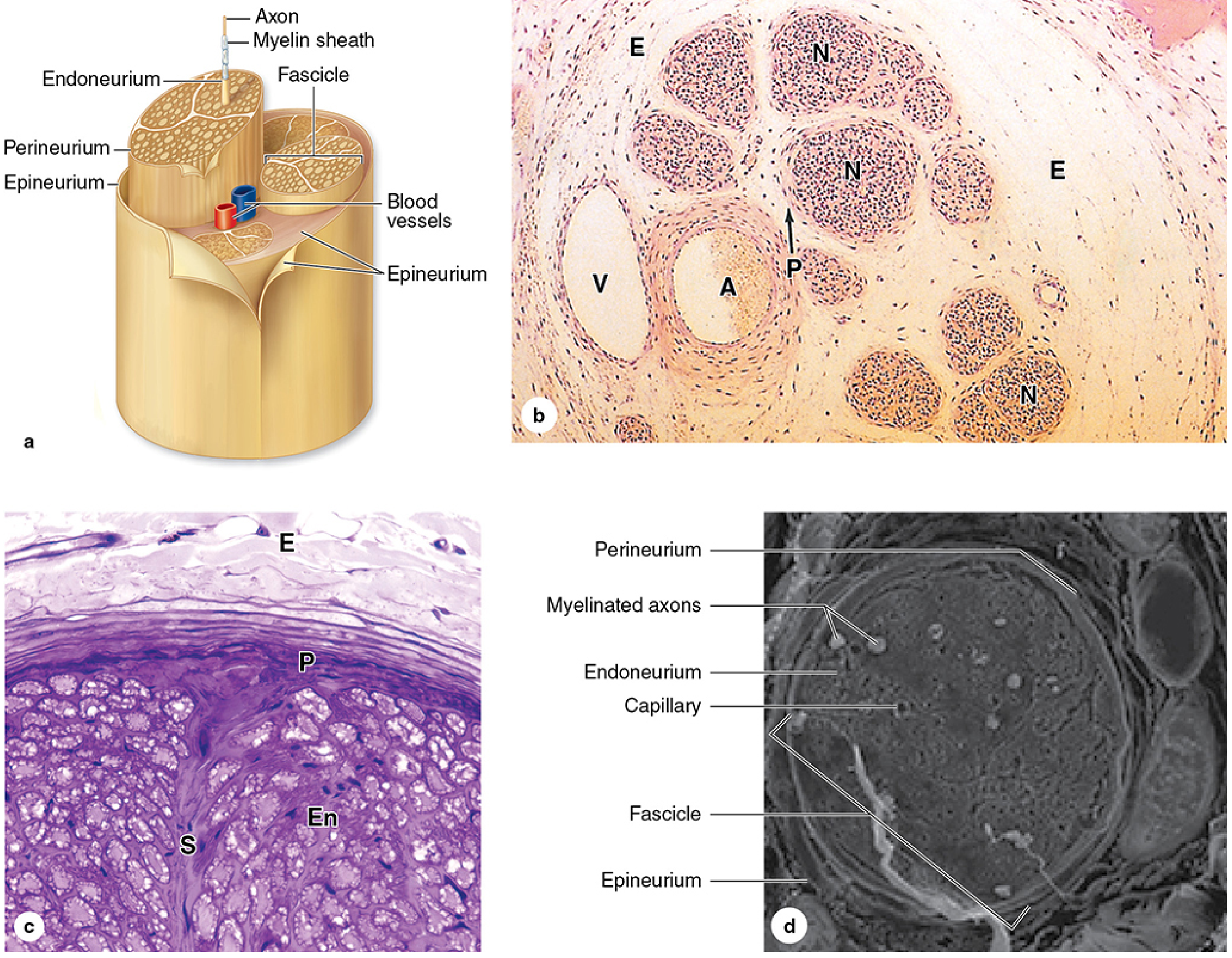
Figure: Epi-, peri-, and endoneurium. (a) 3D diagram; (b) H&E histology showing fascicles (N), perineurium (P), epineurium (E), artery (A), vein (V); (c) perineurial septa (S) within a fascicle; (d) SEM of a single fascicle showing myelinated axons and endoneurium. - Junqueira's Basic Histology, 17e
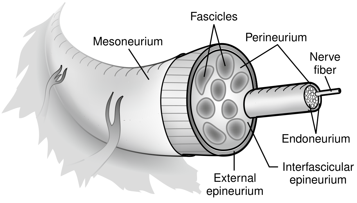
Figure: Full cross-sectional anatomy showing fascicles, perineurium, external epineurium, interfascicular epineurium, endoneurium, nerve fiber, and mesoneurium. - Current Surgical Therapy 14e
3. The Individual Nerve Fiber
Each nerve fiber = axon + Schwann cell sheath.
Myelinated Fibers
- A single Schwann cell rotates around one axon many times, laying down multiple layers of its membrane (containing sphingomyelin) - this is the myelin sheath
- Myelin is an electrical insulator that reduces ion flow ~5000-fold
- The segment of fiber covered by one Schwann cell = internode (0.1-1.8 mm; longer internodes on more heavily myelinated fibers)
- The gap between successive Schwann cells, where the axon membrane is exposed = node of Ranvier (2-3 µm long)
- Action potentials jump from node to node = saltatory conduction - this increases velocity 5-50 fold and is energy efficient
- Guyton & Hall Medical Physiology, p. (Ch. 5)
Unmyelinated Fibers
- Several axons (~10-20) are embedded within the cytoplasmic processes of a single Schwann cell (Remak cell) but are not wrapped
- Conduct impulses slowly by continuous conduction along the membrane
- Unmyelinated fibers outnumber myelinated ~2:1 in a typical nerve trunk
- Sensory and motor nerves have unmyelinated:myelinated ratio of ~4:1
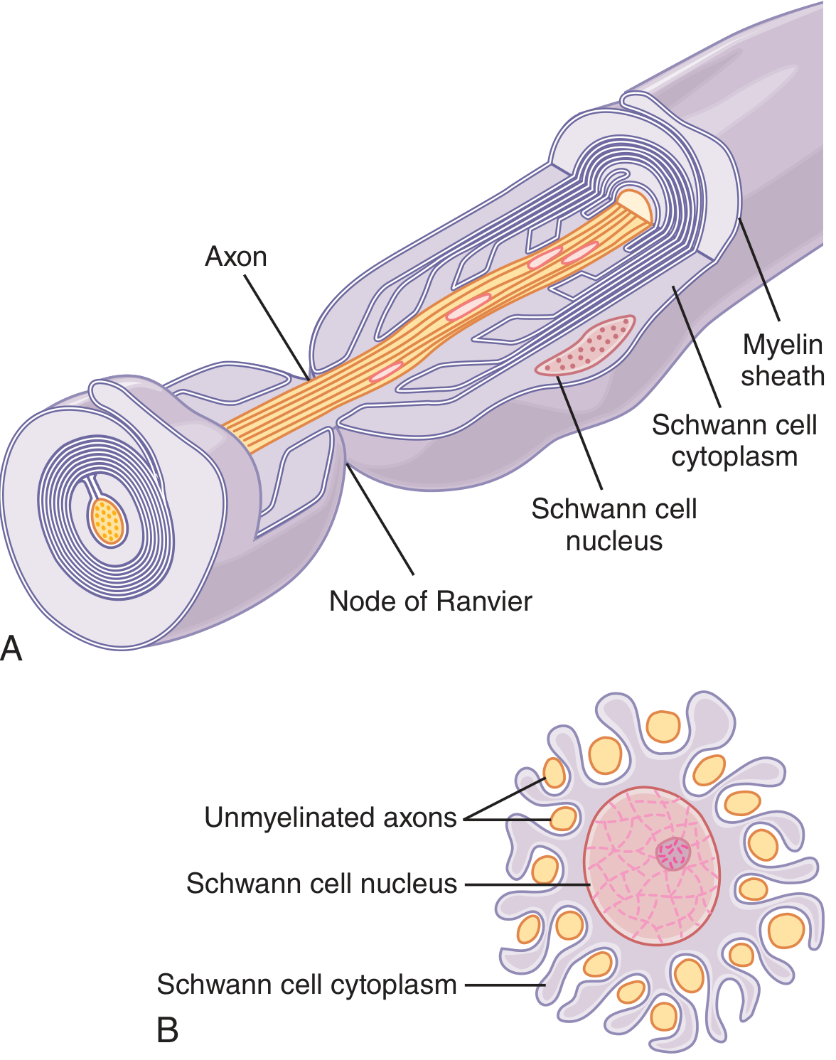
Figure: (A) Single Schwann cell wrapping around one large axon forming myelin sheath with node of Ranvier. (B) One Schwann cell partially wrapping multiple unmyelinated axons (Remak bundle). - Guyton & Hall
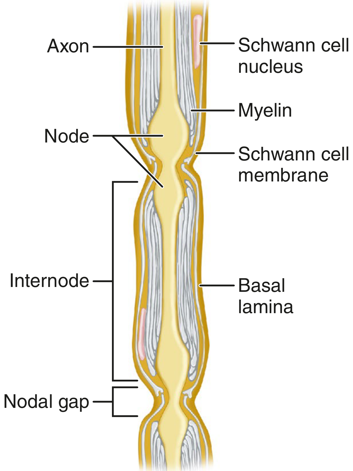
Figure: Axon, myelin, Schwann cell nucleus/membrane, internode, node (nodal gap), and basal lamina. - Campbell's Operative Orthopaedics, 15e
4. Nerve Fiber Classification
Two classification systems are in use - the Erlanger-Gasser (letter) system and the Lloyd-Hunt (Roman numeral) system for sensory fibers only.
Erlanger-Gasser Classification (Ganong's Review of Medical Physiology, 26e)
| Fiber Type | Function | Diameter (µm) | Velocity (m/s) | Myelinated? |
|---|---|---|---|---|
| Aα | Proprioception; somatic motor (α motor neurons) | 12-20 | 70-120 | Yes |
| Aβ | Touch, pressure | 5-12 | 30-70 | Yes |
| Aγ | Motor to muscle spindles (intrafusal fibers) | 3-6 | 15-30 | Yes |
| Aδ | Pain (fast/sharp), temperature, touch | 2-5 | 12-30 | Yes |
| B | Preganglionic autonomic | <3 | 3-15 | Yes |
| C | Slow/aching pain, temperature; postganglionic autonomic | 0.3-1.3 | 0.5-2.3 | No |
Lloyd-Hunt Classification (Sensory Only - Costanzo Physiology, 7e)
| Type | Corresponds to | Example |
|---|---|---|
| Ia | Aα | Muscle spindle primary afferents |
| Ib | Aα | Golgi tendon organ afferents |
| II | Aβ | Muscle spindle secondary afferents; touch, pressure |
| III | Aδ | Touch, pressure, fast pain, temperature |
| IV | C | Slow pain, temperature |
Clinical note: Local anesthetics block unmyelinated C fibers before myelinated A fibers. Conversely, pressure on a nerve selectively blocks large-diameter A fibers (motor, touch, pressure) first, leaving pain (C fibers) relatively intact - the classic "arm fell asleep" pattern.
5. Fascicular Topography
- Proximally, sensory and motor axons intermingle significantly within nerve trunks
- More distally, they segregate into distinct motor and sensory fascicles
- This has direct surgical implications: proximal nerve repairs (e.g. facial nerve within the temporal bone) risk synkinesis/dyskinesis due to poorly differentiated fascicles; distal repairs have better outcomes because the fascicular architecture is more defined
- Current Surgical Therapy 14e, p. 980
6. Blood Supply
- Extrinsic (segmental) supply: enters through the mesoneurium at multiple points along the nerve
- Intrinsic (longitudinal) supply: runs within the epineurium and perineurium; branches penetrate into the endoneurium
- Endoneurial capillaries have tightly joined endothelium - a second component of the blood-nerve barrier
- This dual supply means peripheral nerves can tolerate moderate stretching or positional changes without ischemia
Summary: Structural Hierarchy
Nerve trunk
 └── Epineurium (dense irregular CT)
      └── Fascicles (grouped by interfascicular epineurium)
           └── Perineurium (tight-junction sealed fibrocytes)
                └── Individual nerve fibers
                     └── Endoneurium (loose CT)
                          └── Axon + Schwann cell (± myelin)
Sources: - Junqueira's Basic Histology, 17e | Histology: A Text and Atlas (Ross), 8e | Guyton & Hall Medical Physiology | Ganong's Review of Medical Physiology, 26e | Costanzo Physiology, 7e | Campbell's Operative Orthopaedics, 15e | Current Surgical Therapy, 14e
This is a shared conversation. Sign in to Orris to start your own chat.