
Femoral lateral open wedge osteotomy
Reading File
Now let me search the medical library and PubMed in parallel for femoral lateral open wedge osteotomy.
Finding Sources
Finding Sources
Searching PubMed
"lateral open wedge osteotomy" femoral
The key section is "LATERAL OPENING WEDGE DISTAL FEMORAL OSTEOTOMY" in Campbell's. Let me read that section along with the broader DISTAL FEMORAL OSTEOTOMY section.
Reading File
Reading File
Reading File
I now have all the key content. Let me also fetch the relevant surgical diagram image.
Loading Image
Reading File
I now have everything needed for a comprehensive answer. Let me compile the full response.
Femoral Lateral Open Wedge Osteotomy (Distal Femoral Osteotomy)
Overview and Indications
The lateral opening wedge distal femoral osteotomy (LOWDFO) is a supracondylar corrective procedure that opens the lateral cortex of the distal femur to produce a varus correction at the knee. It is indicated for:
- Genu valgum (lateral compartment osteoarthritis) where the valgus deformity exceeds 12-15 degrees, or the joint line deviation from horizontal is more than 10 degrees
- Younger active patients (typically under 65 years) who are not yet candidates for total knee arthroplasty (TKA)
- Lateral compartment overload - shifts mechanical axis from the lateral to the medial compartment
A comparative study found that functional outcomes after supracondylar osteotomy do not differ significantly from TKA in appropriately selected patients, making it a valid joint-preserving alternative in active patients younger than 65 with valgus angulation ≤15 degrees.
Reported success rates for distal femoral osteotomies for osteoarthritis range from 71% to 86% good or excellent results. Both closing wedge and opening wedge techniques show good clinical outcomes with no significant differences in patient-reported outcome measures.
Lateral Opening Wedge vs. Medial Closing Wedge
| Feature | Lateral Opening Wedge | Medial Closing Wedge |
|---|---|---|
| Approach | Lateral | Lateral/medial |
| Bone loss | None - widens gap | Removes wedge of bone |
| Graft needed | Yes (bone graft) | No |
| Hinge fracture risk | Medial cortex | Lateral cortex |
| Leg length | Slightly increases | Slightly decreases |
| Conversion to TKA | Easier hardware removal if locking plate used | May be more complex |
| Technical difficulty | More demanding | More established |
A 5-year follow-up study of 18 opening wedge osteotomies showed a cumulative survival of 80%, comparable to closing wedge, but the technique is described as technically demanding with higher reoperation rates.
Preoperative Planning
- Full-length mechanical axis radiographs are mandatory
- Characterize the type and degree of deformity
- Determine the size of the lateral opening wedge required
- Patient-specific printed cutting guides (3D-planned PSI) can significantly reduce operative and fluoroscopy time
Surgical Technique (Campbell's Technique 10.14)
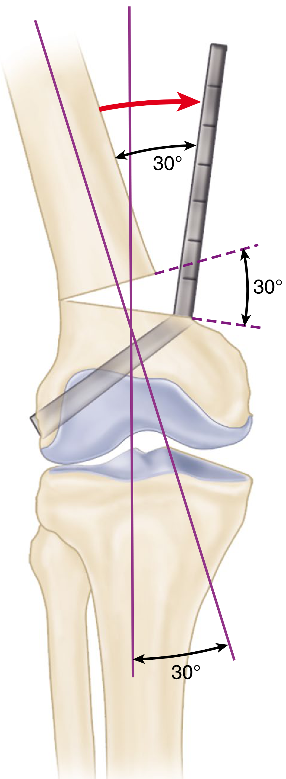
Figure: Distal femoral osteotomy showing the correction angle (30°) measured on preoperative radiograph, with blade plate/nail trajectory into femoral metaphysis.
Step-by-step:
-
Positioning: Supine on a radiolucent table. Hip bump to place the knee perpendicular to the table. A radiolucent foam wedge may assist lateral fluoroscopy views. Sterile tourniquet may be used.
-
Incision: Lateral approach - incision from the lateral epicondyle proximally along the lateral femur. Incise the iliotibial band in line with its fibers. Dissect vastus lateralis off the intermuscular septum. Coagulate perforating vessels carefully.
-
Retraction: Place a malleable retractor posteriorly behind the femoral condyle (confirmed on fluoroscopy, positioned medially enough to protect neurovascular structures). A Bennett or smooth Hohmann retractor anteriorly to retract quadriceps.
-
Kirschner wire guide: Insert a 1.6 mm K-wire along the distal femoral diaphysis toward the adductor tubercle under fluoroscopic guidance. Stop the pin at least 1 cm lateral to the medial cortex (to preserve the medial cortical hinge).
-
Osteotomy: Use a sagittal saw. The cut must be perpendicular to the long axis of the femur. Protect quadriceps anteriorly and posterior neurovascular structures posteriorly. Stop the osteotomy at least 1 cm lateral to the medial cortex - this preserved medial cortex acts as the hinge.
-
Distraction: Slowly distract using a stacked osteotome technique, then use a bone-spreading device (e.g., TomoFix, DePuy-Synthes). Slow distraction is essential - it takes advantage of the viscoelastic nature of bone and minimizes the risk of hinge fracture.
-
Open to templated amount: Remove spreading device, hold open with lamina spreaders.
-
Bone grafting: Place graft prior to removing lamina spreaders to maintain correction. Options include:
- Precut Evans wedges
- Tricortical iliac crest allograft
- Femoral neck allograft (of appropriate size)
- Remainder filled with cancellous chips, femoral head allograft, or demineralized bone matrix (DBM)
-
Fixation: Apply locking plate to lateral femur. Confirm plate placement fluoroscopically (AP and lateral views). Preferred technique: locking screws only (minimizes loss of correction during plate application).
- Place distal screws first (avoid intercondylar notch)
- Then proximal screws
- Use alignment rod to confirm adequate correction
Postoperative Care
Similar to proximal tibial osteotomy:
- Protected weight bearing initially
- Progressive rehabilitation with range-of-motion exercises
- Hardware removal may be required before TKA conversion, though supracondylar locking plates allow percutaneous distal screw removal, facilitating later TKA instrumentation without full plate removal
Contraindications and Poor Prognostic Factors
- Rheumatoid arthritis - consistently poor outcomes
- Inadequate pre-operative knee motion - poor outcomes after distal femoral osteotomy
- Valgus deformity >15 degrees (relative)
- Obesity, medial compartment disease
Conversion to TKA
TKA after distal femoral osteotomy can be complicated by:
- Exposure difficulties from scarring
- Hardware removal challenges
- Blade plates and supracondylar compression screws usually require removal before distal femur preparation
- Locking plate advantage: distal femoral instrumentation can proceed after percutaneous locking screw removal using a short intramedullary guide rod and extramedullary alignment
Key Evidence
- Diaz et al. (2023) - Systematic review and meta-analysis in AJSM: outcomes of DFO for valgus malalignment, closing vs. opening wedge techniques (cited in Campbell's 2026)
- Overall survival rates 71-86% for osteoarthritis indication
- 5-year opening wedge survival ~80%, comparable to closing wedge
Source: Campbell's Operative Orthopaedics, 15th Edition 2026, Chapter 10 (Technique 10.14)
This is a shared conversation. Sign in to Orris to start your own chat.