
I suffered a road traffic acciden causing me TBI resulting me in Diffuse Axonal Injury with right sided weakness, before 2.5 years
What would you like to know or get help with regarding your TBI/DAI?
"diffuse axonal injury" AND prognosis AND recovery
"diffuse axonal injury" AND rehabilitation
"traumatic brain injury" AND "motor recovery" AND "hemiparesis"
"traumatic brain injury" AND "long-term outcome" AND "motor"
PMID: 40141847
diffuse axonal injury recovery after 2 years prognosis right hemiparesis rehabilitation
Traumatic Brain Injury with Diffuse Axonal Injury (DAI) and Right-Sided Weakness
1. Understanding Your Condition
What is Diffuse Axonal Injury?
DAI Grades (Pathological Classification)
| Grade | What is injured | Severity |
|---|---|---|
| Grade I | White matter of cerebral hemispheres only | Mild-Moderate |
| Grade II | Cerebral white matter + corpus callosum | Moderate-Severe |
| Grade III | All of the above + brainstem | Severe |
What happened at the microscopic level
- The initial trauma stretched and tore axons, forming characteristic "retraction balls" (club-shaped swellings visible under microscopy)
- Over the first 2-3 weeks, injured axons continued degenerating even if they initially appeared functional
- After weeks to months, microglial clusters formed, followed by astrocytic scarring and demyelination in the affected white matter tracts
2. Recovery Outlook and Prognosis at 2.5 Years
Good news for you: You have survived past a critical window
- Only 10/30 had good recovery at 6 months
- This improved to 12/30 at 1+ year, showing recovery can continue beyond the first year
- Younger age was strongly associated with better outcomes (mean age of good-outcome group: 24 years vs. poor-outcome group: 40 years)
- DAI grade was a significant predictor
What "recovery" looks like at your stage
- Early phase (0-18 months): Brain plasticity drives spontaneous recovery - injured pathways rewire
- Later phase (18 months onward): Improvement comes mainly from rehabilitation-driven compensation - learning new strategies, strengthening remaining pathways, and adapting function
3. Rehabilitation and Therapy Options
Physiotherapy for Right-Sided Weakness
- Goal-directed physical therapy - task-specific training for the arm and leg
- Ankle-foot orthosis (AFO) - if foot drop is present, an adjustable AFO helps during recovery phase; a rigid AFO is used once a plateau is reached
- Gait training - balance is the best predictor of ability to walk after acquired brain injury ([Miller's Review of Orthopaedics])
- Robot-assisted therapy (Lokomat/exoskeletons) - shown to improve gait symmetry and functional mobility in TBI patients
- Constraint-induced movement therapy (CIMT) - forces use of the weaker right arm by constraining the stronger left
Spasticity Management (if applicable)
- Botulinum toxin (Botox) injections - reduces focal muscle overactivity; useful for planning further treatment
- Oral medications - baclofen, tizanidine
- Surgery (tendon lengthening, transfers) - only if plateau has been reached and spasticity prevents function; requires adequate cognition and motivation
Non-Invasive Brain Stimulation
- Transcranial Magnetic Stimulation (rTMS) - variable evidence for motor and cognitive improvement
- Transcranial Direct Current Stimulation (tDCS) - modest cognitive and motor gains in some TBI studies
Cognitive Rehabilitation
4. Symptoms You May Still Experience and How to Manage Them
Common Ongoing Symptoms After DAI + Right-Sided Weakness
| Symptom | Why it happens | Management |
|---|---|---|
| Right arm/leg weakness or stiffness | Corticospinal tract damage | Physiotherapy, splinting, AFO, Botox if spastic |
| Fatigue | Brain requires more effort for all tasks after axonal injury | Pacing strategies, sleep hygiene, avoid overexertion |
| Memory and concentration problems | Diffuse white matter injury disrupts neural networks | Cognitive therapy, memory aids, structured routines |
| Headaches | Post-traumatic headache is extremely common | Neurologist review; preventive or acute medications |
| Mood changes / depression / irritability | Emotional regulation networks (frontal, limbic) often injured in DAI | Psychological support, antidepressants if indicated |
| Sleep disturbances | A 2024 study (Ann Clin Transl Neurol, PMID 39031956) linked axonal injury to sleep and memory problems | Sleep hygiene; specialist review if severe |
| Balance problems | Cerebellar or brainstem pathways | Vestibular physiotherapy |
| Personality changes | Frontal lobe involvement is common in DAI | Neuropsychological support, counseling for family |
Long-Term Risks to Watch For
Practical Steps for Your Situation Right Now
- See a physiatrist (rehabilitation medicine specialist) if you are not already under one - they coordinate the full rehabilitation team
- Ask your neurologist for an updated MRI (SWI/susceptibility-weighted sequence is most sensitive for DAI lesions) to understand the current state
- Continue / re-engage with physiotherapy - even at 2.5 years, targeted rehab produces functional gains
- Request neuropsychological testing to map current cognitive strengths and weaknesses so therapy is targeted appropriately
- Screen for and treat depression - it is very common post-TBI and actively worsens recovery if untreated
- Avoid alcohol and contact sports - the injured brain is more vulnerable to second-hit injury
Currently I on Tav. Brivgard 50mg Before that after discharge I was on Levipill 500 for 1 year after discharge
brivaracetam AND levetiracetam AND "traumatic brain injury"
PMID: 41669569
PMID: 39606308
Your Medications: Levetiracetam (Levipill 500) and Brivaracetam (Brivgard 50mg)
Why You Are on These Medications
- Year 1 post-discharge: Levipill 500 (Levetiracetam 500mg) - standard first-line prophylaxis
- Now (2.5 years post-injury): Brivgard 50mg (Brivaracetam 50mg) - a next-generation upgrade of the same drug class
How These Drugs Work - The Mechanism
| Feature | Levetiracetam (Levipill) | Brivaracetam (Brivgard) |
|---|---|---|
| SV2A affinity | Standard | 10-30x higher affinity |
| Sodium channel effect | None | Yes - additional blockade |
| Half-life | 6-8 hours | 7-8 hours |
| Protein binding | Very low | <20% |
| Behavioral side effects | More common | Less common |
| Metabolism | Mostly renal | Hydrolysis + CYP2C19 (minor) |
Why Your Doctor Switched You from Levetiracetam to Brivaracetam
- Irritability, aggression, anger
- Anxiety and depression
- Insomnia
- Mood swings
What the Latest Research Says About Your Medications and TBI
- Seizure prevention: Both drugs were equally effective (16.6% seizures in BRV group vs. 25.7% in LEV group - not statistically different, but BRV trended better)
- Neurobehavioral outcomes (most important for you): BRV patients had dramatically lower neurobehavioral symptom scores at 14 days (10.7 vs. 40.1, p=0.005) and at 3 months (4.7 vs. 13.2, p<0.001)
- Conclusion: "BRV may offer a more favorable tolerability profile for seizure prophylaxis following TBI"
Your Current Dose: Brivgard 50mg
Important Things to Know About Brivgard (Brivaracetam)
Side Effects to Watch For
- Drowsiness / sedation (most common)
- Dizziness
- Nausea or GI upset
- Psychiatric effects: Still possible - depression, irritability, anxiety (less than levetiracetam, but not absent)
- Rarely: hypersensitivity reactions
Drug Interactions
- Carbamazepine (Tegretol): BRV can increase the active metabolite of carbamazepine, potentially causing carbamazepine toxicity. If you are on both, your doctor needs to monitor this.
- Phenytoin (Eptoin/Dilantin): BRV can increase phenytoin levels.
- Never combine BRV with levetiracetam - they act on the same target (SV2A) and give no added benefit together.
- Strong CYP450 inducers (rifampicin, some antifungals) can lower BRV blood levels.
What You Must NEVER Do
- Do not stop brivaracetam suddenly - abrupt withdrawal can trigger dangerous rebound seizures
- Never skip doses consistently - missed doses reduce blood levels and seizure protection
- Avoid alcohol - it increases CNS depression and lowers seizure threshold
- Inform any doctor or dentist you see about this medication before any procedure or new prescription
How Long Will You Need This Medication?
- After TBI, seizure risk remains elevated for life, but is highest in the first 2 years
- If you have been seizure-free for 2+ years, some neurologists consider slow tapering and discontinuation - but only with close monitoring and EEG assessment
- The decision depends on your EEG findings, MRI, seizure history, and lifestyle (e.g. driving)
- Do not attempt to stop or reduce the dose on your own - this must be done very slowly under medical supervision


I dislocated my shoulder recently, frim negligible disposition of hand does it have any relation to my previous tbi abd DAI
"shoulder dislocation" AND "hemiplegia" AND "brain injury"
shoulder dislocation TBI hemiparesis spasticity muscle imbalance neurological cause
Your Shoulder Dislocation IS Directly Related to Your TBI and DAI
First - What Your X-rays and Prescription Show
- Both confirm a complete anterior (subcoracoid) glenohumeral dislocation of the left shoulder
- The humeral head is displaced anteriorly and inferiorly out of the glenoid socket
- There is a probable Hill-Sachs lesion - a compression dent on the back of the humeral head caused by the glenoid rim during dislocation (visible on AP view)
- No obvious large fracture of the greater tuberosity or clavicle
- Diagnosis: Dislocation of (L) shoulder - this is your LEFT shoulder
- Treatment: Already reduced (put back into joint)
- Medications prescribed:
- Aminofit Sachet (amino acids / collagen support) x 15
- Tab. Tendon Care x 15 (collagen/tendon nutrition)
- Tab. C2 Daily x 10 (likely Vitamin C based supplement)
- Tab. Tolub x 10 (Tolperisone - a muscle relaxant)
The Direct Neurological Connection to Your TBI/DAI
Important clarification first
How TBI Causes Shoulder Vulnerability
1. Upper Motor Neuron Syndrome - The Root Cause
"Weakness of upper extremity muscles is most marked in the deltoid, triceps, wrist extensors, and finger extensors... the affected arm is adducted at the shoulder, and flexed at the wrist and fingers."
- Muscle weakness of the deltoid, rotator cuff, and periscapular muscles
- Spasticity (over-tightness) in the internal rotators and adductors (pectoralis major, subscapularis, teres major, latissimus dorsi)
- Muscle imbalance - the spastic muscles pull the shoulder inward and downward, while the stabilizing muscles (rotator cuff, deltoid) are weak
2. The Shoulder as the Most Vulnerable Joint
- The rotator cuff cannot hold the humeral head centered in the glenoid
- The deltoid (weakened after upper motor neuron injury) fails to support the arm against gravity
- The result is subluxation (partial slip) or full dislocation from forces that would never dislocate a normal shoulder
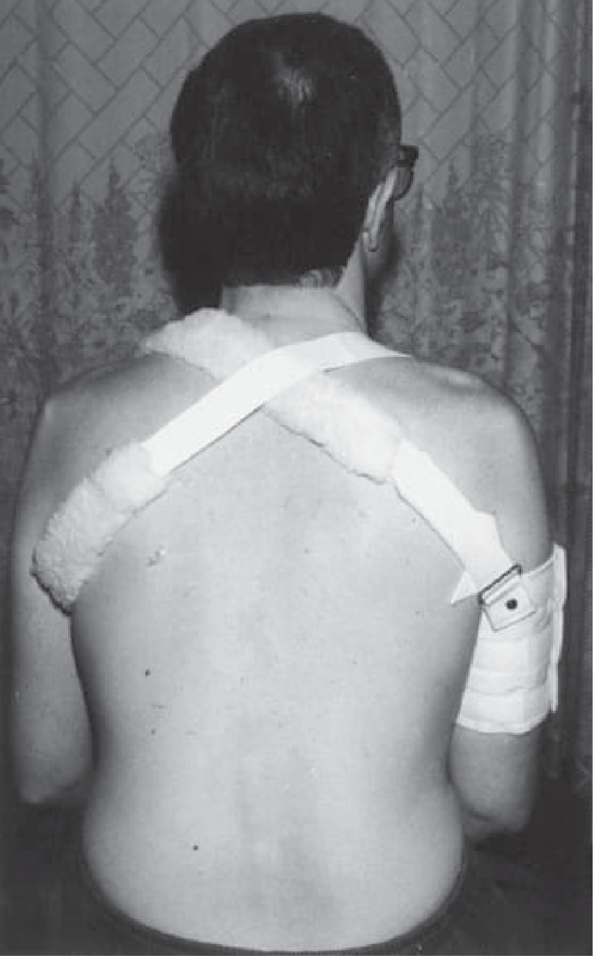
3. Why "Negligible Force" Was Enough
- Spastic internal rotators place the shoulder in a chronically at-risk position
- Weak stabilizers cannot resist even normal movements
- A minor twist, a light fall, reaching overhead, or rolling in bed can be enough
What This Means Going Forward
Risk of Recurrence is HIGH
What You Need Urgently
-
Tell your neurologist and physiatrist about this dislocation - it must be documented and managed as part of your TBI rehabilitation, not just as an isolated orthopaedic event
-
Do not leave the shoulder unsupported - use a shoulder sling or the type of cuff support shown above when walking, especially if your arm hangs with no active muscle control
-
Physiotherapy for the shoulder is essential - specifically:
- Rotator cuff strengthening (supraspinatus, infraspinatus)
- Periscapular muscle training
- Proprioceptive/joint position sense training
- Neuromuscular electrical stimulation (NMES) to the deltoid has evidence for hemiplegic shoulder stability
-
Botulinum toxin (Botox) assessment - if your pectoralis major and internal rotators are spastic and pulling the shoulder into the at-risk position, targeted Botox can reduce this pull and improve joint alignment. [Campbell's Operative Orthopaedics 2026] specifically recommends Botox for spastic hemiplegic shoulder
-
MRI of the shoulder - to check for Bankart labral tear, Hill-Sachs size, and rotator cuff integrity, especially given the high re-dislocation risk
-
Surgery consideration - [Campbell's] notes surgery (fractional lengthening of pectoralis major, latissimus dorsi, teres major) for patients with spastic hemiparesis improves shoulder range of motion and reduces pain, but only when there is reasonable rehabilitation potential and it is done as an adjunct to, not replacement for, physiotherapy
Summary Table
| Factor | How it links to your TBI/DAI |
|---|---|
| Weak deltoid & rotator cuff | Direct UMN injury - loses stabilizing force |
| Spastic internal rotators | Brain injury causes overactivity in pectoralis/subscapularis, pulling shoulder inward |
| Poor proprioception | Damaged sensory pathways reduce joint position awareness |
| Altered posture & gait | TBI-related asymmetric posture increases shoulder loading |
| Negligible force causing dislocation | Because stabilizers are neurologically compromised, trivial forces are sufficient |
But left side is my healthy side I don't have any kind of weakness there so it has become my dominant sid due to right sided weakness
shoulder dislocation overuse compensatory dominant side hemiparesis TBI epilepsy seizure cause
shoulder overuse injury unaffected side stroke TBI hemiplegia crutch walking increased load
Why Your HEALTHY Left Shoulder Dislocated - Still Directly Related to Your TBI
Mechanism 1: Overuse and Overload of the Unaffected Side
"CNS disorders (stroke, TBI, multiple sclerosis) result in mobility impairments that force patients to put a greater burden on unaffected or less affected structures... The development of overuse injuries results from biomechanical issues such as malalignment, poor technique, and muscle imbalance."
- Dressing, bathing, cooking, lifting, reaching
- Supporting body weight when getting up from chairs, bed, floor
- Gripping walls or railings to compensate for gait imbalance
- Compensating during walking/balance with an asymmetric arm swing
- All fine motor tasks that the right hand cannot do
"Hemiplegics have a tendency to use their unaffected shoulders before achieving adequate motor recovery in their affected sides, which results in prolonged overuse."
- Fatigues the rotator cuff muscles
- Causes micro-tears in the capsule and labrum
- Stretches the joint capsule over time
- Leads to a shoulder that looks and feels normal but is actually mechanically compromised
Mechanism 2: Seizure-Related Dislocation (Very Important to Consider)
"In the post-seizure setting, focus the initial exam on injuries... A posterior shoulder dislocation is an injury to specifically look for."
- You are 2.5 years post-TBI - still in a window of risk for post-traumatic epilepsy
- You were on levetiracetam for 1 year, then switched to brivaracetam
- If the medication change involved any gap or dose reduction, breakthrough seizures are possible
- Nocturnal seizures are commonly unwitnessed and cause unexplained injuries that patients notice upon waking
What This All Means - Action Points
Tell your treating doctors immediately:
-
Inform your neurologist that you had a shoulder dislocation from a "negligible" force - they need to know this to:
- Review whether your current brivaracetam dose is adequate
- Arrange an EEG to check for subclinical seizure activity
- Ask specifically about any episodes of confusion, unusual sleep, or involuntary movements around the time of the dislocation
-
Ask your orthopaedic surgeon to document that you are a post-TBI patient on anti-epileptics when planning any further management for the shoulder - this affects surgical options (uncontrolled seizure disorder is a relative contraindication to certain shoulder stabilization surgeries, per [Rockwood and Green's Fractures in Adults, 2025])
-
MRI of the left shoulder - to assess Bankart labral tear and Hill-Sachs lesion properly, since the risk of re-dislocation is very high and you need to know the extent of structural damage
For daily function:
| Problem | What to do |
|---|---|
| Overloading the left shoulder | Learn to use assistive devices - a stick/quad cane reduces arm load |
| Risk of re-dislocation | Ask physio for rotator cuff strengthening program for the left shoulder specifically |
| Possible seizure activity | Do not ignore unexplained injuries - report them to your neurologist |
| Shoulder instability after reduction | Physiotherapy is essential - do not just rely on a sling and the prescribed tablets |
Summary
- Chronic overuse of your healthy side due to 2.5 years of compensating for right-sided weakness - this is a well-recognized overuse injury in all CNS disorders
- Possible seizure - a violent muscular contraction from even a minor or unwitnessed seizure can dislocate a shoulder, and you are on anti-epileptic medication precisely because of your TBI