
Mandibular fracture
I'll look up mandibular fractures in the medical library for a thorough, textbook-based answer.
Searching sources...
Reading File
Now let me search the library and PubMed in parallel for mandibular fractures.
Finding Sources
Searching PubMed
"mandibular fracture"[MeSH Terms] AND management
Good - found a dedicated section. Let me read it and also search for more related content in parallel.
Reading File
Finding Sources
Excellent! Found rich content in Bailey & Love and Schwartz's Surgery. Let me read those in parallel.
Reading File
Reading File
Reading File
I have Bailey & Love content. Let me read more of that section and also find the Schwartz mandible fractures section with the correct offset.
Reading File
Finding Sources
Now let me get the Schwartz mandible fracture section and also fetch the mandible anatomy image.
Finding Sources
Loading Image
Now let me get the Schwartz section and Cummings section on mandible fractures in parallel.
Reading File
Reading File
I now have rich content from multiple authoritative textbooks. Let me read more from Schwartz to get the complete treatment section.
Reading File
I now have comprehensive content from Bailey & Love's, Schwartz's Principles of Surgery, Cummings Otolaryngology, and the Otolaryngology anesthesia text. I have enough to write a thorough, well-organized answer.
Mandibular Fractures
Anatomy and Sites of Fracture
The mandible is the only mobile bone in the facial skeleton and consists of the symphysis, parasymphysis, body, angle, ramus, condyle, subcondylar (condylar neck) region, and coronoid process. The muscles of mastication - masseter, temporalis, medial pterygoid, and lateral pterygoid - attach to the angle, ramus, coronoid process, and condyle, and exert significant displacing forces on fracture fragments.
The three commonest fracture sites are (in decreasing frequency):
- Condylar neck - most common
- Angle of the mandible
- Region of the mental foramen (parasymphysis)
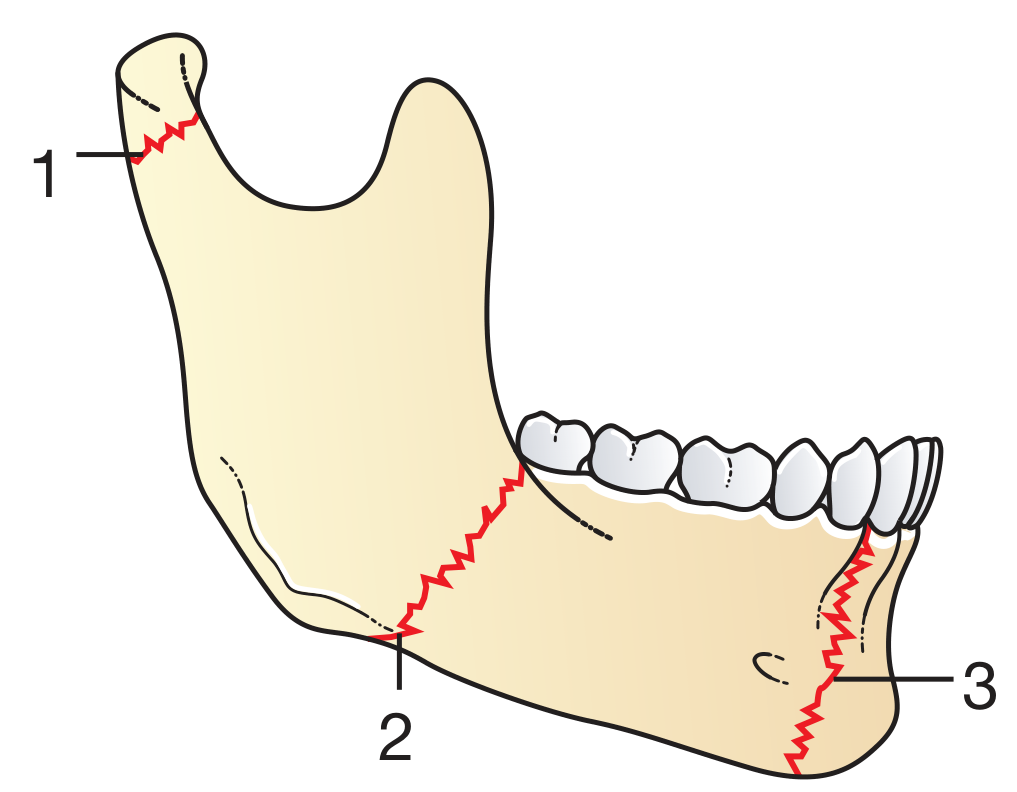
Because of its curved structure, mandibular fractures are frequently multiple - when one fracture is found, always look for a contralateral second fracture. Common paired patterns are:
- Parasymphysis + angle fracture (contralateral sides)
- Parasymphysis + condylar fracture (contralateral sides)
Clinical Features
- Pain and tenderness at the fracture site
- Malocclusion - the most important functional sign; altered dental occlusion results from muscle forces on fracture fragments
- Paraesthesia of the lower lip and chin - due to injury to the inferior alveolar (mental) nerve running within the mandibular canal; must be specifically documented on examination
- Trismus, difficulty chewing, and restricted mouth opening
- Step deformity palpable along the inferior border or at the dental occlusal plane
- Ecchymosis in the floor of the mouth (pathognomonic of mandibular fracture)
- Sublingual haematoma
Classification
By location: Symphysis, parasymphysis, body, angle, ramus, subcondylar (condylar neck), condylar head, coronoid process, alveolar process.
By Angle's classification of occlusion (guides management):
- Class I: Normal occlusion - mesial buccal cusp of maxillary first molar occludes in the intercuspal groove of mandibular first molar
- Class II: Mesial (anterior) positioning of maxillary teeth relative to mandibular
- Class III: Distal (posterior) positioning of maxillary teeth
Favorable vs. unfavorable - historically described by muscle pull on the fracture, but this classification is no longer considered clinically helpful.
Investigations
- Orthopantomogram (OPG/Panorex) - first-line imaging for most mandibular fractures; provides good overall visualization of the entire mandible
- Mandibular series plain films - supplementary
- High-resolution CT with reformats (coronal, sagittal, 3D) - now the standard for complex injuries; 1-mm helical CT has 100% sensitivity vs 86% for OPG. CT is especially useful for comminuted fractures, condylar fractures, and to assess the extent of displacement
- Both OPG and CT are complementary - CT can miss posterior mandibular fractures that OPG identifies; OPG misses comminution visible on CT
Anesthesia for Reduction and Fixation
Complete anaesthesia requires coverage of:
- Mandibular branch of the trigeminal nerve - anesthetized near the foramen ovale
- Maxillary division of the trigeminal nerve - in the pterygopalatine fossa near the foramen rotundum
- Superficial branches of the cervical plexus
Most operative fixation is performed under general anaesthesia (GA).
Treatment
Non-operative (Conservative)
Indicated for:
- Undisplaced or minimally displaced fractures
- Preserved pre-traumatic occlusal relationship
- Normal range of motion
- No significant soft tissue injury
Management: antibiotics, analgesia, soft diet for 4 weeks, with close monitoring for increasing pain or change in occlusion (signs of conservative management failure).
Operative - Open Reduction and Internal Fixation (ORIF)
Most displaced mandibular fractures are treated with ORIF:
- Antibiotics on admission followed by surgical fixation
- Titanium miniplates and screws are the standard implants (Fig. 31.9 - postoperative OPG showing angle + parasymphyseal fixation)
- Ideally performed within 24-48 hours of injury
- For heavily displaced fractures where delay is anticipated, a bridal wire around the teeth can temporarily reduce the fracture, alleviating pain and facilitating oral intake
Plating principles:
- Simple fractures: two 2-mm monocortical screws on each side of the fracture - these are load-sharing plates (fracture reduced, load shared between bone and plate)
- Complex/comminuted fractures: heavy reconstruction plates with bicortical screws as load-bearing fixation
- Angle and body fractures: single plate along the line of maximal tension (Champy technique)
- Parasymphyseal fractures: two plates placed 5 mm apart to resist the torsional forces of the anterior mandibular musculature
- Angle fractures: transbuccal approach (small cheek incision) allows screw placement perpendicular to plate and bone
Maxillomandibular fixation (MMF):
- Arch bars are applied to maxillary and mandibular teeth and wired together to establish and stabilize dental occlusion before fracture fixation
- Alternatives: bone screws placed in maxilla and mandible serving as posts for spanning wires
- Must be released as early as possible post-operatively to prevent TMJ ankylosis
Two schools of thought on fixation rigidity:
- AO/ASIF (Association for Osteosynthesis) - rigid fixation
- Champy technique - less rigid but functionally stable fixation
Condylar / Subcondylar Fractures
- Most condylar fractures are treated conservatively with closed reduction, IMF elastic guidance, soft diet, and analgesia
- Displaced condylar neck fractures with significant loss of mandibular height are increasingly treated with ORIF (transparotid retromandibular approach is most common; endoscopic-assisted fixation is an emerging option)
- Subcondylar fractures (just below the condyle) - typically minimal displacement due to reduced muscle forces and limited bone available for fixation; most often treated with MMF alone
Severely Comminuted Fractures
Intermaxillary fixation (IMF) with wires can be considered, though this is becoming rare due to advances in osteosynthesis techniques.
Complications
| Complication | Notes |
|---|---|
| Infection | More common in compound/open fractures; prophylactic antibiotics mandatory |
| Malocclusion/malunion | Failure to restore normal occlusion |
| Nonunion | Especially with infection or inadequate fixation |
| TMJ ankylosis | Risk increases with prolonged MMF; early range of motion is key |
| Mental/inferior alveolar nerve injury | Paraesthesia of chin and lower lip |
| Facial nerve injury | Especially with approaches near the parotid region |
| Dental fractures | During application of arch bars or instrumentation |
Key Teaching Points (Bailey & Love Summary Box 31.5)
- Always look for a second mandibular fracture - contralateral fractures are common
- Document mental nerve sensation (lower lip/chin paraesthesia) pre- and post-operatively
- Most mandibular fractures are treated with ORIF using titanium miniplates and screws, ideally within 24-48 hours of injury
Sources: Bailey and Love's Short Practice of Surgery 28th Ed., pp. 431-432; Schwartz's Principles of Surgery 11th Ed., pp. 2029-2030; Cummings Otolaryngology Head and Neck Surgery; KJ Lee's Essential Otolaryngology; Current Surgical Therapy 14th Ed.
Classification of mandibular fracture
Searching sources...
Finding Sources
Finding Sources
Reading File
Finding Sources
Finding Sources
Good - found rich content. Let me now read all the key sections in parallel.
Reading File
Reading File
Reading File
Now let me fetch the mandible lines-of-weakness image and get the Schwartz favorable/unfavorable section.
Loading Image
Reading File
Finding Sources
Let me get the favorable/unfavorable content from Schwartz and the Cummings classification section.
Reading File
Reading File
Now let me get the Schwartz favorable/unfavorable diagram and the frequency-by-site data.
Loading Image
Finding Sources
I now have all the material needed for a comprehensive, well-illustrated answer on mandibular fracture classification. Let me compile it.
Classification of Mandibular Fractures
Mandibular fractures are classified across several overlapping schemes - by anatomical location, relationship to the external environment, fracture pattern/morphology, displacement/muscle forces (favorable vs. unfavorable), and dentition status.
1. Classification by Anatomical Location
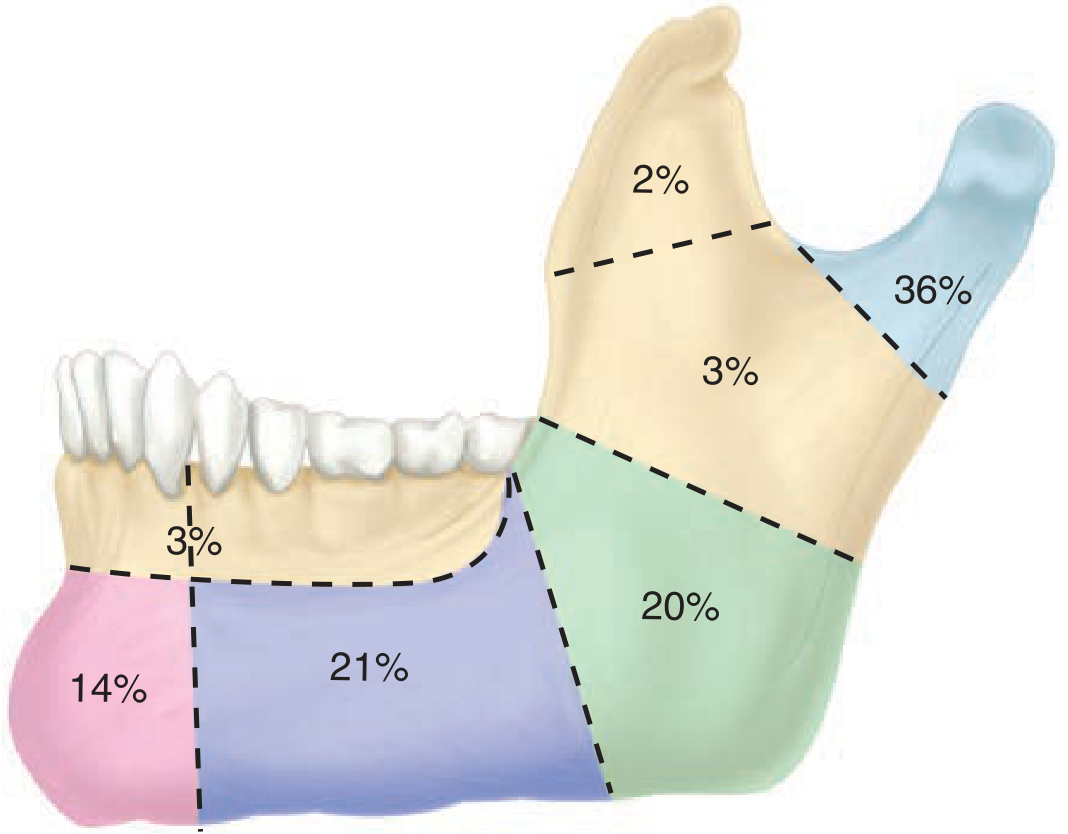
This is the most clinically important and universally used system. The mandible is divided into distinct regions, each with characteristic fracture patterns and treatment implications.
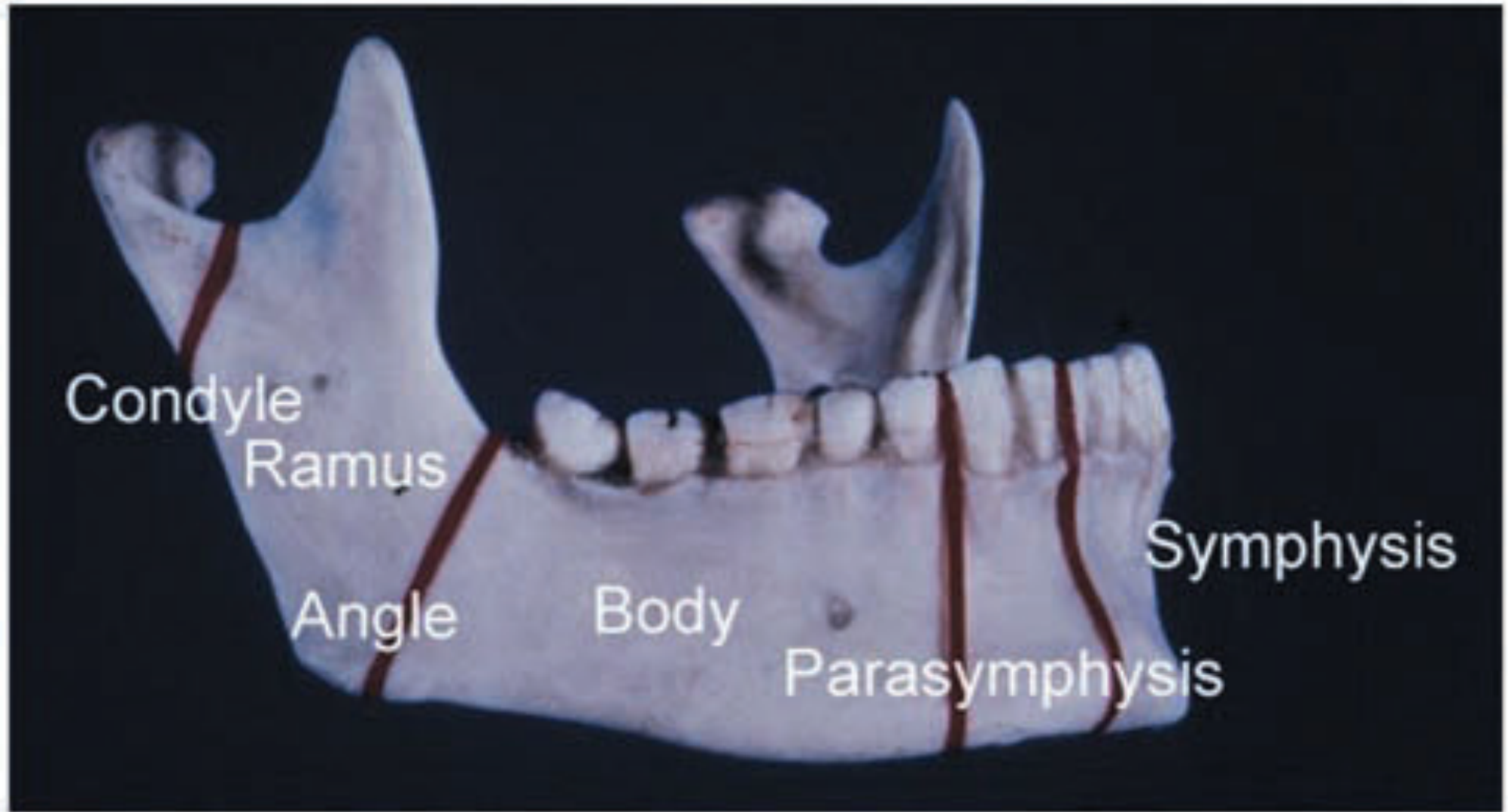
| Region | Description | Frequency |
|---|---|---|
| Condyle (subcondylar / condylar neck) | Most common site overall; slender condylar neck is a structural weak point | ~36% |
| Angle | Junction of body and ramus; weakened by presence of unerupted 3rd molar | ~20% |
| Body | Between mental foramen and angle; at level of 1st/2nd molar | ~21% |
| Parasymphysis | Lateral to midline; weakened by long root of lower canine | ~14% |
| Symphysis | Midline; between canine teeth | ~3% |
| Ramus | Between angle and condylar/coronoid processes; protected by thick bone & muscle | ~3% |
| Coronoid process | Rare; protected by zygomatic arch | ~2% |
| Alveolar process | Tooth-bearing segment only; does not involve the full mandibular body | Variable |
Key points on location:
- The angle is weakened by an unerupted wisdom tooth (partially erupted 3rd molar), which acts like a stress riser
- The parasymphysis is weakened by the long root of the lower canine
- The condylar neck is weakened by its slender anatomy
- Fractures are frequently multiple - when one is found, always look for a second (usually contralateral): e.g., parasymphysis + opposite condyle, or parasymphysis + opposite angle
2. Classification by Relationship to External Environment
| Type | Definition |
|---|---|
| Closed (Simple) | Fracture site does not communicate with external environment or oral cavity |
| Open (Compound) | Fracture communicates with external skin or oral mucosa; most mandibular fractures are open because the mandible is tooth-bearing - presume open until thorough intraoral examination excludes it |
| Comminuted | Bone is shattered into multiple fragments; results from high-velocity trauma; may cause upper airway obstruction due to loss of anterior tongue support |
| Greenstick | Incomplete fracture with one cortex intact; typical in children due to more flexible, pliable bone |
| Pathological | Fracture through diseased bone (tumor, osteomyelitis, osteoradionecrosis, osteoporosis) |
| Avulsive | Bone loss at the fracture site; most severe pattern |
3. Classification by Displacement: Favorable vs. Unfavorable
This classification is based on whether the pull of the masticatory muscles tends to reduce or displace the fracture fragments.
The main displacing forces come from:
- Masseter - pulls the ramus superiorly and medially
- Medial pterygoid - pulls the ramus superomedially
- Lateral pterygoid - pulls the condylar fragment anteromedially
- Mylohyoid, geniohyoid, genioglossus - pull the anterior mandible inferiorly and posteriorly
Favorable fracture - the fracture line runs such that muscle forces compress or reduce the fragments together (fracture surfaces are held in apposition by muscle pull)
Unfavorable fracture - the fracture line runs such that muscle forces pull fragments apart (distract the fracture)
This applies in both the vertical (coronal) and horizontal (sagittal) planes:
| Plane | Favorable | Unfavorable |
|---|---|---|
| Vertical | Fracture line angled so masseter/medial pterygoid compress fragments | Fracture line angled so muscles distract fragments apart |
| Horizontal | Fracture line directed so forces push lingual cortex together | Fracture line directed so forces pull lingual cortex apart |
Note: Cummings Otolaryngology states that the favorable/unfavorable classification is no longer considered helpful in determining the treatment plan and offers no communication or documentation advantages - it is now of historic significance only.
4. Classification by Dentition Status
This is particularly important because it determines the method of maxillomandibular fixation (MMF) available.
| Class | Description |
|---|---|
| Class I | Teeth present on both sides of the fracture line |
| Class II | Teeth present on one side of the fracture line only |
| Class III | No teeth on either side (edentulous); dentures may be used as splints |
Additionally, the mandible as a whole is categorized as:
- Dentulous - teeth present (arch bars can be applied directly)
- Edentulous - no teeth (requires bone screws, Gunning splints, or circummandibular wires)
- Atrophic edentulous - extremely thin, osteoporotic mandible; at high risk of pathological fracture and nonunion; may need reconstruction plates
5. Angle's Classification of Occlusion (Guides Management)
The pre-existing occlusal relationship must be restored:
| Class | Description |
|---|---|
| Class I (Normal) | Mesial buccal cusp of maxillary 1st molar occludes in intercuspal groove of mandibular 1st molar |
| Class II (Mesial) | Maxillary teeth positioned anterior to normal; "overbite" pattern |
| Class III (Distal) | Maxillary teeth positioned posterior to normal; "underbite" pattern |
6. Condylar Fracture Sub-classification
Condylar fractures deserve special attention as they have distinct treatment considerations:
| Type | Location | Features |
|---|---|---|
| Condylar head (intracapsular) | Within the joint capsule | High risk of TMJ ankylosis; conservative management preferred |
| Subcondylar (condylar neck) | Below the condylar head | Most common; typically treated conservatively with closed reduction and IMF |
| High subcondylar | Just below condylar head | |
| Low subcondylar | Near angle of mandible | Displaced fractures increasingly treated with ORIF (transparotid approach) |
| Diacapitular | Through the condylar head | Rare; high risk of ankylosis |
Summary Table
| Classification System | Categories |
|---|---|
| Anatomical site | Condyle, subcondylar, ramus, angle, body, parasymphysis, symphysis, alveolar, coronoid |
| Communication | Closed, open (compound), comminuted, greenstick, pathological, avulsive |
| Muscle forces | Favorable (reduced by muscles), Unfavorable (displaced by muscles) |
| Dentition | Class I (teeth both sides), Class II (one side), Class III (edentulous) |
| Occlusion (Angle) | Class I (normal), Class II (mesial), Class III (distal) |
| Displacement | Non-displaced, displaced, dislocated |
Sources: Tintinalli's Emergency Medicine; Scott-Brown's Otorhinolaryngology Vol. 1; Cummings Otolaryngology Head & Neck Surgery; Schwartz's Principles of Surgery 11th Ed.; Barash Clinical Anesthesia 9th Ed.; Bailey & Love's Short Practice of Surgery 28th Ed.
Body of the mandible fracture and it's extent
Searching sources...
Finding Sources
Finding Sources
Reading File
Reading File
Good - KJ Lee has the anatomical extent definition. Now let me get the Grainger radiology section and search for more on body fracture anatomy, displacement, and nerve injury.
Reading File
Finding Sources
Finding Sources
Reading File
Finding Sources
Finding Sources
Reading File
Now let me get the Rosen/Current Surgical Therapy sections on mandible body fracture management and fetch the panorex image of a body fracture.
Finding Sources
Reading File
Excellent - I now have comprehensive material. Let me fetch the key images and the 3D CT fracture image.
Loading Image
Loading Image
I now have all the material needed for a focused, detailed answer on the body of mandible fracture and its extent. Let me compile the complete response.
Fracture of the Body of the Mandible
Definition and Anatomical Extent
The body of the mandible is the horizontal tooth-bearing segment of the lower jaw. A body fracture is precisely defined as:
A fracture involving the mandible between the mental foramen anteriorly and the anterior border of the masseter muscle (or angle) posteriorly.
- Anterior boundary: The mental foramen (where the mental nerve exits)
- Posterior boundary: The anterior border of the angle of the mandible / anterior attachment of the masseter muscle
This places the body fracture squarely within the dentate (tooth-bearing) segment of the mandible, distinguishing it from:
- Parasymphysis/symphysis fractures - which lie anterior to the mental foramen, between the two canines
- Angle fractures - which begin at or posterior to the 3rd molar region, behind the dentition
- Ramus fractures - which lie entirely posterior to the angle
The body corresponds anatomically to the region of the premolars and molars (1st premolar through 2nd molar). High-velocity trauma (motor vehicle accidents) particularly targets the body at the level of the 1st or 2nd molar, as this is a zone of relative structural vulnerability.
Anatomical Basis of Vulnerability
The body is a point of weakness for several reasons:
- Inferior alveolar canal runs through the body, thinning the medullary bone and creating a stress concentration point
- Tooth roots of the premolars and molars extend deeply, creating further areas of reduced bone stock between the roots and the inferior border
- The body transmits the full bending moment of masticatory forces; during trauma, forces applied to the chin or parasymphysis are distributed around the curved mandible and concentrate at the body
- A common mechanism is high-velocity direct impact (motor vehicle collisions, assault with a weapon), as opposed to fist blows which more commonly fracture the parasymphysis or symphysis
Structures at Risk Within the Extent of the Body Fracture
The most important structure running within the entire length of the mandibular body is the inferior alveolar nerve (IAN) and its terminal branch:
| Structure | Relevance |
|---|---|
| Inferior alveolar nerve (IAN) | Runs in the inferior alveolar canal throughout the body; injury causes anaesthesia of the lower teeth, lower lip and chin |
| Mental nerve | Exits at the mental foramen (the anterior boundary); specific risk at the anterior edge of a body fracture |
| Inferior alveolar artery & vein | Run with the nerve; disruption causes haematoma formation |
| Tooth roots | The fracture line may pass through the tooth socket (making it an open/compound fracture by definition) |
| Oral mucosa/gingiva | Overlying mucosa is easily torn, making most body fractures open (compound) into the oral cavity |
Because the mandibular body is covered by gingival mucosa and is tooth-bearing, virtually all body fractures are presumed open until intraoral examination proves otherwise.
Clinical Features Specific to Body Fractures
The body fracture produces the full constellation of signs of a mobile mandibular fracture:
- Step deformity - palpable externally along the inferior border, or intraorally along the occlusal plane
- Malocclusion - asymmetry of the dental arch; the patient reports the bite "feels wrong"
- Anaesthesia / paraesthesia of the lower lip, chin, and lower teeth on the affected side - from injury to the inferior alveolar or mental nerve
- Pain and paradoxical movement - crepitus on manual distraction of the two fragments
- Sublingual/buccal haematoma - blood-stained saliva; haematoma in the floor of the mouth or buccal sulcus
- Intraoral laceration at the fracture site
- Missing or fractured teeth at the fracture line - always check for aspirated teeth
Displacement: What Moves and Why
The direction of displacement of a body fracture depends on the pull of attached muscles:
| Muscle | Attachment | Effect on Fracture Segments |
|---|---|---|
| Masseter | Outer surface of ramus/angle | Pulls posterior fragment superiorly |
| Medial pterygoid | Inner surface of ramus | Pulls posterior fragment superomedially |
| Mylohyoid | Mylohyoid line of body | Pulls anterior fragment inferomedially |
| Geniohyoid / genioglossus | Genial tubercles (symphysis) | Pull anterior fragment downward and backward |
For a body fracture, the posterior fragment is pulled upward and inward by masseter and pterygoid, while the anterior fragment is pulled downward and backward by the suprahyoid muscles. This creates a vertically unfavorable displacement.
Association with Contralateral Fracture
A body fracture is accompanied by a contralateral fracture in approximately 50% of cases, most commonly of the condylar neck on the opposite side. This is because the mandible is a ring-shaped bone - a single impact sufficient to fracture the body also transmits a countercoup force to the contralateral condyle (the weakest point of the opposite side).
This is why:
- A mandibular fracture should be considered bilateral until proven otherwise
- All imaging must survey the entire mandible, including both condyles
Imaging
- Dental panoramic tomogram (OPG/Panorex) - first-line; demonstrates the body fracture and the tooth-to-fracture-line relationship clearly
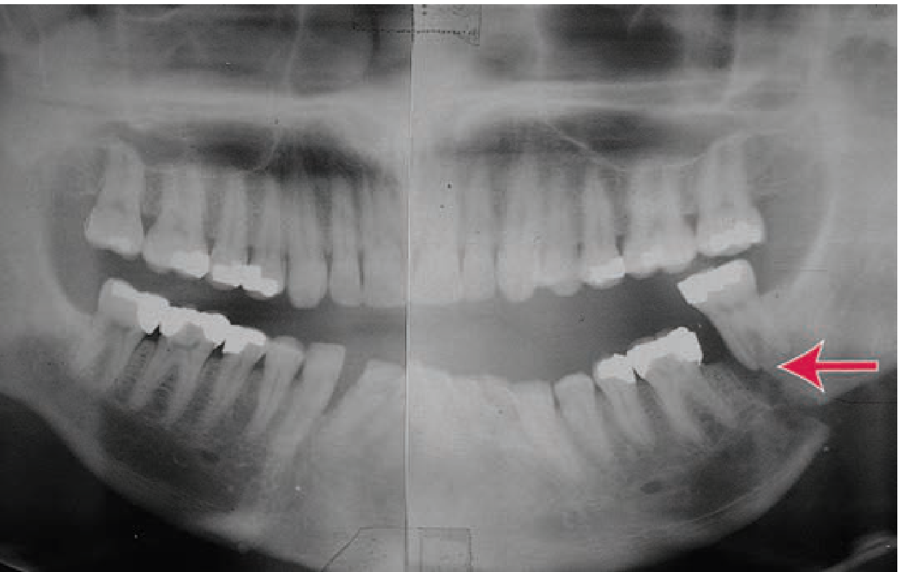
- PA mandible radiograph - paired with OPG as standard
- High-resolution CT (1 mm cuts) with 3D reformats - now preferred for displaced, comminuted, or complex fractures; 100% sensitivity vs 86% for OPG
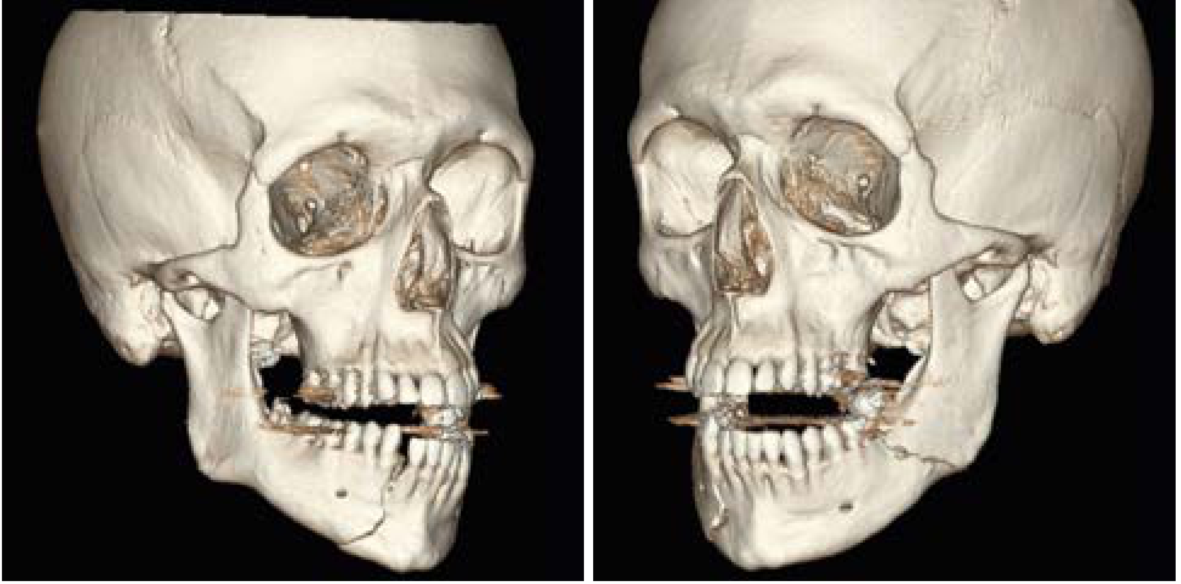
Treatment
Non-displaced / Stable Body Fractures
- MMF (maxillomandibular fixation) alone with a liquid diet for 4-6 weeks
- Patient loses ~10-20 lbs on liquid diet during fixation period
Displaced Body Fractures - ORIF
- ORIF is the standard; performed ideally within 24-48 hours
- Approach: intraoral (transoral) or transcervical (submandibular) incision
- Submandibular incisions must be placed sufficiently below the lower border of the body to protect the marginal mandibular branch of the facial nerve
- Titanium miniplates and screws are applied:
- Single plate along the line of maximal tension (superior/outer cortex) for body fractures - the Champy technique
- Alternatively, two plates may be used for greater stability in comminuted fractures
- Monocortical screws (load-sharing) for simple fractures
- Bicortical reconstruction plates (load-bearing) for comminuted or bone-loss fractures
- The fracture line and tooth roots constrain screw placement - monocortical screws are placed to avoid root damage
Tooth in the Fracture Line
A tooth in the fracture line is a key decision point:
- May act as a lever to aid reduction if it has a good periodontal status - keep it
- Should be extracted if it is mobile, non-restorable, infected, or impedes reduction - leaving an infected tooth dramatically increases infection and nonunion risk
- The molar in the line of an angle fracture often needs extraction if it prevents posterior fragment control (as shown in Scott-Brown's Fig. 107.8)
Postoperative Care
- Chlorhexidine mouthwashes 3-4 times daily
- Soft diet for 4-6 weeks
- Antibiotics (penicillins, cephalosporins, or clindamycin to cover oral organisms)
- Early release from MMF once fixation is confirmed - to prevent TMJ ankylosis
Summary
| Feature | Body Fracture |
|---|---|
| Extent | Mental foramen → anterior border of masseter/angle |
| Frequency | ~21% of all mandible fractures |
| Mechanism | High-velocity direct trauma (MVA); fist/blunt force less common than parasymphysis |
| Open/Compound | Virtually always (tooth-bearing region, covered only by mucosa) |
| Contralateral fracture | ~50%; most often opposite condylar neck |
| Key nerve at risk | Inferior alveolar nerve → mental nerve; anaesthesia of lower lip/chin |
| Imaging | OPG + PA mandible; CT for complex/displaced cases |
| Treatment | ORIF with titanium miniplates within 24-48 h; single plate along tension line (Champy) |
Sources: KJ Lee's Essential Otolaryngology; Grainger & Allison's Diagnostic Radiology; Scott-Brown's Otorhinolaryngology Vol. 1; Current Surgical Therapy 14th Ed.; Barash Clinical Anesthesia 9th Ed.; Bailey & Love's Short Practice of Surgery 28th Ed.
This is a shared conversation. Sign in to Orris to start your own chat.