
Acute Rheumatic Fever
erythema marginatum rheumatic fever rash
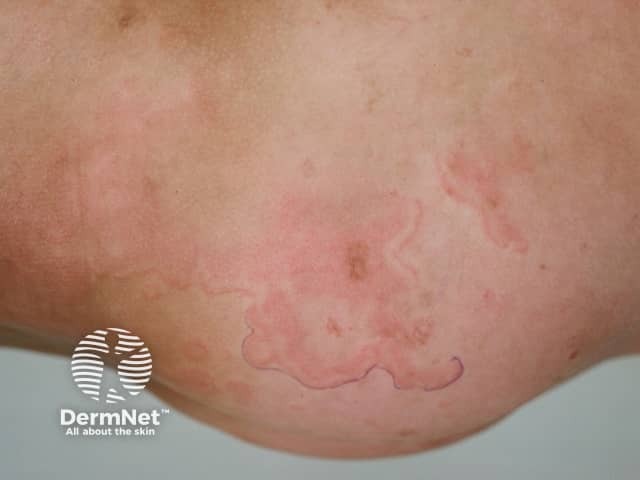
Clinical photograph (dermatology) showing erythema marginatum, an annular, non-itchy rash characteristic of acute rheumatic fever. The image depicts multiple pink to red circular to oval patches on the skin of the lower extremity with sharply demarcated, serpiginous margins and centers that appear pale or clearing. Lesions are non-vesicular, smooth-surfaced, and vary in size, with a tendency to migrate or transiently flare. The distribution favors the trunk and proximal limbs and typically spares the face. On close inspection the patches lack scaling and crusting, consistent with erythema marginatum rather than dermatitis or fungal infection. This cutaneous manifestation is part of the Jones criteria and may correlate with concurrent migratory polyarthritis, fever, and evidence of preceding group A Streptococcus infection. Histologic features (if biopsied) would show superficial perivascular lymphocytic infiltrate and dermal edema with minimal epidermal changes; clinically, the rash is a marker for systemic inflammatory involvement. Differential considerations include urticaria, erythema multiforme, tinea corporis, pityriasis rosea, and other annular dermatoses. Clinically, recognizing erythema marginatum prompts evaluation for acute rheumatic fever and guides management including antibiotic therapy and anti-inflammatory treatment. The image serves educational and diagnostic-reference purposes for dermatology, pediatrics, infectious diseases, and rheumatology training.
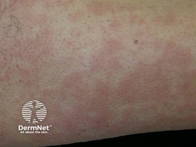
This is a clinical dermatology photograph capturing erythema marginatum on the skin of the lower limb. The modality is Clinical photography; technique: naked-eye, color-balanced image. The observed lesions are erythematous, annular plaques with central clearing and pale, sometimes hypopigmented centers. Lesions appear as rings with slightly raised margins and a uniform pink-to red hue; they range from 0.5 to 2 cm in diameter and may coalesce into larger plaques. The rash is typically non-pruritic and evanescent, with temporary eruption that may migrate or fade over hours to days; distribution commonly involves the trunk and proximal limbs, with sparing of the face. The image may reflect erythema marginatum, a cutaneous manifestation associated with acute rheumatic fever and autoimmune inflammatory processes. In longer-standing presentations, the rash signals preceding or concurrent carditis and migratory arthritis; therefore clinical correlation with recent group A Streptococcus infection, elevated ASO titers, and echocardiography is essential. Differential diagnoses include urticaria, erythema multiforme, pityriasis rosea, and drug-induced eruptions. Clinically, this finding has diagnostic significance as a major criterion in rheumatic fever per Jones criteria; management focuses on antibiotic treatment of streptococcal infection and secondary prophylaxis. This image serves educational purposes for medical students, residents, and researchers studying connective tissue disease and pediatric rheumatology.

A clinical photograph of the lower extremities of a pediatric patient, demonstrating a distinctive cutaneous manifestation. The image shows multiple erythematous, annular (ring-shaped) lesions distributed across the thighs, knees, shins, and ankles. The rash is characterized by raised, pinkish-red borders that form large, asymmetric, and serpiginous patterns, some partially encircling the limbs. These lesions appear papular and lack visible scaling or central crusting, suggesting an inflammatory or migratory process such as Erythema Marginatum or Erythema Migrans. A localized area of yellowish discoloration (bruising or ecchymosis) is visible on the right knee. The morphology of these blanchable, annular lesions is a key diagnostic feature often associated with systemic conditions such as acute rheumatic fever or early-stage Lyme disease. This visual is intended for medical education in dermatology, pediatrics, and infectious diseases to assist in the identification of classic annular rash patterns.
"rheumatic fever" AND diagnosis
acute rheumatic fever 2024 2025 update WHO global burden
Acute Rheumatic Fever (ARF)
Overview
Pathophysiology
- Carditis - antibody binding and T-cell infiltration of the heart (pancarditis), most clinically significant at the mitral valve, then the aortic valve
- Arthritis - immune complex deposition in joints (sterile, lymphocyte-predominant synovial fluid)
- Chorea - antibody binding to the basal ganglia
- Skin findings - delayed hypersensitivity reactions
Clinical Manifestations
| Feature | Approximate frequency |
|---|---|
| Fever | >90% |
| Migratory large-joint polyarthritis | 60-75% |
| Carditis (clinical + subclinical) | ~50-70% |
| Sydenham chorea | ~30% |
| Erythema marginatum | <10% |
| Subcutaneous nodules | <10% |
- Polyarthritis: migratory, affects large joints, dramatic and rapid response to NSAIDs/aspirin - a lack of response should prompt reconsideration of the diagnosis.
- Carditis: pancarditis (endocardium, myocardium, pericardium); presents with new/changing murmurs (mitral regurgitation most common), cardiomegaly, heart failure, or pericarditis.
- Sydenham chorea: involuntary, non-rhythmic, purposeless movements, often one-sided, disappears during sleep.
- Erythema marginatum: pink, non-pruritic, blanching, serpiginous macules/papules on the trunk and proximal limbs.
- Subcutaneous nodules: 0.5-2 cm, painless, over bony prominences/extensor tendons.
- Labs: elevated ESR/CRP, prolonged PR interval on ECG, normocytic anemia, leukocytosis.
Diagnosis: Revised (2015) Jones Criteria
- Initial episode: 2 major criteria, OR 1 major + 2 minor criteria
- Recurrent episode: 2 major, OR 1 major + 2 minor, OR 3 minor criteria
- Carditis (clinical and/or subclinical echocardiographic valvulitis)
- Arthritis - polyarticular only in low-risk populations; mono- or polyarticular in moderate/high-risk populations
- Chorea
- Erythema marginatum
- Subcutaneous nodules
- Low-risk: polyarthralgia; fever ≥38.5°C; ESR ≥60 mm/h and/or CRP ≥3.0 mg/dL; prolonged PR interval
- Moderate/high-risk: monoarthralgia; fever ≥38.0°C; ESR ≥30 mm/h and/or CRP ≥3.0 mg/dL; prolonged PR interval
Treatment
- Eradicate GAS with a standard pharyngitis antibiotic regimen (benzathine penicillin G IM once, or oral penicillin V/amoxicillin, or a macrolide if penicillin-allergic) - even if throat culture is negative at presentation.
- Arthritis/fever: NSAIDs are first-line - traditionally aspirin (80-100 mg/kg/day divided, max 4-8 g/day), with naproxen used successfully as an alternative. Continue until all ARF symptoms resolve (1-2 weeks), not just joint pain.
- Carditis: treat heart failure with standard goal-directed therapy (diuretics, ACE inhibitors/ARBs), fluid restriction, and bed rest. Systemic glucocorticoids are used by some experts in severe carditis, though evidence quality is limited. Valve surgery is rarely needed acutely (e.g., acute leaflet rupture).
- Chorea: usually self-limited, resolving over weeks to months; typically does not require specific treatment.
- Hospitalization is generally recommended for initial diagnosis and management.
Secondary Prophylaxis (Prevention of Recurrence)
- Benzathine penicillin G 1.2 million units IM every 4 weeks (every 3 weeks in high-risk settings) is the most effective regimen; oral penicillin V (250 mg twice daily) or a macrolide are less effective alternatives.
- Duration: minimum 10 years from the last ARF episode or until age 21 (whichever is longer); prolonged into the 3rd-4th decade or lifelong for patients with persistent significant valvular disease, including after valve replacement.
Complications: Rheumatic Heart Disease
Recent Evidence Update (surfaced per literature check)
- A 2024 Cochrane systematic review/meta-analysis (Bray et al., PMID 39312290) evaluated long-term antibiotic prophylaxis for preventing ARF recurrence/RHD progression - broadly supportive of penicillin prophylaxis but highlights persistent gaps in comparative trial quality (oral vs IM, dosing intervals).
- A 2024 systematic review in Lancet Global Health (PMID 38762298) evaluated handheld echocardiography for RHD screening, informing new WHO guidance.
- The WHO published its first global guideline on prevention and diagnosis of rheumatic fever and RHD in October 2024, continuing to endorse the Jones criteria while acknowledging they miss up to ~10% of cases in some endemic settings, prompting interest in simplified clinical criteria for resource-limited areas.
- A 2025 review in The Lancet (Hirani et al.) reaffirms molecular mimicry pathogenesis and underscores that ARF/RHD remain concentrated in Indigenous and low-resource populations globally.