
Hemodynamic disorders: hemorrhage. Etiopathogenesis and morphology
"Hemorrhage"[MeSH Terms] AND "pathology"
petechiae ecchymoses hemorrhage skin purpura
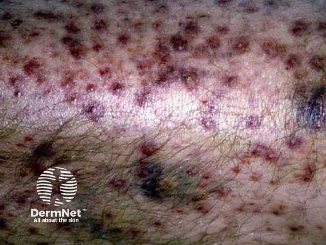
Imaging modality: Clinical close-up photography of cutaneous surfaces. Primary subject: Perifollicular hemorrhage with ecchymoses involving the scalp/adjacent facial skin. Specimen: Human skin. Imaging perspective: High-resolution, macro close-up view aimed at superficial vascular hemorrhage around hair follicles. Anatomical localization: Integumentary system; skin; adnexal structures including hair follicles; regional focus on scalp or perioral/temple skin. Visual features: Numerous small perifollicular purpura/petechiae, dark red to brown ecchymotic patches surrounding follicles, subtle erythema, overlying intact epidermis with no obvious active ulceration; lesions range from pinpoint to confluent clusters; background skin shows hair shafts and follicular openings. Pathology implied: superficial dermal capillary bleeding with perifollicular distribution; ecchymoses reflect extravasation of red blood cells; no proven dermal necrosis. Diagnostic significance: indicative of cutaneous purpura with follicular accentuation; differential includes vasculitis, trauma, coagulopathy, platelet disorders, inflammatory folliculitis, pigmented purpura. Clinical relevance: supports evaluation for systemic bleeding risk or vasculitic processes; warrants laboratory workup and correlation with clinical symptoms; useful for education of dermatologic hemorrhagic presentations and differential diagnosis; potential applications include dermatology clinics, medical education, and literature reviews on perifollicular hemorrhage and ecchymoses. The image can aid in distinguishing perifolliculitis-associated hemorrhage from ecchymotic conditions secondary to systemic disease and guide triage for urgent hematologic or rheumatologic evaluation.
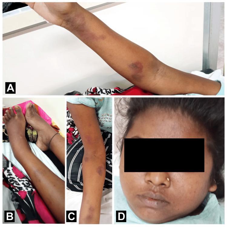
A composite of four clinical photographs (A-D) documenting systemic manifestations in a 19-year-old female patient with a history involving systemic lupus erythematosus and immune thrombocytopenic purpura (ITP). Panels A and C show the left and right upper extremities, respectively, exhibiting multiple purplish-red petechiae and ecchymoses on the forearms and near the antecubital fossa, indicating cutaneous hemorrhage or endothelial dysfunction. Panel B illustrates the lower extremities with prominent bilateral pedal edema and swelling of the legs, characterized by smooth, shiny, and stretched skin suggesting fluid accumulation. Panel D displays a frontal view of the patient's face, highlighting facial puffiness and periorbital edema. Collectively, these images demonstrate a constellation of clinical signs including hematologic (petechiae) and renal/hemodynamic (edema/puffiness) findings relevant to rheumatologic and hematologic specialties, suitable for illustrating multi-organ involvement in autoimmune disease processes.
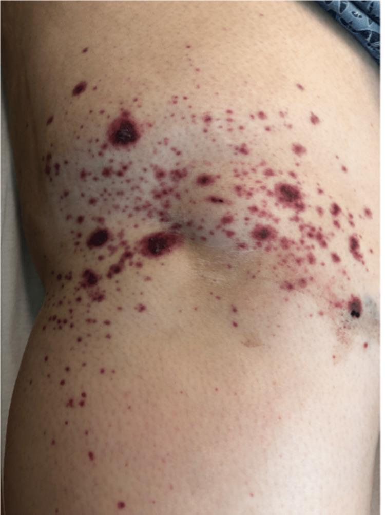
This clinical photograph shows a close-up view of a cutaneous eruption on a human extremity, specifically identified as a purpuric rash. The primary lesions consist of non-blanching, petechiae (pinpoint spots) and larger ecchymoses (purple-red blotches) that range in size from approximately 1 mm to several centimeters. The morphology includes flat macules and slightly raised, palpable papules, characteristic of palpable purpura. The lesions exhibit a range of colors from bright red to dark violaceous and purplish-black, indicating areas of acute hemorrhage and more significant dermal blood accumulation. The distribution is diffuse and irregular across the limb, with several areas showing higher density or clustering of lesions. There is no evidence of active ulceration, crusting, or diffuse erythema in the surrounding skin. This visual presentation is highly suggestive of small-vessel vasculitis, such as IgA vasculitis (Henoch-Schönlein purpura), which often presents with this type of leukocytoclastic vasculitis (LCV) pattern on the lower or upper extremities.
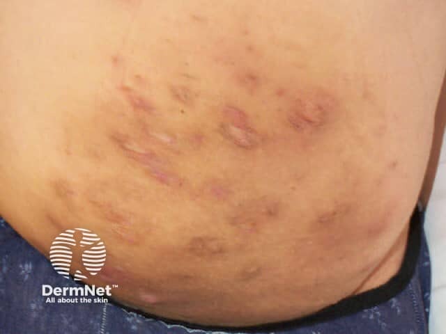
Clinical photograph of the anterior abdominal wall demonstrating multifocal cutaneous ecchymoses and purpuric patches. The lesions are irregular in shape, non-blanching, and range from violaceous to brown with some crusted erosions at the periphery. The distribution is diffuse across the lower abdomen, without a single focal lesion, and variants in size suggest evolving hemorrhage into the dermis and subcutaneous tissue. The skin surface shows mild textural irregularities consistent with superficial trauma or vascular fragility. No apparent vesicles or bullae are evident, and there is no clear involvement of deeper structures in this image. This presentation is compatible with ecchymosis and purpura, which may arise from trauma, coagulopathy, vasculitis, or platelet dysfunction; the color progression implies fresh to resolving bleeding. Given the abdomen’s relatively large surface area, sampling is not possible here, but clinical correlation is essential. The image underscores the diagnostic challenge of distinguishing petechiae, purpura, and contusions, and highlights the need for comprehensive history and laboratory evaluation. Potential clinical scenarios include post-traumatic bruising, dermatologic vasculopathy, drug-induced purpura, and systemic conditions such as infection or hematologic disease. This image is valuable for education on cutaneous hemorrhagic lesions and their differential diagnosis, risk stratification, and management planning.
Hemorrhage: etiopathogenesis and morphology
Definition
Etiopathogenesis
- Trauma, the most common cause.
- Atherosclerosis, or rupture of an aneurysm or aortic dissection.
- Inflammatory erosion, for example peptic ulcer, tuberculous cavity or vasculitis.
- Neoplastic erosion of a vessel wall.
- Chronic venous congestion, where capillary bleeding can occur.
- The bleed may be massive if the damaged vessel is a large artery or vein.
- These are disorders in which bleeding follows a trivial injury or occurs spontaneously.
- The three components of hemostasis must all work properly, so the causes are grouped by component:
- Vessel wall defects: scurvy (vitamin C deficiency), Ehlers-Danlos syndrome, vasculitis, amyloid, and infections such as meningococcemia.
- Platelet defects: thrombocytopenia (ITP, marrow failure, DIC, drugs) or abnormal function (aspirin, uremia, von Willebrand disease).
- Coagulation factor defects: hemophilia A and B, vitamin K deficiency, liver disease, DIC, anticoagulants.
- Broadly, platelet and vessel defects cause mucocutaneous bleeding: petechiae, purpura and epistaxis. Factor defects cause deep bleeding: hemarthrosis and large hematomas.
- Transient vasoconstriction.
- Platelet adhesion through von Willebrand factor, activation and aggregation (primary hemostasis).
- The coagulation cascade, which produces fibrin (secondary hemostasis).
- Counter-regulation by antithrombotic factors, so the clot stays localized.
Morphology
- External bleeding, or accumulation within a tissue as a hematoma.
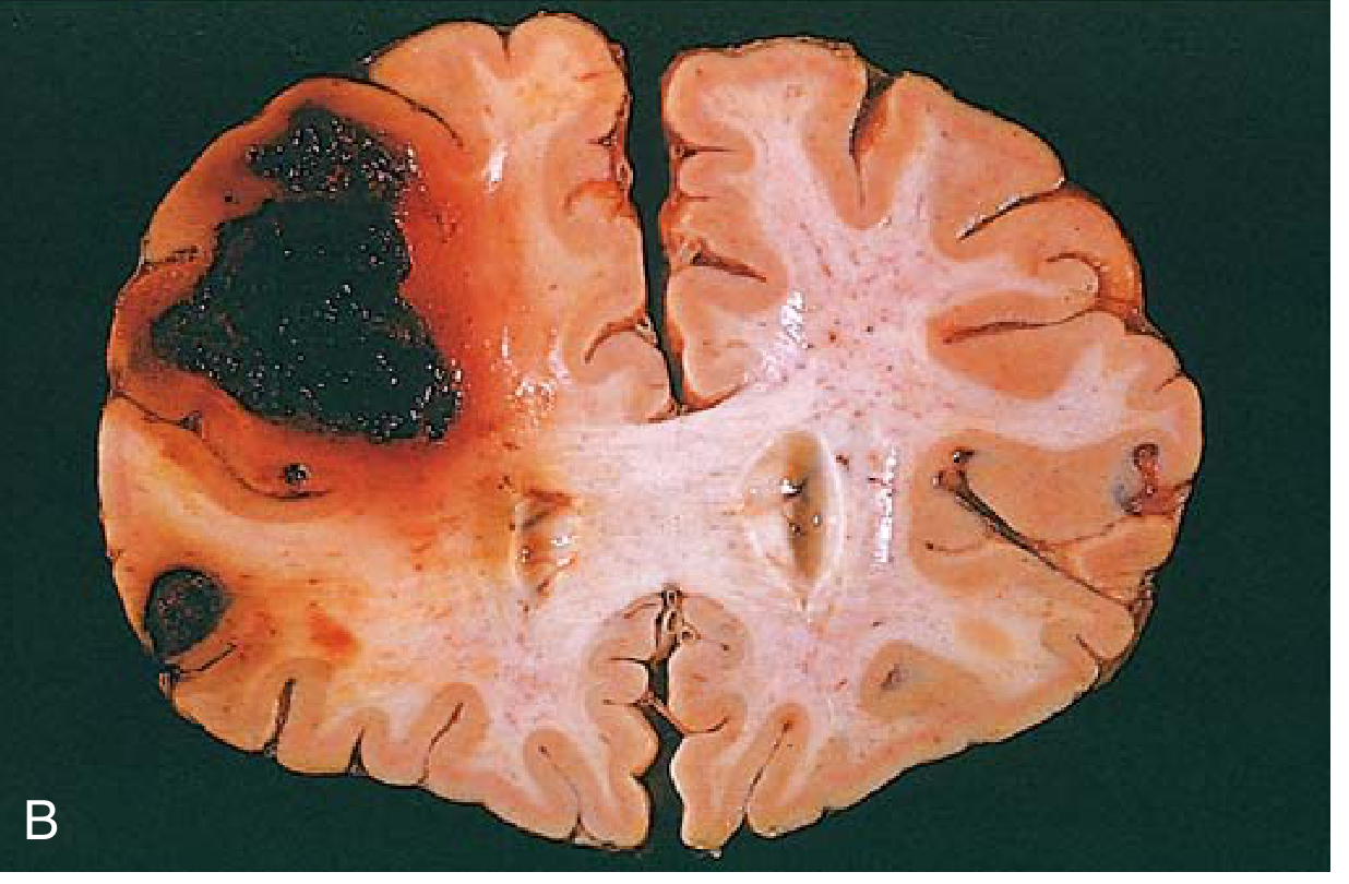
- Hematomas range from trivial (a bruise) to fatal (a massive retroperitoneal hematoma from a ruptured dissecting aortic aneurysm).
- Bleeding into body cavities is named by site:
- hemothorax
- hemopericardium
- hemoperitoneum
- hemarthrosis (joints)
- Large hemorrhages can occasionally cause jaundice, as macrophages break down the red cells and hemoglobin.
| Term | Size | Notes |
|---|---|---|
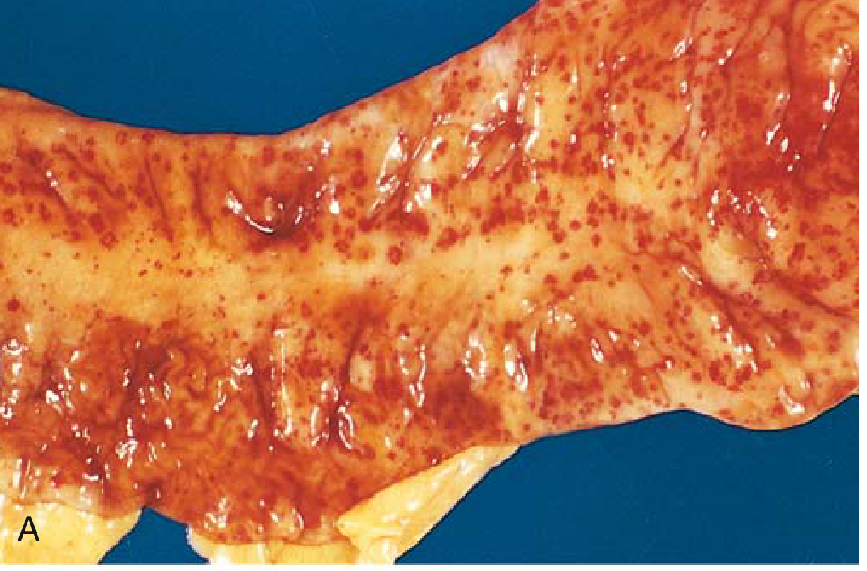
| Petechiae | 1 to 2 mm | Skin, mucous membranes or serosal surfaces. Seen with thrombocytopenia, defective platelet function, and loss of vascular wall support (scurvy). |
| Purpura | 3 to 5 mm | Same causes as petechiae, plus trauma, vasculitis and increased vascular fragility. |
| Ecchymoses | 1 to 2 cm | Subcutaneous hematomas ("bruises"). |
- Macrophages phagocytose and degrade the extravasated red cells.
- Hemoglobin (red-blue) is converted to bilirubin (blue-green) and finally hemosiderin (golden-brown).
- Microscopically, an old hemorrhage shows hemosiderin-laden macrophages. These are the basis of the Prussian blue stain.
Clinical significance
- Volume and rate:
- Rapid loss of up to 20% of blood volume is usually tolerated by healthy adults.
- It can cause cardiovascular decompensation in people with heart or lung disease.
- Greater loss causes hemorrhagic (hypovolemic) shock.
- Site: a small subcutaneous bleed is trivial, but the same bleed in the brain, or in the pericardium (tamponade), can be fatal.
- Chronic or recurrent external blood loss (peptic ulcer, menstruation) leads to iron deficiency anemia.
- Internal bleeding (a hematoma) does not cause iron deficiency, because iron is recycled from phagocytosed red cells.
Recent evidence check
Hemodynamic disorders: hemorrhage. Etiopathogenesis and morphology
hematoma ecchymosis bruise color evolution hemosiderin macrophages Prussian blue
Hemorrhage: etiopathogenesis and morphology
1. Etiopathogenesis
A. Mechanisms of bleeding
| Mechanism | Description | Examples |
|---|---|---|
| Rhexis (rupture) | The vessel wall (artery, vein, capillary or heart) is breached. | Trauma, aneurysm rupture, aortic dissection, ruptured myocardial infarct, hypertensive intracerebral bleed |
| Erosion | The wall is eroded by disease. | Peptic ulcer, tuberculous cavity, tumor invasion, atherosclerotic plaque, inflammation |
| Diapedesis | Red cells leak through an intact but damaged or hypoxic capillary wall. | Chronic venous congestion, severe hypoxia, scurvy, vasculitis, thrombocytopenia |
B. Hemorrhagic diatheses
- Hemorrhagic diatheses are disorders in which bleeding often follows a seemingly insignificant injury. - Robbins & Kumar Basic Pathology
- They arise from inherited or acquired defects in vessel walls, platelets or coagulation factors. All three must function properly for hemostasis.
- Vessel wall defects (purpura with normal platelets and coagulation tests):
- scurvy
- Ehlers-Danlos syndrome
- vasculitis (Henoch-Schönlein purpura)
- amyloid
- meningococcemia
- steroid or senile purpura
- Platelet defects:
- Quantitative: thrombocytopenia from ITP, marrow failure, DIC, TTP or drugs.
- Qualitative: aspirin, uremia, von Willebrand disease, Glanzmann thrombasthenia.
- Uremic platelet dysfunction gives skin bleeding (bruising, ecchymoses) and mucosal bleeding (epistaxis, GI and gum bleeding). It can rarely cause hemopericardium or hemothorax. - Comprehensive Clinical Nephrology, 7th ed.
- Coagulation factor defects:
- hemophilia A and B
- vitamin K deficiency
- liver failure
- DIC
- anticoagulants
- Platelet and vessel disorders give mucocutaneous bleeding: petechiae, purpura, epistaxis, gum bleeding. Bleeding starts immediately after injury.
- Coagulation factor disorders give deep bleeding: hemarthrosis and large hematomas, often delayed after injury.
C. Hemostasis, briefly
- Vasoconstriction.
- Platelets adhere through von Willebrand factor, then activate and aggregate (primary plug).
- The coagulation cascade generates fibrin (secondary stabilization).
- Counter-regulatory antithrombotic mechanisms limit the clot.
2. Morphology
A. Gross patterns
- External bleeding, or accumulation within a tissue as a hematoma. Hematomas range from trivial (a bruise) to fatal (a massive retroperitoneal hematoma from a ruptured dissecting aortic aneurysm).
- Body cavity hemorrhage, named by site:
- hemothorax
- hemopericardium
- hemoperitoneum
- hemarthrosis
- Large hemorrhages can occasionally cause jaundice as macrophages break down the red cells and hemoglobin.
B. Size-based terminology
- Petechiae: 1 to 2 mm, in skin, mucous membranes or serosa. Causes are thrombocytopenia, defective platelet function and loss of vascular wall support (scurvy).
- Purpura: 3 to 5 mm. Causes are the same as for petechiae, plus trauma, vasculitis and increased vascular fragility.
- Ecchymoses: 1 to 2 cm, subcutaneous hematomas ("bruises").
C. Microscopy and fate of extravasated blood
- Extravasated red cells lie outside the vessels in the tissue. Fresh hemorrhage shows RBCs with little or no inflammatory reaction.
- Macrophages phagocytose and degrade the red cells. The bruise changes color as hemoglobin (red-blue) is converted to bilirubin (blue-green) and finally hemosiderin (golden-brown). - Robbins & Kumar Basic Pathology
- Late lesions show hemosiderin-laden macrophages, which stain blue with Prussian blue. Forensic texts note coarse hemosiderin granules in histiocytes at the end of healing, and hematoidin crystals that can persist at the site of a bruise. - Essentials of Forensic Medicine and Toxicology
- A large hematoma may be resorbed, become organized (fibrosis) or calcify. Brain hemorrhages leave a cystic, brown-stained cavity.
3. Consequences
- Volume and rate:
- Rapid loss of up to 20% of blood volume is usually tolerated by healthy adults.
- It can cause cardiovascular decompensation in people with heart or lung disease.
- Greater loss causes hemorrhagic (hypovolemic) shock.
- Site: a trivial subcutaneous bleed can kill if it is intracranial. Hemopericardium can cause tamponade.
- Chronic or recurrent external blood loss (peptic ulcer, menstruation) leads to iron deficiency anemia.
- Internal bleeding (a hematoma) does not cause iron deficiency, because iron is recycled from phagocytosed red cells.
4. Summary
- The mechanisms are rhexis, erosion and diapedesis.
- Causes are either local vessel injury or a systemic hemorrhagic diathesis (vessel, platelet or factor defect).
- Morphology depends on size: petechiae (1 to 2 mm), purpura (3 to 5 mm), ecchymoses (1 to 2 cm), hematoma or cavity bleeding.
- Resolution runs through macrophage removal and hemosiderin.
- The outcome depends on volume, rate, site and host reserve.
- The rhexis, erosion and diapedesis classification and the specific diathesis lists come from standard teaching and my own knowledge, not a textbook page I quoted.
- The image search for bruise color evolution returned nothing.
- In my earlier screen of recent systematic reviews (PMIDs 39826291, 40508117, 38472247), none changed these basic pathology concepts.