
Chronic suppurative otitis media
chronic suppurative otitis media tympanic membrane perforation
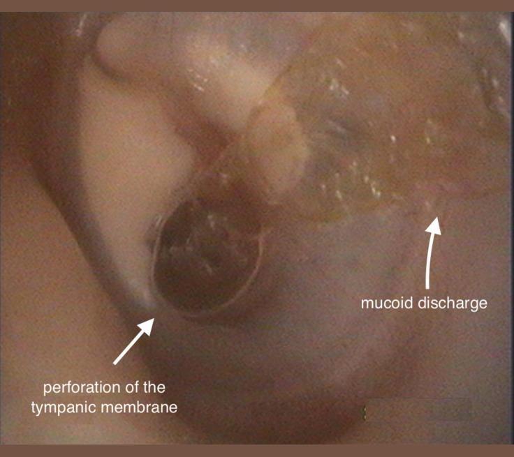
This clinical otoscopic photograph of a left ear demonstrates signs of chronic otitis media (COM). The image clearly shows a large, central perforation of the tympanic membrane, appearing as a dark, circular void where the membrane should be intact. Adjacent to this perforation, there is visible mucoid discharge, which presents as a translucent, glistening, and somewhat viscous fluid resting on the membrane surface. Additionally, white, opaque areas consistent with tympanosclerotic plaques are visible on the remaining portions of the tympanic membrane, indicating previous inflammation or scarring. The external auditory canal appears mildly erythematous. This visual is characteristic of chronic suppurative otitis media, an important educational example for distinguishing types of membrane perforations and associated middle ear pathology. The presence of discharge and structural changes in the membrane are key diagnostic markers used by otorhinolaryngologists to assess disease chronicity and planning for potential tympanoplasty.
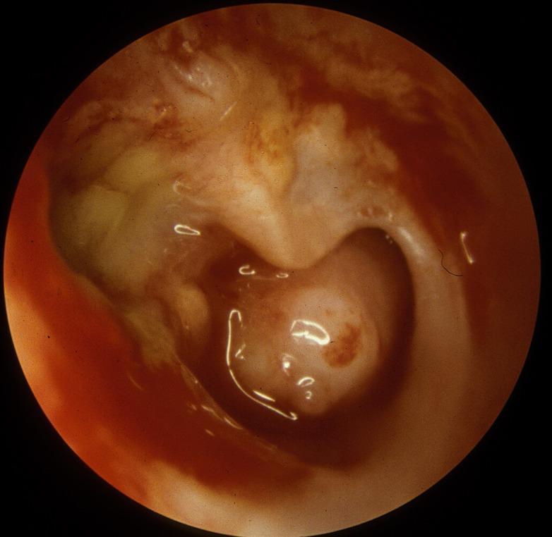
This endoscopic clinical photograph depicts the right tympanic membrane and middle ear cavity, demonstrating hallmark features of Chronic Suppurative Otitis Media (CSOM). A large subtotal perforation is visible, through which the structures of the middle ear are exposed. The middle ear mucosa appears erythematous and thickened, indicative of chronic inflammation. There is evidence of mucoid drainage, presenting as a viscous, translucent fluid within the cavity. Crucially, the long process of the incus shows significant erosion, a common sequela of long-standing inflammatory processes in the middle ear. The remaining tympanic membrane remnants appear thickened and opaque. This image serves as a classic educational example of middle ear pathology and the destructive potential of chronic infectious processes on the ossicular chain.
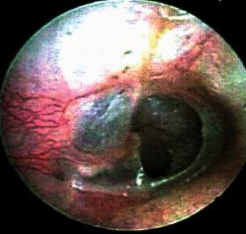
This clinical photograph, captured via otoendoscopy, displays a preoperative view of a human tympanic membrane (eardrum) with a significant central perforation. The perforation is characterized by a well-defined, dark circular void, revealing the underlying middle ear space. The remaining tympanic membrane tissue exhibits marked erythema and increased vascularity, particularly evident in the posterior-superior quadrant, indicating active inflammation or chronic myringitis. The mucosal surface appears thickened and irregular in texture, losing its normal pearly-gray translucency. This image serves as a clinical representation of chronic suppurative otitis media or traumatic perforation, demonstrating the typical morphological changes that necessitate surgical intervention, such as tympanoplasty. The visual highlights key diagnostic features including the size and location of the defect and the health of the surrounding residual membrane, which are critical for surgical planning in otolaryngology.
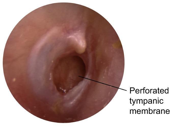
This clinical photograph shows an otoscopic view of the right ear, demonstrating a large, subtotal perforation of the tympanic membrane located primarily in the posterior and inferior quadrants. The perforation is patent, revealing the underlying middle ear space, which appears dark and recessed. The remaining tympanic membrane tissue is erythematous and thickened, suggesting chronic inflammation, with a loss of the normal translucent, pearly-gray appearance and light reflex. The handle of the malleus is visible but obscured by the retracted and inflamed tissue. The external auditory canal appears relatively normal without signs of stenosis or active otorrhea at the time of imaging. This presentation is characteristic of chronic suppurative otitis media (CSOM), a condition defined by persistent tympanic membrane perforation and a history of intermittent or continuous discharge.
"chronic suppurative otitis media" management treatment
Chronic Suppurative Otitis Media (CSOM)
Definition
Classification
| Feature | Tubotympanic (Safe) | Atticoantral (Unsafe) |
|---|---|---|
| TM perforation | Central (pars tensa) | Marginal / attic (pars flaccida) |
| Cholesteatoma | Absent | Present (common) |
| Discharge | Mucoid, non-offensive | Scanty, foul-smelling |
| Bone erosion | Rare | Frequent |
| Complications | Less common | Common, serious |
- The tubotympanic type shows retraction of the TM confined to the pars tensa
- The atticoantral type involves pars flaccida retraction and cholesteatoma is commonly present
Epidemiology & Risk Factors
- Most common in low socioeconomic settings with limited healthcare access
- Peak age: children under 2 years old
- Risk factors include:
- Multiple prior episodes of acute otitis media (AOM)
- AOM in the first months of life
- Chronic secretory otitis media
- Eustachian tube (ET) dysfunction
- Genetic predisposition (higher incidence in Inuit, Native Americans, Aboriginal Australians/New Zealanders)
- Nasopharyngeal reflux
Pathophysiology
- ET dysfunction → decreased middle ear aeration → nitrogen absorption by mastoid cells → negative middle ear pressure
- Negative pressure → TM retraction (pars flaccida most susceptible) → effusion (serous or purulent) → mucosal oedema
- Bacterial infection → purulent effusion → inflammatory mediators → chronic mucosal changes, submucosal gland metaplasia (secretory mucosa perpetuates effusion)
- Granulation tissue forms as bacterial toxins disrupt the basement membrane; inflammatory cells extrude, fibroblasts and angiogenic factors are recruited → polyp formation
- TM weakening - enzymes in granulation tissue and effusion break down the collagen skeleton of the TM; combined with negative pressure → retraction pockets deepen → contact with underlying mucosa → perforation
- Cholesteatoma genesis - deep retraction pockets and perforations allow keratinizing squamous epithelium to invade the middle ear, leading to cholesteatoma
Microbiology
| Organism | % of cultures |
|---|---|
| Pseudomonas aeruginosa | 31.1% |
| Staphylococcus aureus | 19.1% |
| Proteus mirabilis | 7.7% |
| Escherichia coli | 1.4% |
| Klebsiella pneumoniae | 1.0% |
| Peptococcus (anaerobe) | 12.4% |
| Bacteroides sp. (anaerobe) | 12.4% |
| Peptostreptococcus (anaerobe) | 4.8% |
Clinical Features
- Intermittent or continuous otorrhoea (sometimes foul-smelling with anaerobes)
- Conductive hearing loss (the most common functional deficit)
- Otalgia and headache are uncommon - their presence should raise suspicion for intracranial involvement or malignancy
- Vertigo should raise suspicion for labyrinthitis or labyrinthine fistula
- Otoscopy/otomicroscopy: TM perforation, retraction pockets, atelectasis; otorrhoea may obscure the TM
- Inspect for: scutal erosion, ossicular erosion, granulation tissue, aural polyps
- An aural polyp should be considered cholesteatoma until proven otherwise
- White keratin debris in the attic region = cholesteatoma (primary acquired type - see below)
Otoscopic Images
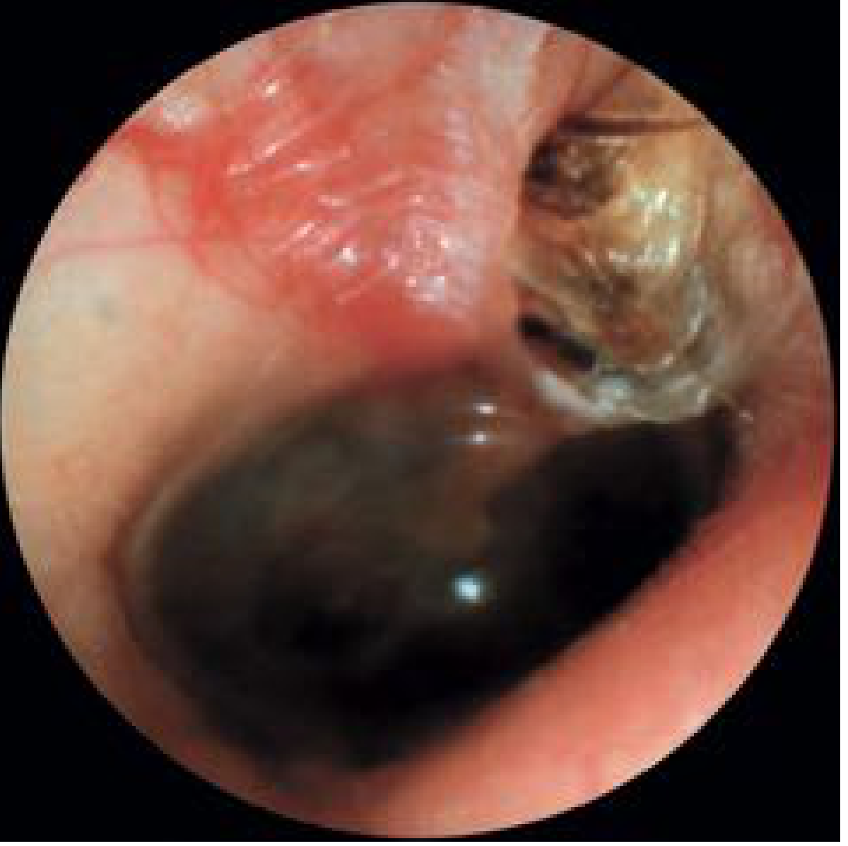
Cholesteatoma
- Congenital (rare, present behind intact TM in young children)
- Primary acquired - keratinizing epithelium migrates through a pars flaccida retraction pocket
- Secondary acquired - epithelium migrates through a pre-existing TM perforation
Diagnosis
- Otomicroscopy with pneumatic insufflation - identifies retraction pockets, mobility, cholesteatoma
- Audiometry - conductive hearing loss is typical (low frequency); mixed hearing loss if sensorineural damage has occurred. A conductive loss >30 dB suggests ossicular erosion
- High-resolution CT temporal bones - indicated for surgical planning, complicated CSOM, and identified or suspected cholesteatoma
- MRI with contrast - for suspected intracranial complications
- Biopsy of granulation tissue unresponsive to topical therapy - to exclude malignancy (squamous carcinoma of temporal bone is associated with long-standing CSOM)
- Ear swab for culture - guides antibiotic selection
Management
Primary Goal
Medical Treatment
- Ciprofloxacin ear drops - the most evidence-supported agent; fluoroquinolones are preferred because they are non-ototoxic and active against Pseudomonas
- A 4-6 week course following debridement
- 2025 Cochrane evidence: Three Cochrane meta-analyses published June 2025 confirm topical antibiotics are more effective than systemic antibiotics or topical antiseptics for CSOM. Topical antiseptics (acetic acid, Burow's solution) remain an alternative.
- Biofilms are frequently resistant; high concentrations from topical delivery partially overcome this
Indications for Surgery
- Cholesteatoma combined with medically refractory CSOM = near-absolute indication
- Failure of multiple courses of medical treatment
- Symptoms suspicious of complications: vertigo, facial weakness, headache
- Deep retraction pockets threatening ossicular chain
- Persistent conductive hearing loss
Surgical Options
| Procedure | Description | Indication |
|---|---|---|
| Myringoplasty | TM repair alone (patch graft) | Simple perforation, no middle ear/mastoid disease |
| Tympanoplasty | TM repair ± ossicular chain reconstruction | Perforation with ossicular involvement |
| Canal wall-up (CWU) mastoidectomy | Mastoid exenteration preserving posterior EAC wall | Limited cholesteatoma; patient preference for normal anatomy |
| Canal wall-down (CWD) mastoidectomy | Posterior EAC wall removed; open cavity created | Extensive cholesteatoma; revision surgery; unresectable matrix |
- CWD provides better surgical access and potentially lower recurrence, but results in an open cavity requiring lifelong routine follow-up and debridement
- CWU preserves normal anatomy and dramatically reduces post-op convalescence, but higher disease recurrence risk for cholesteatoma
- Anatomical position of cholesteatoma is a stronger predictor of recurrence than choice of technique
Complications
Intracranial Complications
- Meningitis
- Brain abscess (temporal lobe, cerebellar)
- Epidural/subdural abscess
- Sigmoid sinus thrombophlebitis
- Otitic hydrocephalus
Extracranial Complications
- Labyrinthine fistula (most commonly horizontal semicircular canal) - presents with vertigo and SNHL
- Facial nerve paralysis - acute (infection) or insidious (cholesteatoma expansion through fallopian canal)
- Coalescent mastoiditis / subperiosteal abscess
- Petrositis (Gradenigo syndrome: otorrhoea, ipsilateral abducens palsy, deep facial pain)
- Ossicular erosion (most commonly the long process of the incus) → conductive hearing loss
- Tegmen tympani/mastoideum erosion → CSF leak, brain herniation
Key Points Summary
- CSOM = TM perforation + chronic otorrhoea >6 weeks; tubotympanic (safe) vs atticoantral (unsafe/cholesteatoma)
- Core pathophysiology: ET dysfunction → negative middle ear pressure → TM retraction → perforation/cholesteatoma
- Most common pathogen: Pseudomonas aeruginosa; mixed aerobic/anaerobic flora
- Foul-smelling discharge = anaerobes/cholesteatoma; malodour is a red flag for the atticoantral type
- An aural polyp = cholesteatoma until proven otherwise
- Otalgia/headache/vertigo/facial weakness = possible complications; urgent evaluation needed
- Topical ciprofloxacin + aural toilet is first-line medical treatment (2025 Cochrane evidence confirms superiority over systemic antibiotics)
- Surgery (tympanoplasty ± mastoidectomy) is indicated for cholesteatoma, treatment failure, or complications
- Cholesteatoma is aggressive: erodes ossicles, otic capsule, fallopian canal, tegmen - treat surgically without delay
- Long-term surveillance is mandatory after CWD mastoidectomy
Middle ear disease salah mansour book
Salah Mansour "Middle Ear Disease" book otology
Available ENT Books in This Library
| Book | Publisher |
|---|---|
| Cummings Otolaryngology Head & Neck Surgery | Elsevier |
| KJ Lee's Essential Otolaryngology | McGraw-Hill |
| Scott-Brown's Otorhinolaryngology Head & Neck Surgery (Vol 1, 2, combined) | CRC Press |
| Shambaugh Surgery of the Ear | PMPH |
About Salah Mansour's Book
- Authors: Salah Mansour, Jacques Magnan, Karen Nicolas, Hassan Haidar
- Publisher: Springer (2018)
- eBook ISBN: 978-3-319-72962-6
- Hardcover ISBN: 978-3-319-72961-9
- Otosclerosis
- Acute Otitis Media and Acute Coalescent Mastoiditis
- Otitis Media with Effusion
- Chronic Suppurative Otitis Media (CSOM)
- Cholesteatoma
- And further chapters on tympanoplasty, ossiculoplasty, and related topics
What I Can Do Instead
Branchial cyst
branchial cyst neck lateral cervical
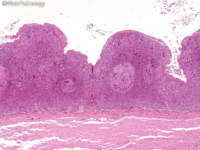
Light microscopy of a formalin-fixed neck specimen stained with Hematoxylin and Eosin reveals a cystic lesion in the lateral cervical region. The cyst is lined by squamous epithelium or ciliated columnar (respiratory-type) epithelium, reflecting branchial apparatus derivatives. Beneath the epithelial lining lies dense lymphoid tissue with prominent germinal centers, consistent with subepithelial lymphoid aggregates typical of branchial cleft remnants. The cyst cavity may contain proteinaceous material and occasionally mucin, with secondary inflammatory changes if infected. In lower neck variants, adnexal structures such as sebaceous or mucous glands can be present within the wall or adjacent tissues. A paucity of atypia supports a benign developmental lesion rather than a malignant process; rare cases show squamous cell carcinoma arising in long-standing branches, but this is uncommon. The combination of lateral neck localization, epithelial lining type, and subepithelial lymphoid tissue is characteristic and helps distinguish branchial cleft cysts from cystic metastasis, cystic hygroma, epidermoid or dermoid cysts, and thyroglossal duct cyst. Clinically relevant implications include confirming diagnosis preoperatively, guiding complete surgical excision, and counseling on infection risk and recurrence. This histology is essential for differential diagnosis, educational purposes, and research into branchial apparatus remnants and neck mass pathology.
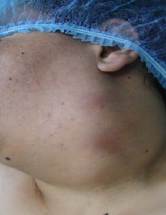
Clinical photograph of a patient's left lateral neck demonstrating a large, voluminous swelling consistent with a branchial cleft cyst (tonsillar cyst). The mass is located in the anterolateral cervical region, extending vertically from the inferior aspect of the ear/mastoid process down toward the supraclavicular area. Visually, the swelling is rounded and significant enough to cause visible asymmetry and contour deformity of the neck. The overlying skin appears slightly erythematous and mildly inflamed, with visible superficial vascularity and areas of uneven pigmentation. The patient is shown wearing a blue surgical cap, indicating a perioperative or clinical setting. This image illustrates the typical clinical presentation of a congenital neck cyst in an adult, highlighting the anatomical distribution and the localized inflammatory response that can occur with such masses.
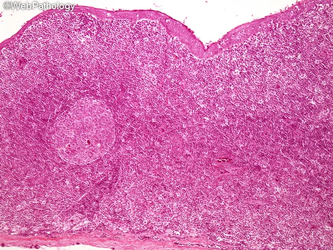
This is a histopathology micrograph (Hematoxylin and Eosin stained) illustrating a branchial cleft cyst of the lateral neck. The cyst lining consists of squamous epithelium or ciliated columnar epithelium, with a subepithelial zone of abundant lymphoid tissue containing germinal centers. The cyst wall may harbor adnexal glands, such as serous, mucinous, or sebaceous elements, particularly in lesions arising in the lower neck. The lumen often contains proteinaceous debris or keratinous material, and the surrounding stroma may show chronic inflammatory infiltrates; secondary bacterial infection is possible. Epithelium generally preserves architecture and shows no significant cytologic atypia or invasion, consistent with benign branchial apparatus remnants. Rarely, malignant transformation to squamous cell carcinoma can occur in long-standing lesions, but this is uncommon. The constellation of epithelial lining (squamous or respiratory-type), deep lymphoid tissue with germinal centers, and a cervical cystic cavity is diagnostic of branchial cleft cysts and helps distinguish them from other neck cysts such as thyroglossal duct cysts, cystic lymphangioma, epidermoid cysts, or cystic metastases. Clinically relevant correlations include lateral neck masses in children or adults, often associated with recurrent infections. Imaging and histology together guide management, surgical excision, and prognosis.
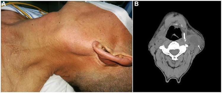
This composite image consists of a clinical photograph and a diagnostic CT scan illustrating a branchial cleft cyst in the neck. (A) A clinical photograph showing the left side of a patient's neck, where a large, prominent mass is visibly protruding from the skin in the upper cervical region. The overlying skin appears intact without obvious erythema. (B) An axial non-contrast CT scan of the neck at the level of the cervical vertebrae. It reveals a well-circumscribed, hypodense, ovoid mass (indicated by a white arrow) located on the left lateral aspect of the neck, deep to the platysma and lateral to the carotid sheath. The low-density center is characteristic of fluid-filled cystic structures. The lesion is located inferior to the parotid gland and anterior to the sternocleidomastoid muscle. The case illustrates the classic presentation of a branchial cleft cyst, which in this clinical context was diagnosed as a primary branchial cleft carcinoma (squamous cell carcinoma).
"branchial cyst" diagnosis management
Branchial Cyst
Definition
- Cyst = no communication with body surface (skin or pharynx)
- Sinus = tract communicates with only one surface (skin or pharynx, not both)
- Fistula = complete tract communicating with both the skin and pharynx
Embryology
- The 1st cleft gives rise to the external auditory canal
- The 2nd arch overgrows the 2nd, 3rd, and 4th clefts, burying them in a transient structure called the cervical sinus
- The cervical sinus then obliterates; failure of this obliteration traps ectoderm → branchial remnant
Classification
| Type | Frequency | Location | Fistula Tract |
|---|---|---|---|
| 1st BCA | 10-18% | Near ear lobule, along mandible, EAC | EAC skin → medial or lateral to facial nerve |
| 2nd BCA | ~65-70% | Anterior border of SCM, level II/III | Skin → between ICA & ECA → above CN IX & XII → tonsillar fossa |
| 3rd BCA | Rare | Anterior border of SCM, level III | Skin → posterior to both carotids → between CN IX & XII → piriform sinus |
| 4th BCA | Very rare | Lower neck; closely associated with thyroid | Skin → above CN XII → posterior to common carotid → below aortic arch (left) → piriform sinus |
First BCA - Work's Classification
| Work Type | Description |
|---|---|
| Type I | Duplication of ectodermal EAC only; cyst near EAC below level of tragus |
| Type II | Duplication of ectodermal + mesodermal elements; cyst near tail of parotid; close to or passing through the facial nerve trunk |
Pathology (Histology)
- Well-circumscribed cystic lesion, 2-5 cm in diameter
- Lined by stratified squamous or pseudostratified columnar (respiratory-type) ciliated epithelium
- Fibrous cyst wall contains dense lymphoid tissue with prominent germinal centres - this is pathognomonic
- Cyst contents: clear watery or mucinous fluid, desquamated cells, and granular/keratinous debris
- Secondary inflammatory infiltrates and wall thickening if previously infected
Clinical Features
- Second BCCs (the vast majority) typically present between ages 10-40 years
- Often diagnosed in early adulthood after an incidental finding or acute infection
- Usually unilateral; bilateral cases are rare and often familial
- Smooth, fluctuant, non-tender lateral neck mass
- May transilluminate (early/thin-walled cysts)
- Can be very soft and difficult to palpate when small
- Often swells and becomes tender during upper respiratory infections (antigen stimulation of wall lymphoid tissue)
- Intermittent drainage if a sinus is present
- At the anterior border of the sternocleidomastoid (SCM) muscle, at the junction of the upper and middle thirds - just inferior to the angle of the mandible
- Superficial to the carotid sheath, posterior to the submandibular gland
Surgical Anatomy of the 2nd BCA Tract
- From the external opening at the anterior border of the SCM in the lower/middle neck
- Through the platysma
- Between the internal and external carotid arteries at the carotid bifurcation (level of thyroid cartilage = C3/C4)
- Superficial to CN IX (glossopharyngeal) and CN XII (hypoglossal)
- Deep to the posterior belly of digastric, stylohyoid ligament
- The vagus (CN X) and spinal accessory (CN XI) lie posterior to the tract
- Terminates at the internal opening in the tonsillar fossa (anterior wall of the posterior faucial pillar)
Investigations
Ultrasound (first-line)
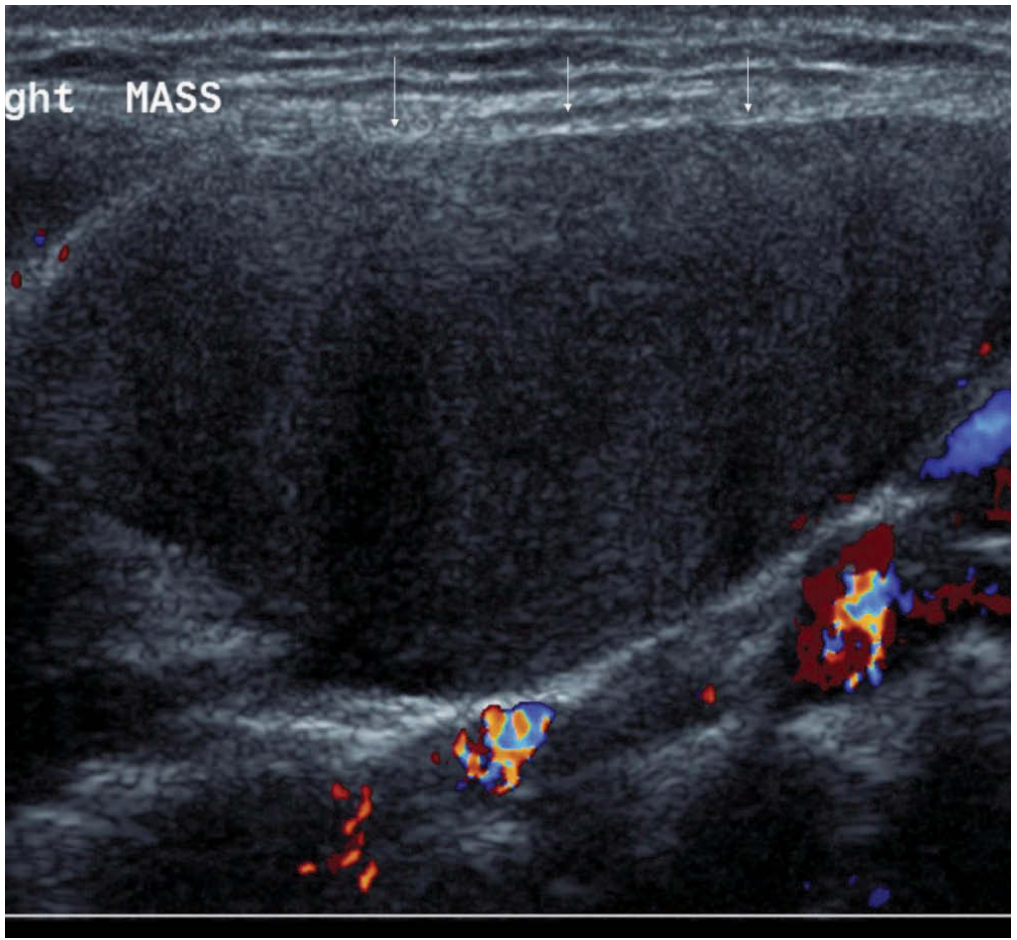
- Classic appearance: anechoic simple cyst with posterior acoustic enhancement, avascular on Doppler
- If previously infected: heterogeneous, thick-walled, with internal debris and septae (pseudosolid appearance)
- If a medially-directed "beak" is seen → suggests sinus/fistula → proceed to CT/MRI
CT (contrast-enhanced)
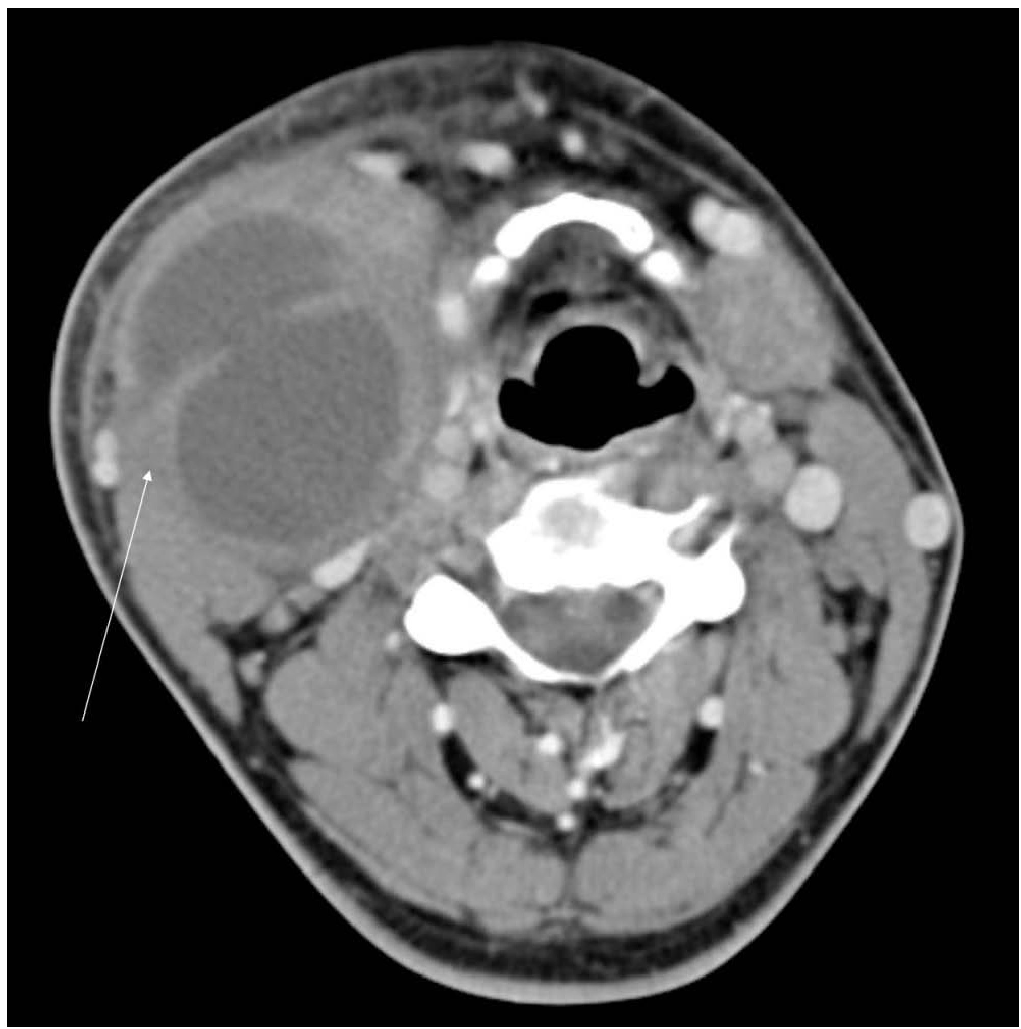
- Classic: homogeneous low-attenuation (fluid density, 0-20 HU), thin smooth wall, displaces submandibular gland anteriorly and lies deep to SCM
- Infected: thick-walled, septated, heterogeneous, ill-defined
MRI
- High T2 signal, low T1 signal (simple fluid)
- Infected: mixed T1/T2 signal, thick irregular wall
FNAC (Fine-Needle Aspiration Cytology)
- Yields thick, turbid, "motor oil"-like fluid
- Cytology: squamous cells, lymphocytes, cholesterol crystals
- Used diagnostically and therapeutically (aspiration)
- Mandatory in patients >35 to exclude cystic SCC metastasis
Differential Diagnosis
| Condition | Distinguishing Features |
|---|---|
| Cystic metastatic SCC (most important) | Patient >35-40 years, smoker/drinker; primary tumour in pharynx/tonsil; HPV-positive oropharyngeal SCC |
| Cystic papillary thyroid carcinoma (PTC) | Intrathyroidal or paratracheal; thyroid mass or nodule |
| Lymphangioma/cystic hygroma | Usually children; soft, multilocular, transilluminates brilliantly; often crosses compartments |
| Thyroglossal duct cyst | Midline, moves with swallowing and tongue protrusion |
| Dermoid/epidermoid cyst | Midline or floor of mouth; doughy texture; no lymphoid wall |
| Reactive/infective lymphadenopathy | Multiple nodes; tender; febrile illness; resolves |
| Reactive cervical lymph node | Solid on US; hilum preserved |
| Carotid body tumour | Pulsatile; bruit; splays carotid bifurcation on imaging |
| Plunging ranula | Originates from floor of mouth; dumbbell shape |
Critical rule: In any patient over 35 years with a lateral cystic neck mass, malignancy must be excluded before assuming benign BCC. If FNAC is not diagnostic and no primary tumour is found on CT/MRI, PET-CT should be considered (especially if smoker/alcohol history). This is a CUP (carcinoma of unknown primary) investigation protocol.
Management
Conservative
- Asymptomatic small cysts in young patients may be observed
- Aspiration for symptomatic relief (high recurrence rate without excision)
Medical
- Acute infection: antibiotics (oral or IV depending on severity) to render the lesion quiescent before definitive surgery
- Surgery is best performed when the lesion is quiescent - operating on an infected cyst is associated with higher rates of incomplete excision, recurrence, and nerve injury
Surgical Excision (Definitive Treatment)
- Complete surgical excision is the standard of care
- Approach: transverse cervical skin crease incision ± "step-ladder" incisions for fistulas
- For a fistula, the entire tract must be excised to prevent recurrence - often requires two transverse incisions (upper and lower) to avoid longitudinal scars
- Key structures to identify and protect:
- CN VII (especially for 1st BCA)
- CN IX (glossopharyngeal)
- CN XII (hypoglossal)
- CN XI (spinal accessory)
- Internal and external carotid arteries
- Internal jugular vein
Recurrence
- Incomplete excision (especially if operating on an infected cyst or failing to trace the entire fistula tract) → recurrence
- Multiple previous incisions/infections create fibrosis and distort anatomy → higher surgical difficulty
Branchial Cyst Carcinoma (Branchiogenic Carcinoma)
- No primary elsewhere in the aerodigestive tract
- Histological appearance consistent with origin from branchial remnant
- Long clinical history of a benign cyst before malignant change
All patients >35 years with lateral cystic neck masses should enter a CUP investigation protocol even if FNAC is not suggestive of metastatic SCC.
Key Points
- Second BCC = 95% of cases; presents at anterior border of SCM, ages 10-40
- Classic triad: lateral neck swelling + fluctuant + enlarges with URTIs
- Histology: squamous/respiratory epithelium + lymphoid tissue with germinal centres (pathognomonic)
- US first-line: anechoic, avascular; CT if sinus/fistula or surgical planning needed
- FNAC: turbid fluid + squamous cells + lymphocytes
- >35 years = exclude malignancy first (cystic SCC metastasis from HPV oropharyngeal cancer)
- Treatment = complete surgical excision when quiescent
- Fistula tract must be completely excised - two-incision technique
- Protect CN VII (1st BCA), CN IX, CN XII during surgery
- Third/fourth BCAs → arise from piriform sinus; may present as suppurative thyroiditis