

Screenshot_2026-07-14-13-54-38-10_c37d74246d9c81aa0bb824b57eaf7062.jpg
I am preparing for my first year university exam of anatomy.
Reading File
I can see your anatomy exam question paper. These are all upper limb anatomy topics. Let me search the medical library for answers to these questions in parallel.
Finding Sources
Finding Sources
Finding Sources
Finding Sources
Gray's Anatomy for Students is the best source for this exam. Let me now read the relevant sections in parallel.
Reading File
Finding Sources
Finding Sources
Finding Sources
Reading File
Finding Sources
Finding Sources
Reading File
Reading File
I now have enough from the textbook to compile comprehensive answers. Let me also fetch the key diagram for the brachial plexus.
Loading Image
Loading Image
I now have all the key information. Let me compile the complete exam answers.Here are comprehensive answers for all 17 questions on your exam paper. All content is sourced from Gray's Anatomy for Students.
Upper Limb Anatomy - Exam Answers
Q1. Attachments of Rotator Cuff Muscles (3M)
The rotator cuff consists of 4 muscles that attach the scapula to the humerus and stabilize the shoulder joint:
| Muscle | Origin (Scapula) | Insertion (Humerus) |
|---|---|---|
| Supraspinatus | Supraspinous fossa | Greater tubercle (superior facet) |
| Infraspinatus | Infraspinous fossa | Greater tubercle (middle facet) |
| Teres minor | Lateral border (upper part) | Greater tubercle (inferior facet) |
| Subscapularis | Subscapular fossa | Lesser tubercle |
Memory aid: SITS (Supraspinatus, Infraspinatus, Teres minor, Subscapularis)
The tendons blend with the fibrous capsule of the glenohumeral joint, reinforcing it on all sides except inferiorly (where dislocations most commonly occur).
Q2. Attachments, Actions and Nerve Supply of Deltoid (3M)
Attachments:
- Origin (U-shaped):
- Lateral 1/3 of clavicle (anterior part)
- Acromion (middle part)
- Spine of scapula (posterior part)
- Insertion: Deltoid tuberosity of the humerus (V-shaped impression on lateral shaft)
Actions:
- Anterior fibres: Flexion and medial rotation of arm
- Middle fibres: Abduction of arm (primary abductor after supraspinatus initiates 0-15°)
- Posterior fibres: Extension and lateral rotation of arm
Nerve Supply: Axillary nerve (C5, C6) - from the posterior cord of brachial plexus. The nerve wraps around the surgical neck of humerus.
Q3. Structures Under Cover of Deltoid Muscle
The deltoid overlies the following structures:
- Shoulder joint (glenohumeral joint)
- Subacromial (subdeltoid) bursa
- Rotator cuff tendons (supraspinatus, infraspinatus, teres minor, subscapularis)
- Tendon of long head of biceps brachii
- Axillary nerve and posterior circumflex humeral vessels (passing through quadrangular space)
- Greater and lesser tubercles of humerus
- Intertubercular sulcus (bicipital groove)
Q4. Attachment of Pectoralis Major Muscle (2M)
Origin (broad):
- Clavicular head: Medial half/anterior surface of clavicle
- Sternocostal head: Anterior surface of sternum + costal cartilages of ribs 1-6
- Small contribution from the aponeurosis of external oblique
Insertion:
- Lateral lip of intertubercular sulcus (bicipital groove) of humerus
- The tendon is folded on itself - clavicular fibres insert lower, sternocostal fibres insert higher (creating a twist)
Q5. Attachments, Actions and Nerve Supply of Serratus Anterior (4M)
Attachments:
- Origin: Outer surfaces and superior borders of upper 8-9 ribs (by digitations)
- Insertion: Anterior (costal) surface of the medial border of scapula (the entire medial border and inferior angle)
Actions:
- Protracts scapula (pulls it forward around the thorax - essential for pushing/punching) - hence called "boxer's muscle"
- Rotates the scapula so the glenoid faces superiorly - essential for raising arm above 90° (works with trapezius)
- Holds the medial border of scapula against the thoracic wall
Nerve Supply: Long thoracic nerve (C5, C6, C7) - nerve of Bell. Runs on the outer surface of serratus anterior.
Clinical: Damage to the long thoracic nerve causes "winged scapula" - the medial border lifts away from the thoracic wall.
Q6. Boundaries and Contents of Carpal Tunnel + Carpal Tunnel Syndrome
Boundaries:
- Floor (posteriorly): Carpal bones (concave arch of carpus)
- Roof (anteriorly): Flexor retinaculum (transverse carpal ligament) - stretches between pisiform & hook of hamate (medially) and scaphoid tubercle & trapezium (laterally)
Contents (9 tendons + 1 nerve):
- Median nerve
- Flexor pollicis longus tendon (1)
- Flexor digitorum superficialis tendons (4)
- Flexor digitorum profundus tendons (4)
Note: The flexor carpi radialis runs in a separate tunnel within the flexor retinaculum. The ulnar nerve and artery pass superficial to the retinaculum (in Guyon's canal).
Carpal Tunnel Syndrome:
- Compression of the median nerve within the carpal tunnel
- Causes: tenosynovitis, pregnancy, hypothyroidism, rheumatoid arthritis, acromegaly
- Symptoms: Pain and tingling in lateral 3½ fingers, thenar wasting, weak opposition of thumb
- Positive Tinel's sign (tapping over wrist reproduces symptoms) and Phalen's test (wrist flexion for 60 sec reproduces symptoms)
Q7. Nerve Supply and Actions of Interossei of Hand
Dorsal interossei (4 muscles):
- Nerve: Deep branch of ulnar nerve (C8, T1)
- Action: Abduction of fingers (DAB - Dorsal ABduct), also flex MCP joints and extend IP joints
Palmar interossei (4 muscles, some say 3):
- Nerve: Deep branch of ulnar nerve (C8, T1)
- Action: Adduction of fingers (PAD - Palmar ADduct), also flex MCP joints and extend IP joints
Memory: PAD and DAB
Clinical: In ulnar nerve palsy, loss of interossei causes "claw hand" (hyperextension at MCP + flexion at IP joints), more pronounced in ring and little fingers.
Q8. Diagram and Explanation of Brachial Plexus (5M)
Root values: Anterior rami of C5, C6, C7, C8, T1

Trunks:
- Superior trunk: C5 + C6
- Middle trunk: C7 alone
- Inferior trunk: C8 + T1
Divisions: Each trunk divides into anterior and posterior divisions (6 total)
Cords (named by relation to 2nd part of axillary artery):
- Lateral cord: Anterior divisions of superior + middle trunks
- Medial cord: Anterior division of inferior trunk
- Posterior cord: All 3 posterior divisions
Terminal branches (remember: M.U.R.A.M):
- Musculocutaneous nerve (C5,6,7) - from lateral cord
- Ulnar nerve (C7,8,T1) - from medial cord
- Radial nerve (C5,6,7,8,T1) - from posterior cord
- Axillary nerve (C5,6) - from posterior cord
- Median nerve (C6,7,8,T1) - from both lateral AND medial cords
Key branches from roots/trunks:
- Dorsal scapular nerve (C5)
- Long thoracic nerve (C5,6,7)
- Suprascapular nerve (C5,6) from superior trunk
- Nerve to subclavius (C5,6) from superior trunk
Q9. Anatomical Basis of Deformity in Erb's Paralysis
Erb's point: Junction of C5 and C6 roots (superior trunk)
Mechanism: Forceful separation of head from shoulder (e.g., birth injury - traction on neck during delivery, or motorcycle accident)
Muscles paralyzed (C5, C6 roots):
- Deltoid (axillary nerve) - no abduction
- Biceps brachii (musculocutaneous) - no flexion, no supination
- Brachialis (musculocutaneous) - no flexion
- Supraspinatus/infraspinatus (suprascapular nerve) - no abduction/lateral rotation
- Subscapularis, teres major - unopposed, cause medial rotation
Resulting deformity - "Waiter's tip position":
- Adduction (deltoid paralyzed, pectorals intact)
- Medial rotation (lateral rotators paralyzed, medial rotators intact)
- Extension at elbow (biceps/brachialis paralyzed)
- Pronation (supinator paralyzed, pronators intact)
- Hand/wrist relatively spared
Q10. Pronation and Supination (2M)
Pronation - palm faces posteriorly/downward:
- Muscles: Pronator teres (C6,7 - median nerve) and Pronator quadratus (C8,T1 - anterior interosseous nerve)
- Mechanism: Radius rotates over ulna, crossing it
Supination - palm faces anteriorly/upward:
- Muscles: Supinator (C6 - deep branch of radial nerve/posterior interosseous nerve) and Biceps brachii (C5,6 - musculocutaneous nerve)
- Biceps is the most powerful supinator, especially when elbow is flexed
- Mechanism: Radius uncrosses to lie parallel to ulna
Q11. Median Nerve in Hand and Forearm (4M)
Root value: C6, C7, C8, T1 (from both lateral and medial cords)
In the forearm:
- Enters between the two heads of pronator teres
- Runs between FDS (superficially) and FDP (deeply)
- Gives off the anterior interosseous nerve (supplies FPL, lateral half of FDP, pronator quadratus)
- Enters hand through carpal tunnel
Muscles supplied in forearm (LOAF - not quite, but...):
- All flexors EXCEPT FCU and medial half of FDP (those are ulnar)
- Pronator teres, FCR, Palmaris longus, FDS, FPL, lateral FDP, pronator quadratus
In the hand (enters via carpal tunnel):
- Recurrent (thenar) branch: Supplies 3 thenar muscles - Abductor pollicis brevis, Flexor pollicis brevis (superficial head), Opponens pollicis
- Digital branches: 1st and 2nd lumbricals
- Sensory: Lateral 3½ fingers (palmar surface) + dorsal tips of same fingers
Injury in forearm ("Ape hand" if high lesion):
- Loss of opposition and abduction of thumb
- Thenar wasting
- Loss of sensation in lateral 3½ fingers
Q12. Musculocutaneous Nerve (3M)
Root value: C5, C6, C7 (from lateral cord of brachial plexus)
Course:
- Pierces coracobrachialis muscle
- Passes between biceps brachii (anteriorly) and brachialis (posteriorly)
- Exits lateral to biceps tendon at elbow as the lateral cutaneous nerve of the forearm
Muscles supplied:
- Coracobrachialis (C6, C7)
- Biceps brachii (C5, C6) - flexion and supination
- Brachialis (C5, C6) - main flexor of the elbow
Cutaneous supply: Lateral cutaneous nerve of the forearm - supplies lateral aspect of forearm (both anterior and posterior)
Injury: Weakness of elbow flexion (especially in supinated position), loss of supination, sensory loss over lateral forearm.
Q13. Origin, Insertion and Distribution of Radial Nerve
Root value: C5, C6, C7, C8, T1 (posterior cord)
Origin: Posterior cord of brachial plexus
Course:
- Enters arm posterior to brachial artery
- Winds in the radial groove (spiral groove) of humerus with profunda brachii artery
- Passes through lateral intermuscular septum to enter anterior compartment
- Enters cubital fossa between brachialis and brachioradialis
Distribution/muscles supplied:
- In arm: Triceps brachii, brachioradialis, ECRL, part of brachialis
- In forearm (via deep branch / posterior interosseous nerve): ECRB, supinator, EDC, EDM, ECU, APL, EPL, EPB, EIP
- Superficial branch (sensory): Dorsum of lateral 3½ fingers (proximal phalanges only)
Q14. Root Value, Muscles Supplied and Clinical Anatomy of Radial Nerve
Root value: C5, C6, C7, C8, T1
All muscles supplied:
| Location | Muscles |
|---|---|
| Arm | Triceps brachii (all 3 heads), Anconeus |
| Elbow region | Brachioradialis, ECRL |
| Forearm (deep branch) | ECRB, Supinator, EDC, EDM, ECU, APL, EPL, EPB, EIP |
Clinical Anatomy:
-
Injury in axilla (Saturday night palsy, crutch palsy): All muscles paralyzed - loss of triceps + wrist/finger extensors. Sensory loss over posterior arm, forearm, dorsum of hand.
-
Injury in radial groove (midshaft humerus fracture): Triceps spared (branch arises before the groove), but wrist drop (loss of wrist extensors), loss of supination. Wrist drop is the classic sign.
-
Injury at lateral epicondyle / posterior interosseous nerve: Finger drop without wrist drop (ECRL spared). No sensory loss (pure motor).
Q15. Parts, Course and Branches of Axillary Artery (3M)
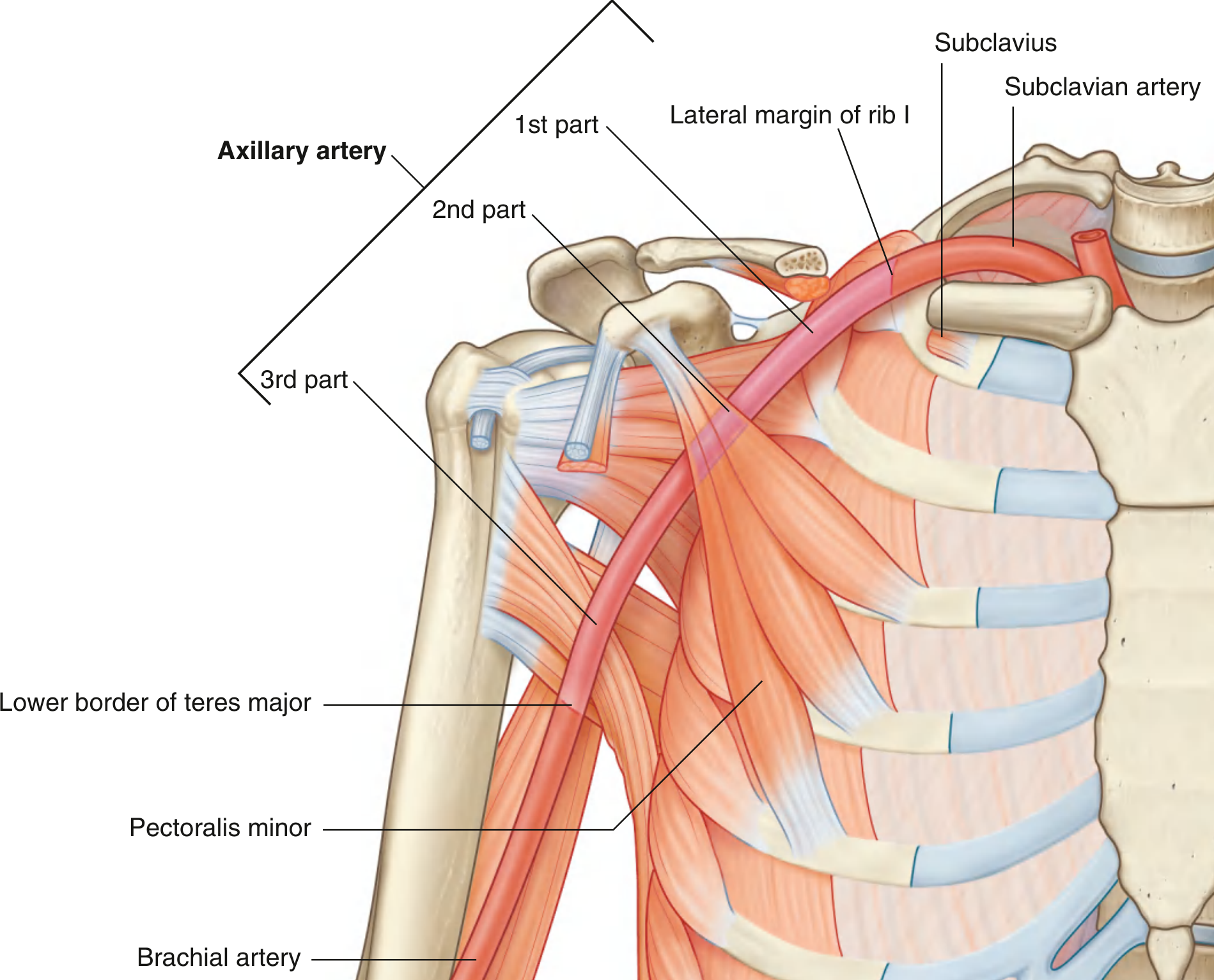
The axillary artery is the continuation of the subclavian artery from the lateral margin of rib I to the lower border of teres major (where it becomes the brachial artery). It is divided into 3 parts by pectoralis minor:
Memory: 1 branch, 2 branches, 3 branches (total = 6)
| Part | Relation to Pec. Minor | Branches |
|---|---|---|
| 1st part | Proximal (medial) | Superior thoracic artery (1) |
| 2nd part | Posterior to pec. minor | Thoraco-acromial artery, Lateral thoracic artery (2) |
| 3rd part | Distal (lateral) | Subscapular artery, Anterior circumflex humeral a., Posterior circumflex humeral a. (3) |
Thoraco-acromial artery divides into: Pectoral, Deltoid, Clavicular, Acromial branches.
Subscapular artery (largest branch) divides into: Circumflex scapular + Thoracodorsal arteries.
Q16. Cubital Fossa (7M)
Definition: Triangular depression anterior to the elbow joint.
Boundaries:
- Superiorly (base): Imaginary horizontal line between medial and lateral epicondyles of humerus
- Medially: Pronator teres (medial epicondyle origin)
- Laterally: Brachioradialis (lateral supraepicondylar ridge)
- Floor: Brachialis muscle (mainly) and supinator
- Roof: Deep fascia + skin, reinforced by bicipital aponeurosis
Contents (lateral to medial - "TAN" or "TMBN"):
- Radial nerve - under brachioradialis lip, divides into superficial and deep branches (not strictly "inside")
- Tendon of biceps brachii - central, inserts into radial tuberosity
- Brachial artery - bifurcates into radial and ulnar arteries at the apex
- Median nerve - most medial, exits by passing between heads of pronator teres
Mnemonic (lateral to medial): Really Need Beer More (Radial nerve, biceps tendon, brachial artery, Median nerve)
Note: The ulnar nerve does NOT pass through the cubital fossa - it passes posterior to the medial epicondyle.
The bicipital aponeurosis covers and protects the brachial artery and median nerve anteriorly.
Clinical significance:
- Site for venepuncture (median cubital vein in roof)
- Brachial artery palpation / blood pressure measurement
- Site for elbow joint aspiration
Q17. Axilla - Boundaries, Contents and Applied Anatomy (3M)
The axilla is a pyramidal space between the arm and thoracic wall.
Boundaries:
| Wall | Structure |
|---|---|
| Anterior wall | Pectoralis major, pectoralis minor, subclavius, clavipectoral fascia |
| Posterior wall | Subscapularis (upper), teres major + latissimus dorsi (lower) |
| Medial wall | Serratus anterior + upper 4 ribs with intercostals |
| Lateral wall | Intertubercular sulcus of humerus (narrowest wall) |
| Apex (inlet) | Bounded by clavicle, 1st rib, superior border of scapula |
| Base (floor) | Axillary fascia + skin of armpit |
Contents:
- Axillary artery (and its 6 branches)
- Axillary vein
- Brachial plexus (cords and branches)
- Axillary lymph nodes (5 groups: anterior/pectoral, posterior/subscapular, lateral, central, apical)
- Long thoracic nerve (on medial wall)
- Intercostobrachial nerve
- Axillary fat and loose connective tissue
Applied Anatomy:
- Axillary lymph nodes are surgically important in breast cancer - they receive 75% of breast lymph drainage. Axillary clearance is part of breast cancer surgery; it risks damage to the long thoracic nerve (winged scapula) and thoracodorsal nerve (loss of latissimus dorsi)
- Crutch palsy (radial nerve compression in axilla) - wrist drop
- Brachial plexus injuries - occur at inlet of axilla
- Axillary vein thrombosis - can result from hyperabduction
All answers based on Gray's Anatomy for Students (Elsevier). Good luck with your exam!All 17 questions are covered above with exam-ready answers. Here is a quick orientation to help you use the notes:
High-mark questions to prioritize:
- Q16 (Cubital fossa - 7M): Memorize boundaries, contents in order (lateral to medial), and the mnemonic "Really Need Beer More"
- Q8 (Brachial plexus - 5M): Learn the diagram - Roots (C5-T1) → Trunks (Superior/Middle/Inferior) → Divisions → Cords (Lateral/Medial/Posterior) → Terminal nerves (M.U.R.A.M.)
- Q5 (Serratus anterior - 4M): Origin on ribs, insertion on medial border of scapula, long thoracic nerve C5,6,7, winged scapula
- Q11 (Median nerve - 4M): Two-compartment answer - forearm muscles + hand muscles (LOAF: Lumbricals 1&2, Opponens pollicis, Abductor pollicis brevis, Flexor pollicis brevis)
Key mnemonics to remember:
- Rotator cuff: SITS
- Cubital fossa contents: Really Need Beer More (Radial, biceps tendon, Brachial artery, Median nerve - lateral to medial)
- Interossei: PAD (Palmar = ADduct) and DAB (Dorsal = ABduct)
- Brachial plexus terminal nerves: M.U.R.A.M. (Musculocutaneous, Ulnar, Radial, Axillary, Median)
This is a shared conversation. Sign in to Orris to start your own chat.