
Anatomy of, अ, meninges.
meninges anatomy diagram dura mater arachnoid pia mater
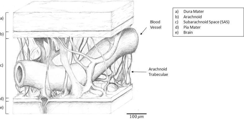
This medical anatomical diagram illustrates the three-dimensional structure of the cranial meninges and the pia-arachnoid complex (PAC). The cross-sectional drawing is labeled with five distinct layers: (a) the thick, outermost dura mater; (b) the thinner arachnoid mater; (c) the subarachnoid space (SAS); (d) the pia mater; and (e) the underlying brain tissue. A primary focus is the subarachnoid space, which contains a network of fibrous arachnoid trabeculae and a large, cylindrical blood vessel. The trabeculae are shown as thin, branching collagenous strands that bridge the space between the arachnoid and pia mater, providing mechanical stability. The diagram highlights the spatial relationship between the leptomeninges (arachnoid and pia) and the underlying neural parenchyma, with a scale bar indicating a 100 µm reference. This illustration is intended for educational use in neuroanatomy and pathophysiology to demonstrate the mechanical protection of the brain and the distribution of cerebrospinal fluid pathways.
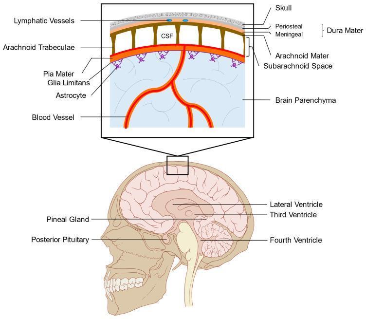
This medical anatomical diagram illustrates the layers of the human meninges and the organization of the subarachnoid space. The lower panel displays a sagittal view of the head, identifying internal structures such as the lateral, third, and fourth ventricles, alongside circumventricular organs including the pineal gland and posterior pituitary. The upper panel provides a high-magnification cross-sectional view of the cranial protective layers. From superficial to deep, it depicts the skull, the dura mater (consisting of periosteal and meningeal layers containing lymphatic vessels), the arachnoid mater, and the subarachnoid space filled with cerebrospinal fluid (CSF). The arachnoid trabeculae are shown spanning the subarachnoid space to connect with the pia mater. Below the pia mater, the glia limitans is illustrated, formed by astrocytic foot processes that interface with the underlying brain parenchyma and cortical blood vessels. This diagram is designed to explain leptomeningeal anatomy and the fluid dynamics between the ventricular system, subarachnoid space, and the blood-brain barrier interface.
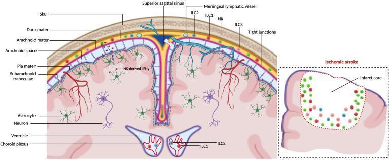
Anatomical and Pathophysiology Diagram illustrating the immunological landscape of the central nervous system (CNS) borders during steady-state and ischemic stroke. The primary cross-section depicts the spatial arrangement of the meninges: the skull, dura mater (containing meningeal lymphatic vessels and superior sagittal sinus), arachnoid mater, arachnoid space with trabeculae, and the pia mater overlying the brain parenchyma. The diagram highlights the distribution of innate lymphoid cells (ILCs), showing NK cells, ILC1, ILC2, and ILC3 residing primarily within the dural meninges. NK-derived IFN-γ is shown diffusing across the tight-junction-restricted arachnoid and pia mater to interact with astrocytes. In the deeper brain, the choroid plexus within the ventricles is shown to harbor ILC1 and ILC2 subsets. An inset titled 'Ischemic stroke' demonstrates the recruitment and accumulation of various ILCs at the border of an infarct core, emphasizing their role in neuroinflammatory responses. The illustration serves as an educational tool for neuroimmunology, detailing the interface between the peripheral immune system and the CNS.
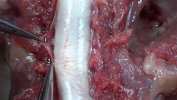
This clinical photograph captures a microsurgical dissection of the spinal meninges, demonstrating the anatomical differentiation between the dura mater and the arachnoid mater. In the center of the frame, the spinal cord appears as a prominent, whitish, cylindrical structure with visible fine vascularization on its surface. Surgical forceps are used to retract and distinguish two distinct layers: the dura mater (labeled '1') and the arachnoid mater (labeled '2'). The dura mater (1) is visualized as a thicker, more opaque, and slightly reddish fibrous membrane, reflecting its dense collagenous composition and relative resistance to manipulation. In contrast, the arachnoid mater (2) is depicted as a thinner, more translucent, and whiter membrane underlying the dura. The surrounding surgical field contains exposed paraspinal muscles and subcutaneous tissues, appearing reddish and fleshy with a glistening texture due to the presence of biological fluids. This image serves as an educational reference for neurosurgical anatomy, specifically the layered structure of the spinal meninges and the plane of dissection required to access the subarachnoid space.
Meninges - Anatomy (मेनिंजेस)
- Dura mater (outermost)
- Arachnoid mater (middle)
- Pia mater (innermost)
Diagram - Cranial Meninges (Gray's Anatomy for Students)
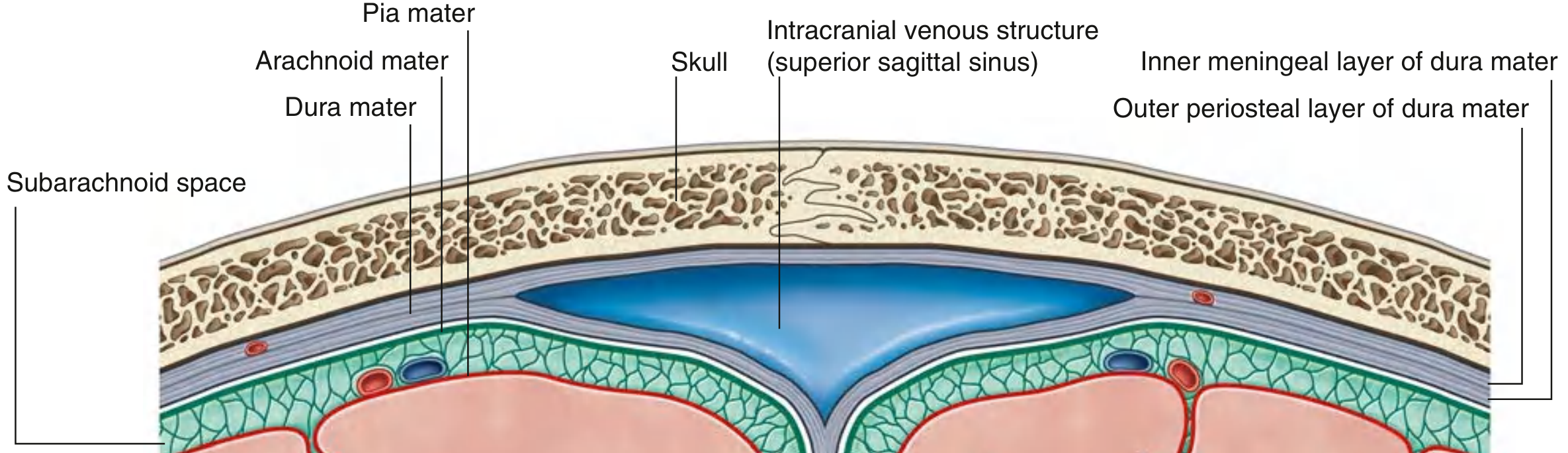
1. DURA MATER ("Hard Mother")
| Layer | Features |
|---|---|
| Outer periosteal layer | Firmly adherent to inner surface of skull; acts as periosteum of cranial cavity; contains meningeal arteries; continuous with periosteum of outer skull at foramina |
| Inner meningeal layer | Lies against arachnoid; continuous with the spinal dura mater through the foramen magnum |
(A) Dural Partitions (Reflections / Septa)
| Partition | Description | Attachments |
|---|---|---|
| Falx cerebri | Crescent-shaped, sickle-like vertical fold; lies between the two cerebral hemispheres | Anteriorly: crista galli of ethmoid + frontal crest; Posteriorly: blends with tentorium cerebelli |
| Tentorium cerebelli | Horizontal tent-like fold; separates cerebellum (posterior fossa) from posterior cerebral hemispheres; has a free medial edge = tentorial notch (through which midbrain passes) | Posteriorly: occipital bone along transverse sinus grooves; Laterally: superior border of petrous temporal bone; ends at anterior & posterior clinoid processes |
| Falx cerebelli | Small midline fold in posterior cranial fossa; lies between two cerebellar hemispheres | Posteriorly: internal occipital crest; Superiorly: tentorium cerebelli; anterior edge is free |
| Diaphragma sellae | Horizontal shelf over hypophyseal fossa of sella turcica; has a central opening through which the infundibulum passes | Covers pituitary gland in sphenoid bone |
(B) Intracranial Venous Sinuses
Arterial Supply of Dura Mater
- Anterior cranial fossa - anterior meningeal arteries
- Middle cranial fossa - middle meningeal artery (largest; most clinically significant) + accessory meningeal artery
- Posterior cranial fossa - posterior meningeal artery + other branches
Nerve Supply of Dura Mater
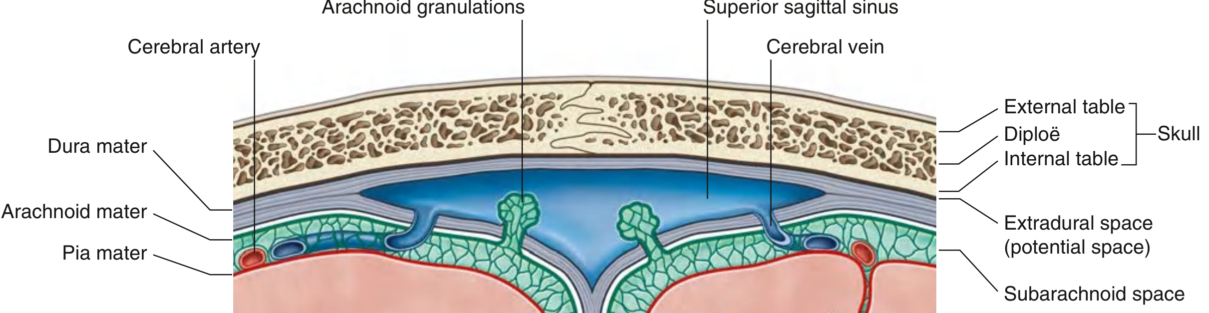
Diagram - Meningeal Spaces with Arachnoid Granulations
2. ARACHNOID MATER ("Spider Web")
- Thin, avascular membrane that lines the inner surface of the dura but is not adherent to it
- Its appearance and consistency resemble a spider web (Greek: arachne = spider)
- It does not follow the gyri and sulci of the brain (except the longitudinal fissure between hemispheres)
- From its inner surface, thin fibrous strands called arachnoid trabeculae extend across the subarachnoid space and connect to the pia mater below
- Arachnoid granulations (villi) - tufted protrusions of arachnoid membrane that project into the dural venous sinuses (especially the superior sagittal sinus). They are the primary site of CSF reabsorption back into the venous system.
3. PIA MATER ("Tender Mother")
- Thin, delicate, highly vascular membrane
- Closely invests the surface of the brain, following every gyrus, sulcus, groove, and fissure
- Closely applied to roots of cranial nerves at their origins
- The subarachnoid space between pia and arachnoid is a real (not potential) space filled with CSF and containing the major cerebral arteries and veins
Meningeal Spaces - Summary
| Space | Nature | Location | Clinical Relevance |
|---|---|---|---|
| Epidural (Extradural) | Potential space | Between periosteal dura and bone | Epidural hematoma - usually arterial; rupture of middle meningeal artery |
| Subdural | Potential space (not a true anatomical space - involves dural border cell layer) | Between inner meningeal dura and arachnoid | Subdural hematoma - usually venous; tearing of bridging veins |
| Subarachnoid | Real/actual space | Between arachnoid and pia | Contains CSF; SAH from ruptured aneurysm; lumbar puncture accesses this space |
4. SPINAL MENINGES
| Feature | Cranial | Spinal |
|---|---|---|
| Dura mater layers | Two layers (periosteal + meningeal) | One layer only (the meningeal layer; no periosteal layer as the vertebral periosteum is separate) |
| Epidural space | Potential | Real, fat-filled space (clinically important for epidural anaesthesia) |
| Dura-arachnoid adherence | Tightly adherent | Less adherent (theoretical subdural plane) |
| Dura inferior extent | Lines cranial cavity | Extends to S2 vertebral level |
| Subarachnoid space inferior extent | - | Also ends at S2 |
Denticulate (Dentate) Ligament
- A flat, continuous sheet of pia mater running midway between the anterior and posterior nerve roots
- Sends tooth-like projections (denticulations) through the arachnoid to attach onto the inner surface of the dura
- Function: anchors and positions the spinal cord within the subarachnoid space, preventing lateral displacement
5. Meningeal Image Gallery
Quick Summary Table
| Layer | Consistency | Vascularity | Adherence | Space below |
|---|---|---|---|---|
| Dura mater | Tough, leathery | Vascular (meningeal vessels in periosteal layer) | Adherent to skull (periosteal layer) | Subdural (potential) |
| Arachnoid mater | Delicate, web-like | Avascular | Lines inner dura but not adherent | Subarachnoid (real, CSF-filled) |
| Pia mater | Very thin, delicate | Highly vascular | Tightly adherent to brain/spinal cord surface | - |
- Gray's Anatomy for Students, Fig. 8.31, 8.32, 8.35 (Chapter 8, Head)
- Bradley and Daroff's Neurology in Clinical Practice, Chapter on Meninges
- Neuroscience: Exploring the Brain, 5th ed., Chapter 7