
My grandfather having ocular pressure of 34mmhg in right eye and having blurred vision since 1 week and he is not a diabetic patient so what is management
ocular hypertension 34mmhg glaucoma management treatment guidelines 2024
ocular hypertension glaucoma treatment IOP lowering
glaucoma optic nerve cupping IOP elevated fundus
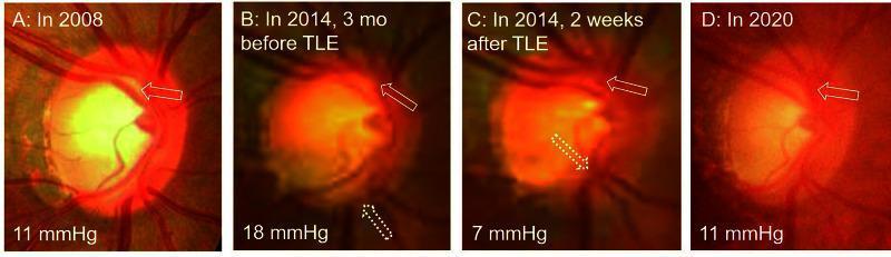
A series of four clinical fundus photographs (A-D) documenting optic disc morphological changes in a glaucoma patient over a 12-year period (2008–2020). The images illustrate the dynamic relationship between intraocular pressure (IOP) and optic nerve cupping. Panel A (2008) shows an oval optic disc with an IOP of 11 mmHg and a vertical cup-to-disc (C/D) ratio of 0.72. Panel B (2014) displays disease progression prior to trabeculectomy (TLE), characterized by an increased IOP (18 mmHg), thinning of the superior and inferior neuroretinal rims, a circular disc shape (H/V ratio = 1.0), and enlarged cupping (C/D ratio = 0.78). Panel C (two weeks post-TLE) demonstrates rapid structural reversal after IOP reduction to 7 mmHg, showing decreased cupping (C/D = 0.61) and centripetal shifting of the retinal vasculature. Panel D (2020) shows long-term stabilization at 11 mmHg, where the disc has largely regained its baseline oval morphology and rim area. This sequence serves as a clinical example of 'cup reversal' or optic disc remodeling following surgical intraocular pressure reduction.
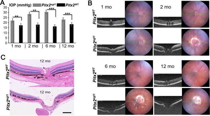
This composite educational resource illustrates an early-onset glaucoma phenotype in a mouse model (Pitx2egl1) compared to wild-type (Pitx2WT). Panel A contains a bar graph showing longitudinal intraocular pressure (IOP) data at 1, 2, 6, and 12 months. Pitx2egl1 mice exhibit significantly elevated IOP (peaking around 30 mmHg at 6 months) compared to the stable ~18 mmHg in controls. Panel B displays longitudinal diagnostic imaging, combining fundus photography and Optical Coherence Tomography (OCT). In mutants, fundus images reveal a characteristic partial white ring around the optic cup, while OCT B-scans demonstrate progressive optic nerve cupping and excavation starting as early as 2 months. Panel C presents H&E-stained histological sections of the optic nerve head at 12 months. The mutant section shows severe glaucomatous cupping (excavation) and thinning of the inner retinal layers, specifically the retinal nerve fiber layer (RNFL), compared to the healthy morphology of the wild-type. This visual summarizes the clinical signs of hypertensive glaucoma including elevated IOP, structural optic disc changes, and neurodegeneration.
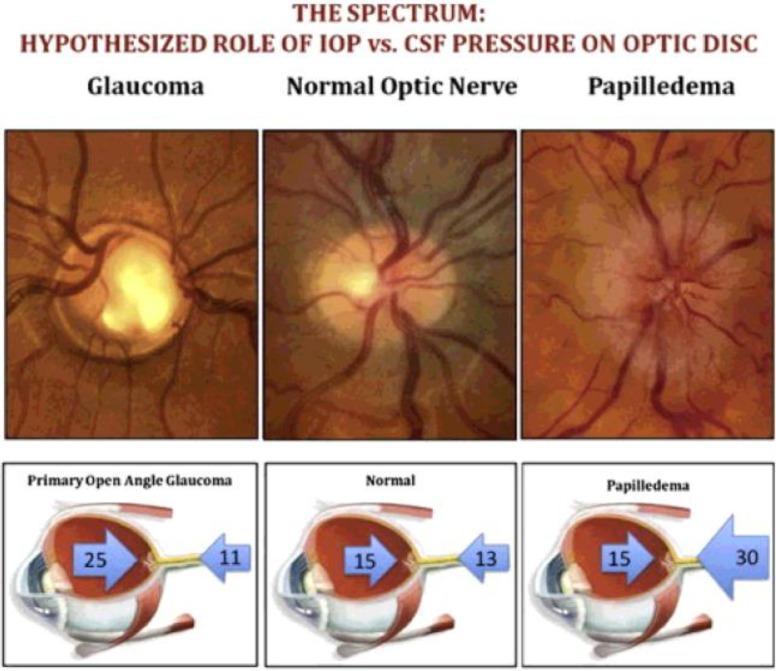
This educational graphic illustrates the 'Cerebrospinal Fluid (CSF) Theory' of the optic nerve head, comparing Glaucoma, a Normal Optic Nerve, and Papilledema through a combination of fundus photographs and cross-sectional ocular diagrams. The top row features three clinical fundus images: Glaucoma displays a pale, enlarged optic cup with bayoneting vessels; the Normal Optic Nerve shows healthy pink neuroretinal rim and distinct margins; and Papilledema exhibits a hyperemic, swollen optic disc with blurred margins and tortuous vessels. The bottom row consists of three corresponding anatomical diagrams showing the Translaminar Pressure Gradient (TLPG). Large blue arrows represent Intraocular Pressure (IOP) pushing posteriorly, while opposing blue arrows represent Intracranial Pressure (ICP) pushing anteriorly. In Primary Open Angle Glaucoma, high IOP (25) outweighs low ICP (11), leading to posterior bowing (cupping). In the Normal state, pressures are balanced (IOP 15 vs. ICP 13). In Papilledema, high ICP (30) outweighs IOP (15), causing anterior displacement and swelling. This comparison highlights how the relationship between IOP and ICP contributes to optic nerve pathology.
Your Grandfather's Presentation - Likely Diagnosis
Step 1: Urgent Ophthalmology Workup First
- Measure IOP multiple times (diurnal curve) - single readings can vary
- Gonioscopy - to confirm open angle (rule out angle-closure glaucoma, which is an emergency)
- Optic disc exam - look for cupping, notching, disc hemorrhage (DDLS score)
- Visual field testing (Humphrey 24-2) - check for arcuate scotomas or nasal steps
- Optical coherence tomography (OCT) - retinal nerve fiber layer (RNFL) thickness
- Central corneal thickness (CCT) - thin corneas (<555 µm) overestimate IOP risk; thick corneas can falsely elevate readings
- Blood pressure check - ocular perfusion pressure matters
At IOP >30 mmHg with IOP asymmetry (fellow eye should be checked), treatment should be started even without confirmed structural damage (Wills Eye Manual, p. 555).
Step 2: Treatment - The IOP Reduction Goal
Step 3: Medical Treatment (First Line)
A. Prostaglandin Analogues (First-line preferred)
| Drug | Example | IOP Reduction |
|---|---|---|
| Latanoprost 0.005% | Xalatan | ~25-32% |
| Bimatoprost 0.03% | Lumigan | ~25-33% |
| Travoprost 0.004% | Travatan | ~25-30% |
| Tafluprost | Saflutan | ~25% |
Per AOA 2024 guidelines: "Prostaglandin analogs should be considered as initial therapy in patients with ocular hypertension or POAG, unless contraindicated."
B. Beta-Blockers (Adjunct or alternative)
| Drug | Example |
|---|---|
| Timolol 0.5% | Timoptic |
| Betaxolol 0.5% | Betoptic |
C. Carbonic Anhydrase Inhibitors (Add-on therapy)
- Topical: Dorzolamide (Trusopt) or Brinzolamide (Azopt) - 3x/day
- Oral (for acute spikes): Acetazolamide 250 mg orally - very useful for rapid IOP reduction in the short term, especially with blurred vision. Can be given as a loading dose 500 mg stat then 250 mg QID.
D. Alpha-2 Agonists (Add-on)
- Brimonidine 0.2% (Alphagan) - twice or three times daily
- Reduces aqueous production and increases uveoscleral outflow
- Caution: Can cause drowsiness/fatigue in elderly; avoid in those on MAOIs
E. Fixed Combination Drops (when monotherapy insufficient)
- Latanoprost + timolol (Xalacom)
- Dorzolamide + timolol (Cosopt)
- Bimatoprost + timolol (Ganfort)
Step 4: Laser Treatment
Selective Laser Trabeculoplasty (SLT)
- Now recommended as first-line alongside medications by NICE (2022) and European Glaucoma Society
- Applies laser energy to trabecular meshwork to improve aqueous drainage
- ~25-30% IOP reduction, effects may last 3-5 years
- Can be repeated
- Minimal side effects - suitable for elderly patients who struggle with eye drop compliance
Argon Laser Trabeculoplasty (ALT) - older alternative to SLT
Step 5: Surgical Treatment
- Trabeculectomy - creates a new drainage channel under the conjunctiva (most common filtration surgery)
- Glaucoma Drainage Devices (e.g., Ahmed valve, Baerveldt tube) - indicated when trabeculectomy fails
- Minimally Invasive Glaucoma Surgery (MIGS) - iStent, Hydrus - suitable for mild-moderate glaucoma
- Diode laser cyclodestruction - last resort to reduce aqueous production by ablating ciliary body
Practical Immediate Steps for Your Grandfather
- See an ophthalmologist today or within 24-48 hours - IOP of 34 mmHg with visual symptoms is urgent, not a "wait and see" situation.
- If angle-closure is ruled out: Start latanoprost 0.005% one drop right eye at bedtime as first prescription.
- If IOP is very high and symptoms are acute: Short course of oral acetazolamide may be added for rapid lowering while awaiting longer-term therapy to kick in.
- Check the left eye - glaucoma is often bilateral.
- Follow-up in 4-6 weeks to reassess IOP response.
- Monitor with visual fields and OCT every 6-12 months once stable.
Important Notes
- Non-diabetic status is actually favorable here - diabetes is not required for glaucoma; POAG is the most common cause of elevated IOP in the general population.
- Elderly age is itself a major risk factor for POAG.
- IOP of 34 mmHg is significantly elevated (nearly 1.6x the upper limit of normal) and places him at high risk of progressive optic nerve damage if untreated.
- Blurred vision at this IOP may also indicate corneal edema from acute IOP spike - the ophthalmologist should assess this.