
Gross features of large intestine and small intestine for my anatomy discussion topic
Reading File
Now let me search the medical library for gross anatomy of large and small intestine simultaneously.
Finding Sources
Finding Sources
Gray's Anatomy for Students is the best source for gross anatomy. Let me read both intestine sections simultaneously, plus search for specific gross features.
Reading File
Finding Sources
Finding Sources
Reading File
Finding Sources
Reading File
Reading File
Excellent content. Let me read more of the colon section from Gray's and more from the surgery text on small intestine gross anatomy.
Reading File
Reading File
Now let me get the rectum/anal canal section and also the duodenum details and key images.
Reading File
Reading File
I now have comprehensive content from both sources. Let me fetch a couple of the key anatomy diagrams to include in the answer.
Loading Image
Loading Image
I have all the material needed. Here is a thorough, discussion-ready overview.
Gross Features of the Small Intestine and Large Intestine
SMALL INTESTINE
Overview
The small intestine extends from the pylorus to the ileocecal valve and measures approximately 5-7 m (16-23 ft) in adults (200-300 cm in newborns). It makes up more than 60% of the total length of the alimentary tract. It is divided into three regions: the duodenum, jejunum, and ileum.
1. Duodenum
- Length: ~20-30 cm (the shortest, widest, and most fixed part)
- Peritoneal relations: Mostly retroperitoneal (secondarily), except the first 2.5 cm (the "cap" or bulb) which is intraperitoneal
- Divided into four parts:
| Part | Description | Length |
|---|---|---|
| 1st (Superior/Cap/Bulb) | Attached to pylorus; smooth mucosa; site of ~90% of duodenal ulcers | ~5 cm |
| 2nd (Descending) | Contains the ampulla of Vater (major papilla, ~7-10 cm from pylorus) where the common bile duct and main pancreatic duct (duct of Wirsung) enter; also has minor papilla (duct of Santorini) | ~10 cm |
| 3rd (Horizontal/Transverse) | Crosses the midline at L3 | ~10 cm |
| 4th (Ascending) | Ascends to left of aorta and terminates at the ligament of Treitz (duodenojejunal flexure) | ~5 cm |
- The Kerckring folds (plicae circulares) begin at the junction of the 1st and 2nd parts
- Arterial supply: Upper duodenum - branches of the celiac trunk (gastroduodenal artery); lower duodenum - superior mesenteric artery (SMA)
2. Jejunum and Ileum
- Both are intraperitoneal and suspended by the mesentery from the posterior abdominal wall
- No sharp anatomic boundary between them; by convention: proximal 2/5 = jejunum, distal 3/5 = ileum
- The jejunum begins at the ligament of Treitz (in the left upper abdomen) and the ileum terminates at the ileocecal valve (right lower quadrant)
Key Gross Differences - Jejunum vs. Ileum
| Feature | Jejunum | Ileum |
|---|---|---|
| Diameter | Wider | Narrower (caliber decreases distally) |
| Wall thickness | Thicker | Thinner |
| Plicae circulares (Kerckring's folds) | Prominent, closely spaced | Shorter, less frequent (absent in terminal ileum) |
| Vascularity | More vascular, redder | Less vascular, paler |
| Vasa recta | Long, straight | Short, with greater arborization |
| Arterial arcades | 1-2 tiers | 3-5 tiers |
| Fat in mesentery | Less (mesentery translucent near bowel) | More (mesentery opaque near bowel) |
| Peyer's patches (lymphoid tissue) | Rare, small | Numerous, prominent (especially terminal ileum) |
| Location | Left upper abdomen | Right lower abdomen/pelvis |
- The ileocecal valve prevents reflux of colonic contents back into the small intestine (though 70-90% are functionally incompetent on contrast studies)
- Blood supply: Both jejunum and ileum are supplied by the SMA, which forms arcades within the mesentery before the vasa recta penetrate the bowel wall
LARGE INTESTINE
Overview
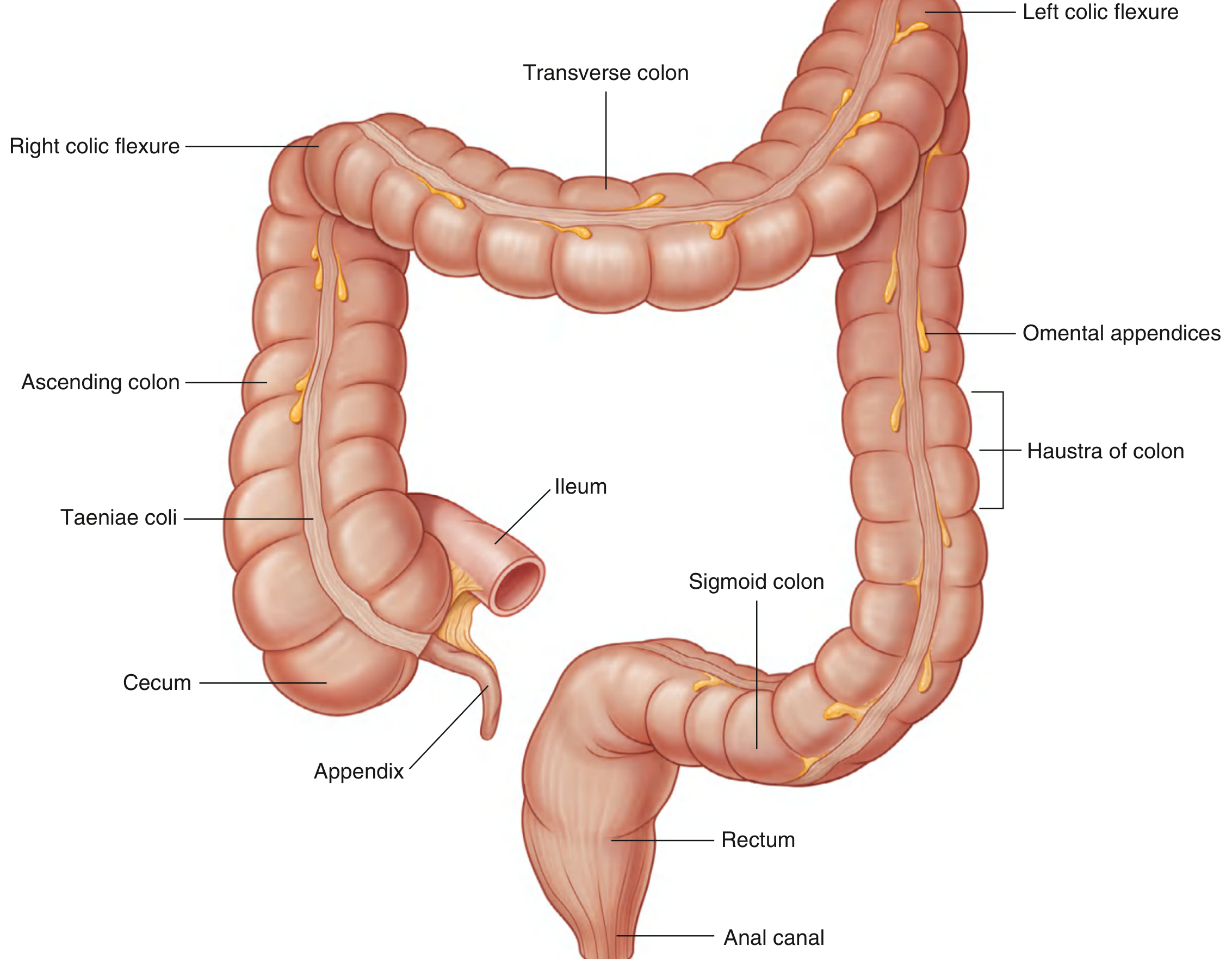
The large intestine extends from the distal ileum to the anus, approximately 1.5 m in adults. Its primary function is to absorb fluids and salts from gut contents and form feces. It consists of the cecum, appendix, colon, rectum, and anal canal.
Distinguishing Gross Features of the Large Intestine (vs. Small Intestine)
- Larger internal diameter compared to the small intestine
- Taeniae coli - three narrow bands of longitudinal muscle running along the wall (most visible on cecum and colon; less so on rectum); the free taenia leads directly to the base of the appendix
- Haustra of the colon - sacculations (pouches) of the colon wall between the taeniae
- Omental appendices (epiploic appendages) - peritoneum-covered fat tags attached to the outer surface of the colon
Parts of the Large Intestine
1. Cecum
- Lies in the right iliac fossa, inferior to the ileocecal opening
- Generally intraperitoneal (mobile, no mesentery)
- The first part of the large intestine; may descend into the true pelvis
- The appendix attaches to its posteromedial wall, just inferior to the ileal opening
2. Appendix
- Narrow, hollow, blind-ended tube with a large amount of lymphoid tissue in its walls
- Suspended from the terminal ileum by the mesoappendix (containing the appendicular vessels)
- Variable position: retrocecal (most common), pelvic/descending, subcecal, pre-ileal, post-ileal
- McBurney's point = junction of lateral and middle 1/3 of a line from the ASIS to the umbilicus (surface projection of the appendix base; tender in appendicitis)
3. Colon
| Segment | Peritoneal Relations | Location | Key Feature |
|---|---|---|---|
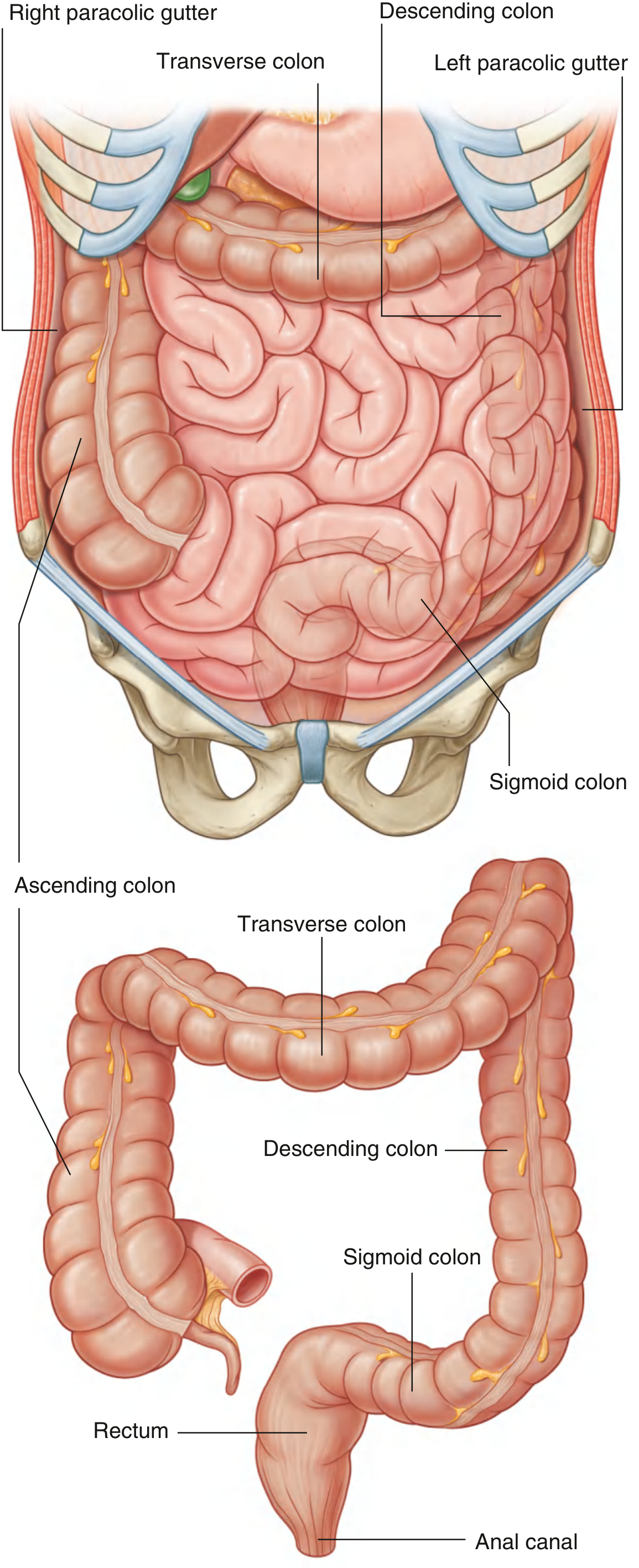
| Ascending colon | Retroperitoneal (secondarily) | Right flank, right iliac fossa to liver | Bends at right colic (hepatic) flexure |
| Transverse colon | Intraperitoneal (has transverse mesocolon) | Crosses abdomen from right to left | Suspended by transverse mesocolon |
| Descending colon | Retroperitoneal (secondarily) | Left hypochondrium to left iliac fossa | Bends at left colic (splenic) flexure (higher and more posterior than hepatic flexure; attached to diaphragm by phrenicolicligament) |
| Sigmoid colon | Intraperitoneal (has sigmoid mesocolon) | Left iliac fossa to S2 level | S-shaped; mobile; ends at rectum |
Blood supply:
- Ascending and transverse colon: Superior mesenteric artery (SMA) - via ileocolic, right colic, and middle colic arteries
- Descending and sigmoid colon: Inferior mesenteric artery (IMA) - via left colic and sigmoid arteries
- Anastomotic connection along the entire colon = marginal artery (of Drummond)
4. Rectum
- Begins at the rectosigmoid junction (level of S3 or end of sigmoid mesocolon)
- Retroperitoneal structure (no mesentery)
- Follows the sacral curvature; wider ampulla stores feces before defecation
- Blood supply: Superior rectal artery (from IMA), middle rectal artery (from internal iliac), inferior rectal artery (from internal pudendal)
5. Anal Canal
- Terminal part of the large intestine; continuation of rectum inferiorly
Summary Comparison Table: Small vs. Large Intestine
| Feature | Small Intestine | Large Intestine |
|---|---|---|
| Length | ~5-7 m | ~1.5 m |
| Diameter | Smaller (~2.5-3 cm) | Larger (~6-8 cm) |
| Taeniae coli | Absent | Present (3 bands) |
| Haustra | Absent | Present |
| Omental appendices | Absent | Present |
| Plicae circulares | Prominent (esp. jejunum) | Absent |
| Peyer's patches | Present (esp. ileum) | Absent |
| Mesentery | Yes (jejunum + ileum); retroperitoneal (duodenum) | Mixed (see above) |
| Function | Digestion and absorption | Water/salt absorption; feces formation |
Sources:
- Gray's Anatomy for Students, pp. 379-385 (Large Intestine section)
- Mulholland and Greenfield's Surgery: Scientific Principles and Practice, 7e, pp. 2362-2368 (Small Intestine Gross Anatomy and Embryology)
This is a shared conversation. Sign in to Orris to start your own chat.