
Free radicals definition generation function disease
I'll look this up in the medical library right away.
Searching sources...
Reading File
Now I'll search the library in parallel for all aspects of free radicals.
Finding Sources
Finding Sources
Finding Sources
Excellent - found rich sources. Now I'll read the key sections from Robbins Pathology (most comprehensive), Basic Medical Biochemistry, and Tietz in parallel.
Reading File
Reading File
Reading File
Now let me get the Robbins sections on generation, removal, and pathologic effects, plus the diseases table from Basic Medical Biochemistry.
Reading File
Reading File
Reading File
I now have comprehensive content from Robbins Pathology, Basic Medical Biochemistry, and Tietz. Let me also fetch the key diagrams and get the diseases table.
Reading File
Loading Image
Loading Image
I now have all the content needed for a thorough answer. Let me also fetch the Robbins ROS generation diagram.
Loading Image
Searching Images
free radicals reactive oxygen species ROS generation cell injury diagram
I now have everything needed for a comprehensive, well-illustrated answer. Here it is:
Free Radicals: Definition, Generation, Function, and Disease
1. Definition
A free radical is any chemical species - atom, ion, or molecule - capable of independent existence that contains one or more unpaired electrons in an outer orbital. The unpaired electron makes these species highly reactive; they "attack" and modify adjacent molecules (proteins, lipids, carbohydrates, nucleic acids) and can trigger autocatalytic chain reactions that propagate damage.
"A free radical is a radical that is capable of independent existence. Radicals are highly reactive and initiate chain reactions by extracting an electron from a neighboring molecule to complete their own orbitals."
- Basic Medical Biochemistry: A Clinical Approach, 6e
Reactive Oxygen Species (ROS) are the most important class of free radicals in biology - oxygen-containing compounds that are highly reactive radicals (or are readily converted to them in cells).
Key ROS species:
| Species | Symbol | Notes |
|---|---|---|
| Superoxide anion | O₂⁻ | 1-electron reduction of O₂; still a radical |
| Hydrogen peroxide | H₂O₂ | Not a radical itself, but converts to OH• |
| Hydroxyl radical | OH• | Most reactive; principal ROS causing damage |
| Peroxynitrite | ONOO⁻ | From O₂⁻ + NO; no enzymatic inactivation |
| Nitric oxide | NO• | Radical; can convert to ONOO⁻ |
The stepwise 4-electron reduction of O₂ to water generates these intermediates:
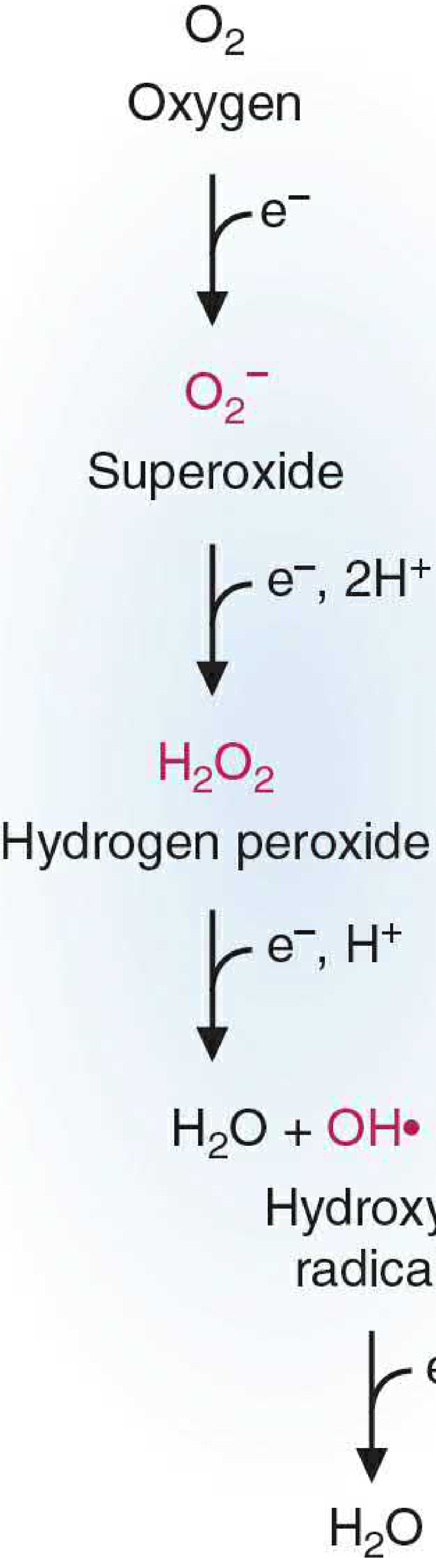
Figure: Four one-electron reduction steps for O₂ progressively generate superoxide → hydrogen peroxide → hydroxyl radical → water (Basic Medical Biochemistry, 6e)
2. Generation of Free Radicals
Free radicals are generated through multiple pathways:
A. Normal Metabolic Processes (Mitochondrial Respiration)
During oxidative phosphorylation, O₂ is reduced by transfer of four electrons to H₂ to generate water. Small amounts of partially reduced intermediates (O₂⁻, H₂O₂, OH•) are produced as unavoidable byproducts. Most intracellular ROS originate in the mitochondria via the electron transport chain.
B. Redox Reactions in the ER, Cytosol, and Peroxisomes
Oxidative enzymes in multiple compartments (e.g., xanthine oxidase, cytochrome P450) generate O₂⁻ during enzymatic cycling. Xanthine oxidase is especially important in ischemia-reperfusion injury - during ischemia it accumulates xanthine, then upon reperfusion generates a burst of H₂O₂ and O₂⁻.
C. Absorption of Radiant Energy
Ionizing radiation (UV light, X-rays) hydrolyzes water into •OH and hydrogen (H•) free radicals.
D. Activated Leukocytes (Respiratory Burst / Oxidative Burst)
Neutrophils and macrophages use NADPH oxidase to generate rapid bursts of O₂⁻ during phagocytosis - an essential weapon for killing bacteria and fungi. Myeloperoxidase further converts H₂O₂ to hypochlorite (OCl⁻), an even more potent bactericidal agent.
E. Transition Metals (Fenton Reaction)
Iron (Fe²⁺) and copper catalyze free radical formation:
H₂O₂ + Fe²⁺ → Fe³⁺ + •OH + OH⁻ (Fenton reaction)
Superoxide helps regenerate Fe²⁺ from Fe³⁺, allowing the cycle to continue. Storage proteins (transferrin, ferritin, ceruloplasmin) normally sequester these metals to limit this reaction.
F. Enzymatic Metabolism of Exogenous Chemicals
Drugs and toxins (e.g., CCl₄ → •CCl₃) are metabolized by cytochrome P450 into reactive radicals that cause direct cellular damage (e.g., centrilobular hepatic necrosis from CCl₄).
G. Nitric Oxide
NO is itself a free radical (one unpaired electron). Macrophages, endothelium, and neurons generate NO via NO synthase. At high concentrations it reacts with O₂⁻ to form the very damaging peroxynitrite (ONOO⁻).
3. Function of Free Radicals (Physiologic Roles)
Free radicals are not merely damaging - they have essential physiologic roles:
| Function | Mechanism |
|---|---|
| Antimicrobial defense | Neutrophils/macrophages use ROS (O₂⁻, OCl⁻, OH•) to kill ingested pathogens (oxidative burst) |
| Signal transduction | ROS act in NF-κB, MAPK, and other signaling pathways regulating immunity, cell growth, and development |
| Gene expression | Free radicals alter gene expression in both protective and deleterious ways (mitogenic effects) |
| Vasodilation | NO (a radical) relaxes vascular smooth muscle, regulates blood pressure |
| Apoptosis signaling | Controlled ROS production can trigger programmed cell death to eliminate damaged or infected cells |
"Free radicals have important functions in the body: defense against micro-organisms, signal transduction (NF-κB pathways), and mitogenic effects."
- Tietz Textbook of Laboratory Medicine, 7e
4. Removal / Defense Against Free Radicals
Cells maintain a steady state where low-level free radicals exist but cause no damage via multiple defense mechanisms:
Antioxidant Enzymes
| Enzyme | Location | Reaction |
|---|---|---|
| Superoxide dismutase (SOD) | Mitochondria (Mn-SOD), Cytoplasm (Cu/Zn-SOD) | 2O₂⁻ + 2H⁺ → H₂O₂ + O₂ |
| Catalase | Peroxisomes | 2H₂O₂ → O₂ + 2H₂O |
| Glutathione peroxidase | Cytosol, mitochondria | H₂O₂ + 2GSH → GSSG + 2H₂O |
| Peroxiredoxins | Cytosol, mitochondria | Inactivate ONOO⁻ → HNO₂ |
The ratio of oxidized glutathione (GSSG) to reduced glutathione (GSH) reflects the cell's oxidative state and antioxidant capacity.
Non-Enzymatic Antioxidants
- Vitamin E (α-tocopherol) - lipid-soluble; protects membranes
- Vitamin C (ascorbic acid) - water-soluble; scavenges ROS in cytosol
- Vitamin A / β-carotene - lipid-soluble
- Glutathione - cytosolic scavenger
- Urate, albumin, transferrin, ceruloplasmin - bind metals and scavenge radicals
- Plant flavonoids
Metal Sequestration
Transferrin, ferritin, lactoferrin, and ceruloplasmin bind iron and copper under normal conditions, preventing Fenton reaction-mediated radical generation.
5. Pathologic Effects of Free Radicals
Three key mechanisms of cellular injury (illustrated below):
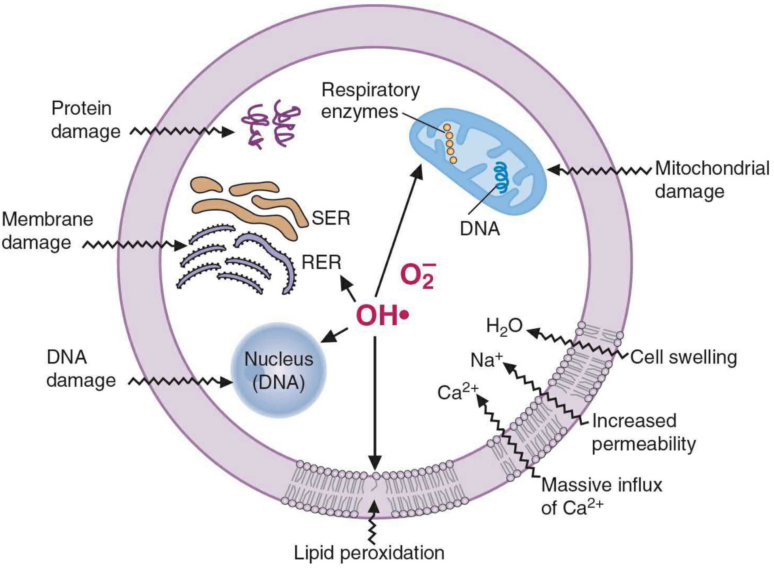
Figure: Free radical-mediated cellular injury showing lipid peroxidation of membranes, protein oxidation, and DNA strand breaks, with Ca²⁺ influx and mitochondrial damage (Basic Medical Biochemistry, 6e)
A. Lipid Peroxidation
Free radicals (especially OH•) attack the double bonds of unsaturated fatty acids in membrane phospholipids. This generates lipid peroxy radicals and peroxides in an autocatalytic chain reaction ("propagation"). Products include malondialdehyde (detectable in blood/urine as a biomarker of oxidative stress), ethane, and pentane. Result: membrane disruption, increased permeability, massive Ca²⁺ influx, and cell death.
B. Oxidative Modification of Proteins
ROS cause:
- Oxidation of amino acid side chains (especially cysteine sulfhydryls)
- Protein-protein cross-linking (disulfide bonds)
- Oxidation of the protein backbone
- Inactivation of enzyme active sites
- Conformational change in structural proteins
- Enhanced proteasomal degradation of misfolded proteins
C. DNA Damage
Free radicals cause:
- Single- and double-strand breaks in DNA
- DNA strand cross-linking
- Formation of base adducts (oxidized bases)
- Oxidative DNA damage implicated in cell aging and malignant transformation
"Evidence of free-radical damage has been described in >100 disease states."
- Basic Medical Biochemistry: A Clinical Approach, 6e
6. Diseases Associated with Free Radicals
Free radical damage (oxidative stress) contributes to a wide spectrum of diseases:
| Disease | Mechanism of Free Radical Involvement |
|---|---|
| Atherosclerosis / Ischemic heart disease | LDL oxidation, endothelial damage, plaque formation |
| Myocardial infarction | Ischemia-reperfusion injury: xanthine oxidase generates burst of ROS upon reoxygenation |
| Cancer | ROS-induced DNA mutations causing malignant transformation |
| Alzheimer disease | Oxidative damage to neurons; ROS-mediated amyloid toxicity |
| Parkinson disease | Dopaminergic neuron vulnerability; inability to convert tyrosine to DOPA |
| Amyotrophic lateral sclerosis (ALS) | Familial form: SOD mutations → impaired superoxide disposal → motor neuron death |
| Diabetes mellitus | Hyperglycemia-driven ROS generation damages vascular endothelium |
| Age-related macular degeneration | Oxidative damage to retinal pigment epithelium (RPE) → progressive vision loss |
| Chronic granulomatous disease | Deficient NADPH oxidase → inadequate ROS for microbial killing → recurrent infections |
| Stroke | Ischemia-reperfusion; phagocytes clearing dead tissue release ROS, damaging surrounding viable neurons |
| Rheumatoid arthritis / inflammatory diseases | Neutrophil/macrophage ROS release during inflammation damages surrounding tissue |
| Chemical/drug toxicity | CCl₄, acetaminophen, etc. metabolized to reactive free radicals causing organ damage |
| Radiation injury | Ionizing radiation generates OH• from water hydrolysis |
| Aging | Cumulative oxidative damage to proteins, lipids, and DNA ("free radical theory of aging") |
"In a number of disease states, free-radical release by neutrophils or macrophages during inflammation contributes to injury in the surrounding tissues. During stroke or myocardial infarction, phagocytic cells that move into the ischemic area to remove dead cells may release excessive ROS."
- Basic Medical Biochemistry: A Clinical Approach, 6e
Summary
| Aspect | Key Points |
|---|---|
| Definition | Chemical species with unpaired electron(s); highly reactive; capable of independent existence |
| Key types | O₂⁻ (superoxide), H₂O₂, OH• (hydroxyl), ONOO⁻ (peroxynitrite), NO• |
| Generation | Mitochondrial respiration, NADPH oxidase (leukocytes), Fenton reaction (Fe²⁺/Cu), radiation, xenobiotics |
| Functions | Microbial killing, signal transduction (NF-κB), apoptosis, vasodilation (NO) |
| Defense | SOD, catalase, glutathione peroxidase; vitamins E/C; metal sequestration proteins |
| Pathologic effects | Lipid peroxidation, protein oxidation, DNA strand breaks |
| Diseases | Atherosclerosis, cancer, neurodegeneration, ischemia-reperfusion, aging, CGD, ALS |
Sources:
- Robbins, Cotran & Kumar Pathologic Basis of Disease (pp. 64-66)
- Basic Medical Biochemistry: A Clinical Approach, 6e (Ch. 25)
- Tietz Textbook of Laboratory Medicine, 7e (pp. 1267-1268)
This is a shared conversation. Sign in to Orris to start your own chat.