


df7538f8-08cf-4a64-8827-5deaa3da3da2.jpeg
+2 more
Fibroadenoma of breast make it a perfect mbbs university exam answer with required subheading answer for 5 marks questions
Here is a complete, exam-ready 5-mark answer on Fibroadenoma of the Breast, structured for MBBS university examinations:
FIBROADENOMA OF THE BREAST
Definition
Fibroadenoma is the most common, benign fibroepithelial neoplasm of the female breast. It shows biphasic proliferation of both epithelial and stromal components, arising from the intralobular stroma.
Epidemiology / Age Group
- Most common in females between 20-30 years of age.
- Growth is hormonally influenced - tumors may enlarge rapidly during pregnancy and regress after menopause.
Clinical Features
- Presents as a palpable, freely movable, well-circumscribed mass.
- The characteristic mobility on examination earns it the name "breast mouse."
- Usually solitary, but may be multiple or bilateral.
- Non-tender, smooth, rubbery consistency.
Morphology
Gross Appearance
| Feature | Description |
|---|---|
| Shape | Solid, round to ovoid |
| Circumscription | Well-circumscribed, not fixed |
| Cut section | Uniform, rubbery, lobulated, glistening grayish-white nodule |
| Slit-like spaces | Present on cut surface |
| Size | Usually 1-4 cm in diameter |
The tumor compresses surrounding breast tissue but does not invade it.
Microscopy
Fibroadenomas contain a mixture of:
-
Neoplastic epithelial component - Ducts lined by a double layer of epithelium ranging from normal lobules to varying degrees of hyperplasia; ducts may be simple/round or elongated/branching.
-
Neoplastic stromal component - Fibrous connective tissue stroma that is uniform, delicate, myxoid, and of low cellularity; resembles normal intralobular (peritubular) stroma; contains spindle cells without atypia.
Growth Patterns (Two Types)
- Pericanalicular pattern: Epithelium forms round or oval tubules with patent lumen; proliferating stromal cells grow around open ducts in a circumferential fashion.
- Intracanalicular pattern: Stromal growth compresses ducts into curvilinear slits (clefts); ducts appear distorted.
Both patterns may coexist and have no prognostic significance.
Hormonal Changes
- Pregnancy: Epithelial component may show lactational changes; glands increase in size.
- Post-menopause: Stroma becomes more fibrous and densely hyalinized; may calcify forming "popcorn" calcifications on mammography.
Variants
| Variant | Key Feature |
|---|---|
| Juvenile fibroadenoma | Most common in adolescent girls; grows rapidly; may reach 20 cm; stroma is more cellular; does not recur after excision |
| Tubular adenoma | Closely packed tubular structures with small lumina; very scanty stroma |
| Lactating adenoma | Occurs during pregnancy/postpartum; composed of normal-appearing breast acini with secretory activity |
Malignant Potential
- Malignant transformation is extremely rare (<0.3%).
- Fibroadenoma is considered a benign lesion with an excellent prognosis.
Treatment
- Surgical excision (lumpectomy) is the treatment of choice.
- Does not recur after complete excision.
Exam Tip: Remember the key triad - young woman + freely mobile breast lump ("breast mouse") + biphasic (epithelial + stromal) histology = Fibroadenoma.
Addison
I'll read the medical library skill first, then pull the best textbook content on Addison's disease for you.
Searching sources...
Reading File
Finding Sources
Finding Sources
Robbins Pathology has a dedicated section. Let me read that plus Costanzo Physiology for clinical/lab content.
Reading File
Reading File
Reading File
I now have comprehensive content from Robbins Pathology and Costanzo Physiology. Let me fetch the histology image too.
Loading Image
Excellent - I have everything needed. Here is your complete exam answer:
ADDISON'S DISEASE (PRIMARY CHRONIC ADRENOCORTICAL INSUFFICIENCY)
Definition
Addison's disease is a primary chronic adrenocortical insufficiency caused by progressive destruction of the adrenal cortex, resulting in deficiency of glucocorticoids (cortisol), mineralocorticoids (aldosterone), and adrenal androgens.
First described by Thomas Addison in 1855 as a syndrome of "general languor and debility, remarkable feebleness of the heart's action, and peculiar changes in the skin."
Etiology / Causes
More than 90% of cases are due to four major causes:
| Cause | Frequency | Notes |
|---|---|---|
| Autoimmune adrenalitis | 80-90% (developed countries) | Autoantibodies to 21-hydroxylase and 17-hydroxylase |
| Tuberculosis | Previously 90%; now less common | Granulomatous destruction; adrenals enlarged and caseous |
| Metastatic carcinoma | Less common | Lung and breast carcinoma commonest sources |
| AIDS / Fungal infections | Growing cause | CMV adrenalitis, histoplasmosis |
| Rare causes | Amyloidosis, sarcoidosis, hemochromatosis, genetic (ABCD1/adrenoleukodystrophy, NROB1 mutations) | - |
Autoimmune Polyendocrinopathy Syndromes (APS)
- APS-1: Adrenalitis + hypoparathyroidism + mucocutaneous candidiasis; caused by AIRE gene mutation (chr 21q22)
- APS-2 (Schmidt syndrome): Adrenal insufficiency + autoimmune thyroiditis ± T1D; starts in 4th decade
- APS-4: Adrenalitis + gastritis/vitiligo/pernicious anemia but no thyroiditis or T1D
Pathogenesis
- Autoimmune destruction of steroidogenic cells in all three zones of the adrenal cortex (zona glomerulosa, fasciculata, reticularis)
- Results in loss of all three classes of adrenocortical hormones
- Low cortisol removes negative feedback on the hypothalamo-pituitary axis → ACTH rises markedly
- ACTH is derived from POMC, which also yields melanocyte-stimulating hormone (MSH) - this causes the characteristic hyperpigmentation
Morphology
Gross
- Autoimmune adrenalitis: Irregularly shrunken glands, difficult to identify within perirenal fat
- Tuberculous / fungal: Adrenals enlarged, architecture effaced by granulomatous inflammation
- Metastatic: Adrenals enlarged, normal architecture obscured by infiltrating neoplasm
Microscopy
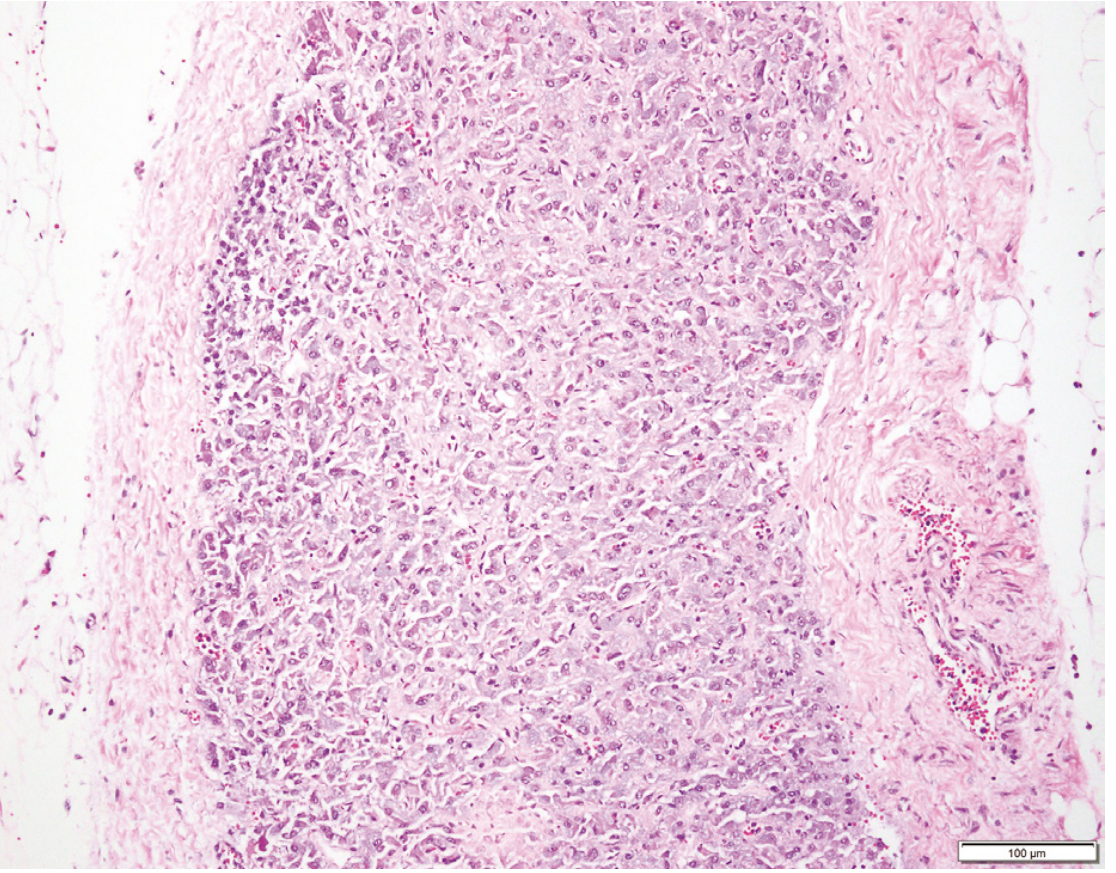
Autoimmune adrenalitis (Robbins Pathology): Adrenal cortex is completely replaced by connective tissue with a collapsed reticulin framework. Only scattered residual cortical cells remain. Lymphoid infiltrate present in the cortex extending to adjacent medulla. The medulla itself is largely preserved.
- Tuberculous cases: Classic caseating granulomas
- Metastatic cases: Neoplastic cells replacing cortical tissue
Clinical Features
Symptoms appear insidiously, only when >90% of cortex is destroyed.
Due to Glucocorticoid (Cortisol) Deficiency
- Progressive weakness and easy fatigability (most common initial symptom)
- Anorexia, nausea, vomiting, weight loss, diarrhea
- Hypoglycemia (loss of gluconeogenic effect)
Due to Mineralocorticoid (Aldosterone) Deficiency
- Hyponatremia and hyperkalemia (Na loss, K retention)
- Metabolic acidosis
- Hypotension and volume depletion
- Salt craving
Due to ACTH / MSH Excess (Pathognomonic)
- Hyperpigmentation - most characteristic sign
- Affects sun-exposed areas, pressure points (neck, elbows, knees, knuckles), nail beds, nipples, areolae, buccal mucosa, and recent scars
- Absent in secondary adrenal insufficiency (pituitary/hypothalamic disease) - key differentiating point
Due to Androgen Deficiency (in females)
- Decreased pubic and axillary hair
- Decreased libido
Laboratory Findings
| Test | Finding |
|---|---|
| Serum cortisol | Decreased |
| Serum ACTH | Markedly elevated (primary) |
| Serum sodium | Low (hyponatremia) |
| Serum potassium | High (hyperkalemia) |
| Blood glucose | Low (hypoglycemia) |
| ACTH stimulation test (cosyntropin test) | No rise in cortisol - confirms primary adrenal failure |
| Autoantibodies | Anti-21-hydroxylase antibodies positive |
Key differentiator: In secondary adrenal insufficiency (pituitary failure), ACTH is low and there is NO hyperpigmentation.
Addisonian Crisis (Acute Adrenal Insufficiency)
- Precipitated by stress (infection, surgery, trauma) in a patient with chronic insufficiency
- Features: Severe hypotension/shock, vomiting, abdominal pain, fever, profound hyponatremia, hyperkalemia
- Medical emergency - treat with IV hydrocortisone immediately
Treatment
- Replacement therapy: Oral hydrocortisone (glucocorticoid) + fludrocortisone (mineralocorticoid)
- Dose must be doubled or tripled during illness/stress ("sick day rules")
- Patient education regarding crisis prevention
Exam Tip - Key triad of Addison's disease: Hyperpigmentation + Hypotension + Hyponatremia/Hyperkalemia. Remember: HIGH ACTH = hyperpigmentation = PRIMARY adrenal failure. Low ACTH = no pigmentation = SECONDARY failure.
Sources: Robbins & Cotran Pathologic Basis of Disease; Costanzo Physiology 7th Edition
This is a shared conversation. Sign in to Orris to start your own chat.