
Investigations in thyroid swelling
"thyroid nodule"[MeSH Terms] AND "diagnosis"
thyroid nodule ultrasound malignant microcalcification
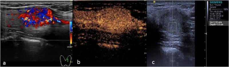
This composite diagnostic image illustrates the multimodal ultrasound evaluation of Papillary Thyroid Carcinoma. Panel (a) is a two-dimensional B-mode and Color Doppler ultrasound showing a hypo-echoic, irregular thyroid nodule containing internal microcalcifications, appearing as punctate hyperechoic foci. Panel (b) presents Contrast-Enhanced Ultrasound (CEUS) findings, demonstrating heterogeneous low enhancement within the lesion compared to the surrounding thyroid parenchyma, suggesting irregular vascularization and complex tissue composition. Panel (c) shows Virtual Touch Tissue Quantification using Acoustic Radiation Force Impulse (ARFI) imaging to measure Shear Wave Velocity (SWV). The panel displays a region of interest (ROI) centered on the nodule to assess tissue stiffness. These imaging modalities are combined to differentiate malignant from benign thyroid nodules by evaluating echogenicity, microcalcification presence, vascular perfusion patterns, and mechanical tissue elasticity (stiffness).
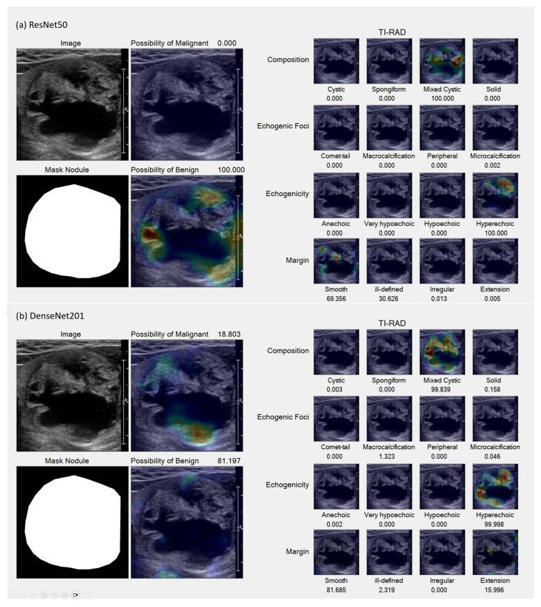
This composite diagnostic image displays ultrasound scans of a thyroid nodule analyzed by two deep learning models, ResNet50 (top) and DenseNet201 (bottom), for TI-RADS classification. The panel includes original grayscale B-mode images, binary nodule masks, and Grad-CAM heatmaps showing regions of interest for benign versus malignant predictions. The nodule is characterized by a mixed solid-cystic composition with hyperechoic regions and largely smooth margins, leading to a TI-RADS 2 (benign) classification. Detailed heatmap sub-panels evaluate specific ACR TI-RADS categories: Composition (Cystic, Spongiform, Mixed Cystic, Solid), Echogenicity (Anechoic, Hypoechoic, Hyperechoic), Margin (Smooth, Ill-defined, Irregular, Extension), and Echogenic Foci (Macrocalcification, Microcalcification). Each sub-panel provides a quantitative confidence score for the feature identification. The Grad-CAM overlays illustrate that the models focus on the internal solid components and the interface of the nodule borders to differentiate benign and malignant features, demonstrating the utility of explainable AI in radiological decision-making for thyroid oncology.
Bethesda System thyroid cytopathology 2023 categories risk of malignancy ATA 2025 thyroid nodule guideline
Investigations in a thyroid swelling
1. Biochemical (thyroid function)
- Serum TSH is the first-line test. If it is abnormal, add free T4 and T3.
- Low TSH suggests thyrotoxicosis, including an autonomous ("hot") nodule.
- High TSH suggests hypothyroidism, such as Hashimoto thyroiditis.
- Free T4 and free T3 confirm and grade dysfunction. Total T3 matters in T3 thyrotoxicosis, where T4 is normal (Schwartz's Principles of Surgery, 11th ed., p. 1661). Total T4 is affected by binding proteins, for example in pregnancy, with oestrogen use or in nephrotic syndrome, so free T4 is more reliable.
- Thyroid autoantibodies (anti-TPO and anti-thyroglobulin) point to autoimmune thyroiditis. About 80% of patients with Hashimoto thyroiditis have raised levels. They do not determine thyroid function (Schwartz, p. 1661).
- TSH receptor antibodies (TRAb/TSI) are used for Graves disease.
- Serum thyroglobulin is not a diagnostic test for a swelling. It is a tumour marker for follow-up of differentiated cancer after total thyroidectomy and radioiodine ablation. Anti-Tg antibodies interfere with the assay, so measure them at the same time (Schwartz).
- Serum calcitonin (CEA is an alternative) is optional. Use it if medullary carcinoma is suspected, for example with a family history, MEN 2, or an FNAC result suggesting MTC.
- Corrected serum calcium is optional. It is useful if parathyroid disease is possible and before surgery.
- Phaeochromocytoma screening (plasma or urinary metanephrines) is needed before surgery if MTC or MEN 2 is suspected.
2. Imaging
- It describes the number, size, shape, margins, vascularity and calcification of nodules.
- It assesses the regional lymph nodes, especially in the lateral neck.
- It guides FNA, which is more accurate than free-hand sampling.
- It involves no ionising radiation and is cheap and non-invasive.
- It cannot see well behind the sternum.
- microcalcification
- irregular border
- increased intranodular flow on Doppler
- hypoechogenicity
- absent halo
- a taller-than-wide shape (from ATA/TI-RADS rather than this box)
- simple cyst with no solid component
- comet-tail artefact (colloid)
- honeycomb or spongiform appearance
- It is indicated mainly when TSH is suppressed, to see whether a nodule is hot (autonomous, rarely malignant) or cold.
- Cold nodules carry a higher malignancy risk, but most cold nodules are benign, so the scan is not a good test for cancer.
- Bailey and Love also list it when a discrete swelling and toxicity coexist.
- It can also show retrosternal goitre and ectopic thyroid tissue.
- retrosternal goitre, to define its extent
- invasive or fixed tumours
- known cancer, to stage nodes and look for mediastinal and lung disease
- some reoperations
3. Cytology and histology
| Category | Typical ROM | Usual action |
|---|---|---|
| I Non-diagnostic | Variable | Repeat FNA under ultrasound guidance |
| II Benign | Low (about 3%) | Clinical and ultrasound follow-up |
| III AUS (atypia of undetermined significance) | Intermediate | Repeat FNA, molecular testing or lobectomy |
| IV Follicular neoplasm | Higher | Molecular testing or diagnostic lobectomy |
| V Suspicious for malignancy | About 74% | Lobectomy or total thyroidectomy |
| VI Malignant | About 97% | Surgery |
- Follicular carcinoma cannot be diagnosed on FNAC, because it needs histology to show capsular or vascular invasion. Any follicular lesion therefore needs diagnostic lobectomy.
- Cytology can diagnose papillary, medullary and anaplastic carcinoma and lymphoma.
- Core biopsy is useful for suspected lymphoma or anaplastic carcinoma.
- Molecular testing on indeterminate cytology (BRAF, RAS, RET/PTC and others) helps with risk stratification.
- Diagnostic hemithyroidectomy gives the final histology when cytology is indeterminate.
4. Practical approach to a thyroid swelling
- Take the history and examine the patient, including the neck nodes. Look for risk factors: neck irradiation, family history, rapid growth, hoarseness, dysphagia, and a hard fixed nodule.
- Measure TSH.
- Low TSH: do a radionuclide scan. A hot nodule is treated as hyperthyroidism and usually does not need FNA.
- Normal or high TSH: do ultrasound, then FNA according to the risk grade and size.
- Add imaging as needed: CXR or CT for a retrosternal goitre or compressive symptoms, and calcitonin if MTC is suspected.
- Do laryngoscopy before surgery.