
Cleft lip and palate repair 20 mark answer
cleft lip and palate repair surgical technique
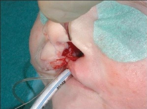
This intraoperative clinical photograph shows an intermediate stage of a cleft lip repair procedure, specifically focusing on the closure of the nasal floor and mucosal flaps. The surgical field encompasses the infant's midface, with the nostrils and upper lip area visible. Rectangular mucosal flaps have been elevated and are being approximated with sutures to reconstruct the nasal vestibule and floor. A surgical instrument, likely a small suction tip or probe, is positioned within the cleft, and a clear endotracheal tube is visible inferiorly, indicating the patient is under general anesthesia. The tissue shows surgical redness and minor bleeding consistent with active dissection. The pedagogical focus of the image is to demonstrate the primary suturing of mucosal layers in the 'lip adhesion' technique for managing complete cleft lip and palate, aiming to reduce tension on subsequent definitive repairs and facilitate nasal platform alignment.
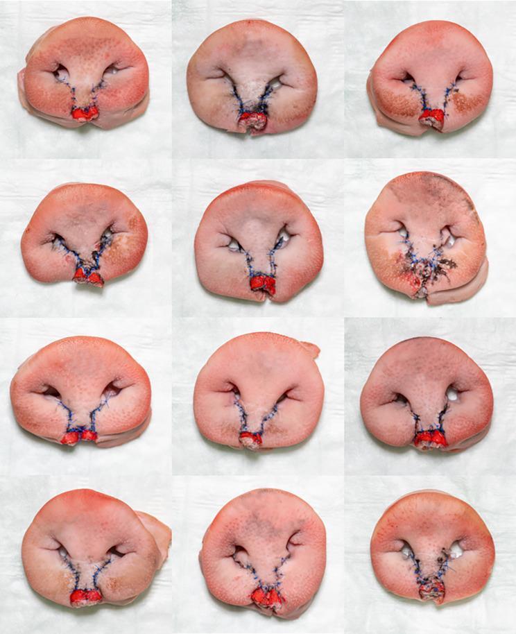
This clinical photograph displays a grid of 12 ex vivo porcine snout specimens, utilized as surgical training models for bilateral cleft lip repair using the Millard (rotation-advancement) technique. Each specimen serves as a biological simulator for human craniofacial surgery, specifically recreating the anatomical challenges of a bilateral cleft lip and palate. The images show the post-operative outcomes following surgical reconstruction. Key features across the models include the central prolabial flap, reconstruction of the vermillion border (highlighted in red), and surgical approximation of the lateral lip elements. Blue non-absorbable sutures are visible along the incision lines, which follow the characteristic Millard pattern to reconstruct the philtral columns and Cupid’s bow. Variations in tissue handling, suture tension, and anatomical symmetry are evident across the samples, demonstrating the range of results in a training cohort. The models illustrate essential concepts in reconstructive plastic surgery, including skin flap rotation, muscle approximation (orbicularis oris), and vermillion border alignment.
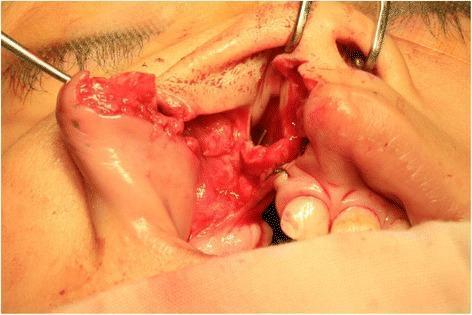
This clinical photograph provides an intraoperative view of a surgical procedure for unilateral cleft lip and palate repair. The image focuses on the reconstruction of the nasal floor and oral lining. Visible is the lateral mucosal flap, pedicled at the root of the pyriform aperture, being rotated medially by 90 degrees to cover the oral side of the alveolar cleft. Surgical retractors and sutures are visible, used to stabilize the elevated mucosal flaps and approximate them to the tissue cuff of the gingivopalatal mucosa. The surgical site demonstrates the anatomical landmarks of the nasolabial region, including the nostril, alveolar segments, and exposed primary palate. This image illustrates a critical step in double-layered nasal floor reconstruction, aimed at closing the communication between the oral and nasal cavities and creating a sound anatomical barrier using local mucosal flaps.
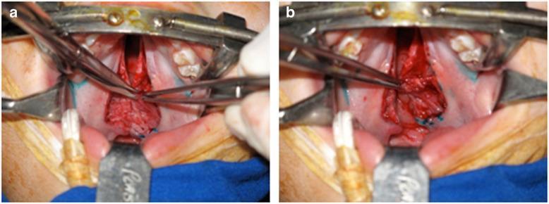
Two intraoperative clinical photographs (a and b) demonstrating a palatoplasty procedure, specifically a modified von Langenbeck repair for cleft lip and palate (CL/P). The images show the oral cavity maintained by a Dingman mouth gag, with the patient in the Rose position and an endotracheal tube visible. Image (a) depicts the exposure and dissection of the levator veli palatini muscle (LVPM), which appears as a distinct reddish muscular bundle being manipulated by surgical forceps. Blue marking ink delineates the planned incision lines on the palatal mucosa. Image (b) shows the surgical field after a small fragment (approximately 3–5 mm³) of the LVPM has been harvested for stem cell culture. The harvest site is visible, with surgical sutures used for intravelar veloplasty or closure of the donor site. These images illustrate the technique of harvesting autologous muscle tissue during secondary palatal reconstruction for use in regenerative medicine research, specifically for isolating levator veli palatini muscle-derived stromal cells (LVPMDSCs).
Cleft Lip and Palate - Repair
1. Introduction
2. Incidence and Distribution
- Cleft lip alone: 15%
- Cleft lip + palate: 45%
- Isolated cleft palate: 40%
3. Aetiology
- Genetic: First-degree family history significantly raises recurrence risk. Mutations affecting IRF6, TP63, BMP4, and FGFR genes are implicated.
- Environmental/maternal factors:
- Maternal smoking
- Alcohol use
- Anticonvulsant drugs (phenytoin, valproate, carbamazepine)
- Folic acid deficiency
- Teratogen exposure in first trimester
4. Classification
- Isolated cleft palate
- Cleft lip with or without alveolus or palate involvement (unilateral or bilateral)
- Laterality (left/right/bilateral)
- Completeness (complete vs. incomplete)
- Involvement (lip only, lip + alveolus, lip + alveolus + palate)
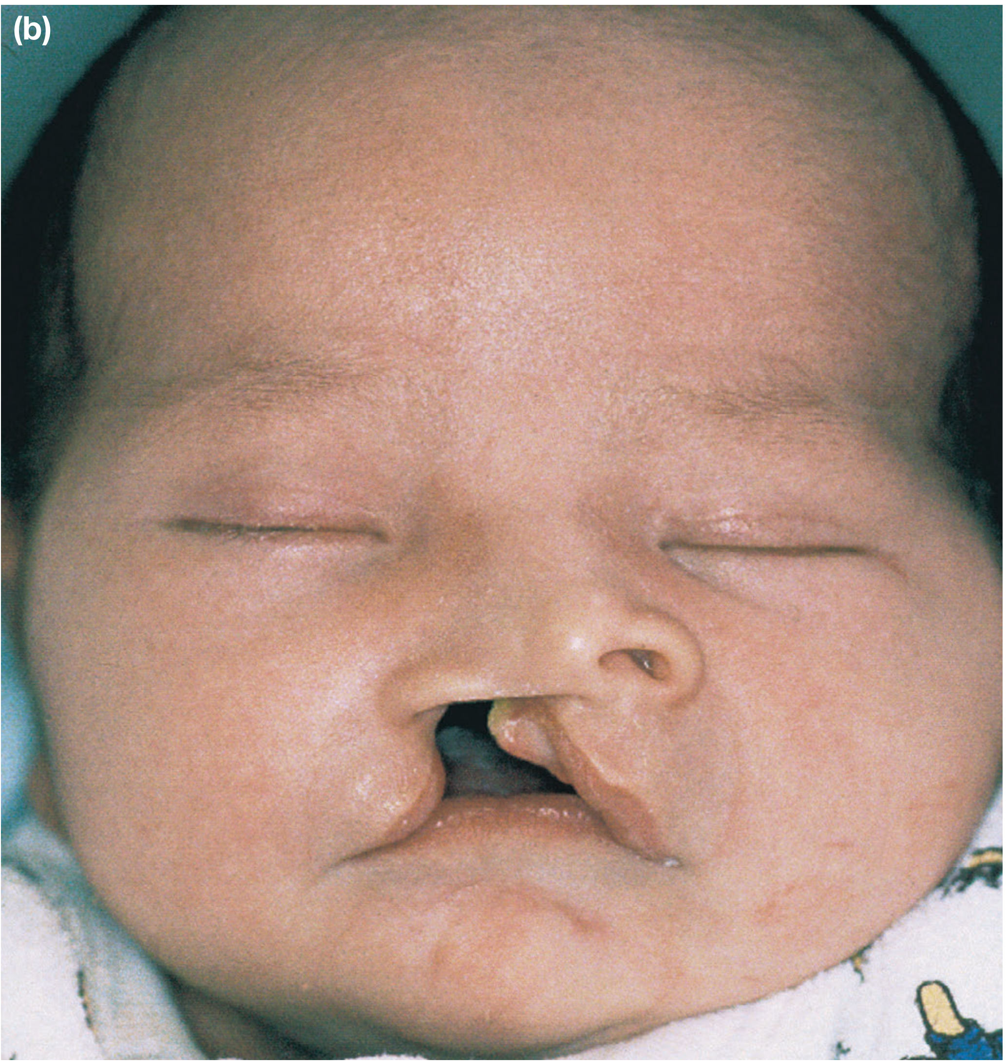
5. Pathological Anatomy
Cleft Lip Anatomy
- The orbicularis oris muscle is discontinuous and inserts abnormally onto the lateral pyriform aperture and columella, rather than crossing the midline
- In unilateral cleft: nasiolabial and bilabial muscle chains are disrupted unilaterally, causing nasal flaring on the affected side
- In bilateral cleft: disruption is symmetric, with a protrusive premaxilla, flared nostrils, and a skin island (the prolabium) in front of the premaxilla that is devoid of muscle
Cleft Palate Anatomy
- The levator veli palatini muscles insert abnormally anteriorly into the hard palate rather than forming a posterior transverse sling
- Normal levator sling is absent, compromising velopharyngeal closure
- Hard palate: three mucosal zones - central palatal fibromucosa (thin, nasal floor), maxillary fibromucosa (thick, contains greater palatine neurovascular bundle), and gingival fibromucosa
- In complete cleft palate, the median palatal vault is absent and palatal fibromucosa is reduced
6. Multidisciplinary Team (MDT)
| Team Member | Role |
|---|---|
| Cleft coordinator/administrator | Organises patient pathway and surgical episodes |
| Clinical Nurse Specialist (CNS) | First contact; feeding, airway, family support |
| Paediatrician | Manages associated medical conditions (cardiac, respiratory) |
| Plastic/maxillofacial surgeon | Primary and secondary surgical repair |
| Speech and Language Therapist (SLT) | Monitors speech development, diagnoses VPI |
| ENT/Audiologist | Hearing screening, management of OME |
| Paediatric dentist | Dental disease prevention |
| Orthodontist | Dentofacial growth monitoring, pre-surgical orthopaedics |
| Psychologist | Psychosocial support for child and family |
| Geneticist | Counselling where syndromic association is suspected |
7. Presurgical Preparation
Feeding
- Specialised squeeze bottles (e.g., Haberman feeder)
- Specially shaped teats
- Nasogastric tube in severe cases
Presurgical Infant Orthopaedics / Nasoalveolar Moulding (NAM)
- An individually moulded oral plate with nasal stents is fitted shortly after birth
- Worn full time, regularly adjusted to reshape nasal cartilages and columella
- Aims to reduce severity of the cleft deformity and improve nasal symmetry before surgery
- Evidence remains mixed, but positive results have been shown for improving nasal symmetry in unilateral CLP patients
- Lip adhesion: taping/surgical approximation of lateral lip segments to the premaxilla - controversial and not strongly supported in literature
8. Staged Surgical Management
| Age | Procedure |
|---|---|
| 3-6 months | Cleft lip repair ± primary nasal repair ± hard palate (vomerine flap) |
| 9-12 months | Cleft palate repair (soft palate ± hard palate) ± ventilation tubes |
| 4-5 years | Secondary speech surgery if VPI persists (pharyngoplasty) |
| 7-9 years | Alveolar bone graft (ABG) |
| 15-18 years | Secondary rhinoplasty |
| 16+ years | Orthognathic surgery if required |
9. Cleft Lip Repair (Cheiloplasty)
- Restore muscular continuity of orbicularis oris
- Reconstruct a symmetrical Cupid's bow with minimal scarring
- Achieve symmetrical nasal sill and philtral columns
- Avoid straight-line scars across the lip
Techniques for Unilateral Cleft Lip Repair
| Technique | Description |
|---|---|
| Millard rotation-advancement (most widely used) | Rotation flap from the philtral column medially + advancement flap from the lateral lip. Scar placed along the philtral column. Flexible, can be modified intraoperatively. |
| Tennison-Randall (triangular flap) | Geometric triangular flap inserted into the medial lip element to gain height. More mathematically precise, less adaptable. |
| Fisher anatomic subunit repair | Uses anthropometric landmarks to recreate philtral subunit anatomy. Increasingly popular in UK. |
| Rose-Thompson straight-line repair | Simple straight-line closure; only suitable for incomplete minor clefts. |
Bilateral Cleft Lip Repair
- More challenging due to absent muscle in prolabium and protrusive premaxilla
- Muscle is brought across from both lateral lip elements and sutured in the midline
- Modified Millard technique is commonly used
- Premaxillary setback (surgical or orthopaedic) may be needed preoperatively to reduce tension
10. Anterior Palate (Hard Palate) Closure
- In complete CLP, the anterior palate (primary palate) is closed at the time of lip repair using a single-layer vomerine mucosal flap
- The vomer flap is raised and sutured to the nasal mucosa of the opposite cleft edge
- Avoidance of denuded bone surfaces is important as periosteal stripping impairs maxillary growth
11. Cleft Palate Repair (Palatoplasty)
- Avoid straight-line closure on the soft palate
- Avoid or minimise lateral relaxing incisions over the hard palate (impairs growth)
- Meticulous technique, often under optical magnification
Techniques
a) Intravelar Veloplasty (IVVP) - Sommerlad technique
- Most common approach in the UK
- Radical dissection of the velar (levator veli palatini) muscles from their abnormal anterior insertions
- Muscles are reorientated transversely and sutured together in the midline to recreate an anatomical levator sling
- Nasal and oral mucosae are dissected free from the muscles and closed separately over the newly formed sling
- Associated with good speech outcomes
b) Furlow Double-Opposing Z-Plasty
- Uses two Z-plasties (one on oral surface, one on nasal surface) oriented in opposite directions
- Results in lengthening of the soft palate and posterior repositioning of levator muscles
- Useful where palatal length is deficient
- Associated with good speech outcomes; some degree of asymmetry
- No difference in audiological outcomes compared to IVVP
c) Von Langenbeck Repair
- Bipedicled mucoperiosteal flaps raised along the hard palate
- Oral and nasal mucosae closed in layers
- Lateral relaxing incisions used
- Risk of maxillary growth restriction due to lateral denuded bone
d) Veau-Wardill-Kilner (VWK) / V-Y Pushback
- Mucoperiosteal flaps are retracted posteriorly to lengthen the palate
- More bone left exposed - greater risk of growth impairment
- Largely abandoned in favour of IVVP
12. Primary Nasal Repair (Rhinoplasty)
- The cleft nose deformity is characterised by: asymmetric nasal tip, deviated septum, asymmetric nasal bones, displaced lower lateral cartilage on the affected side
- Primary rhinoplasty at the time of lip repair: repositions alar cartilages and nasal tip
- Definitive secondary rhinoplasty is deferred to adolescence (15-18 years) after facial skeletal growth is complete
- Evidence supports that primary rhinoplasty achieves stable long-term results with limited growth disturbance
13. Alveolar Bone Grafting (ABG)
- Performed at 7-9 years of age (mixed dentition phase), before eruption of permanent canine teeth
- Cancellous bone is harvested from the iliac crest (occasionally cranium or rib) and packed into the alveolar cleft
- Aims to:
- Provide bony continuity to the alveolar arch
- Support eruption of the permanent canine
- Stabilise the premaxilla in bilateral cases
- Allow later orthodontic treatment
14. Secondary Surgery and Long-Term Management
Velopharyngeal Incompetence (VPI)
- VPI arises when the repaired soft palate fails to achieve adequate velopharyngeal closure
- Presents as hypernasal speech, nasal air escape, and compensatory articulation errors
- Investigated with lateral videofluoroscopy and nasendoscopy
- Managed with:
- Re-repair / secondary palatoplasty (when levator muscle insertion is anterior)
- Pharyngoplasty: creates a partial obstruction at the velopharyngeal port
- Posterior pharyngeal flap: midline flap from posterior pharyngeal wall sutured to soft palate
- Sphincter pharyngoplasty: medial transposition of lateral pharyngeal wall flaps
- Normal resonance achieved in up to 85% of cases
- Main risk: obstructive sleep apnoea (must be counselled preoperatively)
Hearing and OME
- Eustachian tube dysfunction is common with palatal clefts due to abnormal tensor veli palatini function
- High incidence of otitis media with effusion (OME) - "glue ear"
- Regular audiological screening essential
- Ventilation tubes (grommets) placed at the time of palate repair if OME is significant
Dental Anomalies
- Hypodontia or hyperdontia, most commonly involving the maxillary lateral incisor at the cleft site
- Delayed eruption, morphological abnormalities
- Regular paediatric dental review and orthodontic monitoring
Orthognathic Surgery
- Approximately 25% of CLP patients develop midface hypoplasia (Class III skeletal pattern) due to:
- Scar tethering from previous repairs
- Intrinsic growth restriction of the maxilla
- Le Fort I maxillary advancement ± mandibular osteotomy performed at 16+ years after skeletal maturity
15. Complications of Cleft Repair
| Complication | Management |
|---|---|
| Wound dehiscence | Re-repair when healed |
| Oronasal fistula | Secondary fistula repair |
| VPI / hypernasality | Speech therapy, pharyngoplasty |
| Midface hypoplasia | Orthognathic surgery |
| Nasal deformity | Secondary rhinoplasty |
| OME / hearing loss | Grommets, hearing aids |
| Dental anomalies | Orthodontics, prosthetics |
| Hypertrophic scarring | Scar management, revision |
16. Outcomes
- Experience of the cleft surgeon and MDT
- Timing of surgery
- Quality of muscle reconstruction
- Long-term orthodontic and speech follow-up
- Acceptable lip aesthetics
- Normal or near-normal speech
- Functional dentition
- Acceptable nasal appearance after rhinoplasty