

Give details explanation and what should we have to do operate or any other tratment
ureteric stone hydronephrosis ultrasound diagram
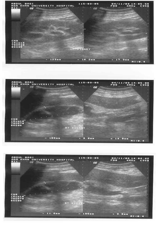
This set of three ultrasound images demonstrates bilateral renal pathology in a 65-year-old male. The top panel shows the left kidney with evidence of hydronephrosis, characterized by an anechoic dilation of the renal pelvis, and an echogenic renal stone. The middle and bottom panels visualize the right kidney, which appears smaller and more echogenic, consistent with chronic scarring. A pigtail percutaneous nephrostomy catheter is visible within the renal collection system as a bright, linear echogenic structure. In the lower panel, a hyperechoic focus with distal acoustic shadowing is identified in the proximal ureter, signifying an obstructive ureteric stone. These diagnostic images illustrate key features of obstructive uropathy, including hydronephrosis, nephrolithiasis, and the use of drainage catheters in the management of renal obstruction. The comparative views highlight the difference between acute obstructive changes in a preserved kidney (left) and chronic changes (scarring) in the contralateral kidney (right).
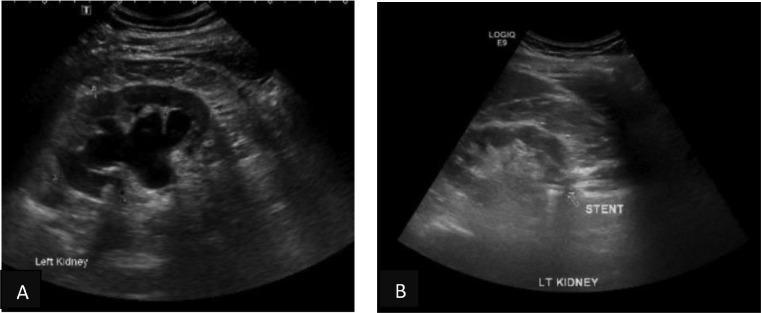
This dual-panel ultrasound image comparison illustrates the diagnostic and therapeutic monitoring of left-sided hydronephrosis. Panel (A) displays a grayscale ultrasound of the left kidney in a longitudinal plane, showing marked pelvicalyceal dilatation. The renal pelvis and calyces are distended with large, centrally located anechoic (fluid-filled) areas, characteristic of significant hydronephrosis. The renal parenchyma appears preserved but displaced peripherally by the dilated collecting system. Panel (B) shows a follow-up ultrasound of the same kidney following the insertion of a Memokath ureteric stent. There is a prominent reduction in the distension of the renal pelvis and calyces, indicating resolution of the hydronephrosis. A highly echogenic, linear structure with acoustic shadowing is visible within the renal pelvis/upper ureter region, labeled as 'STENT'. This educational comparison demonstrates the effectiveness of internal drainage in relieving upper urinary tract obstruction caused by ureteric strictures. The image is relevant for urology and radiology trainees focusing on renal imaging and interventional urology outcomes.
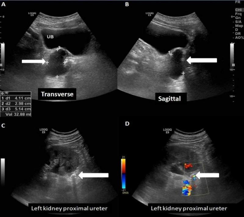
This diagnostic image composite features four ultrasound scans (A-D) demonstrating urological pathology. Panels A (transverse) and B (sagittal) show a well-defined cystic lesion (indicated by white arrows) measuring approximately 4.1 x 3.0 x 5.1 cm with a volume of 32.88 ml. The lesion displays internal echoes/debris and is located on the right posterolateral aspect of the urinary bladder (UB), causing extrinsic compression and indenting the bladder wall. Panels C and D focus on the left kidney and proximal ureter. Panel C reveals a hyperechoic structure within the proximal ureter, identified as an obstructing stone, characterized by strong posterior acoustic shadowing and associated upstream pelvicalyceal dilatation (hydronephrosis). Panel D utilizes color Doppler to highlight a distinct 'twinkling artifact' at the site of the ureteral stone, a characteristic finding for nephrolithiasis. This visual material is used in nephrology and radiology curricula to teach the sonographic identification of cystic pelvic lesions and obstructive uropathy secondary to ureteric calculi.
Ultrasound Report Analysis - Sayma Firdous, 32Y/F
What the Report Shows
- Left-sided upper ureteric calculus with moderate hydroureteronephrosis
- Changes of cystitis (bladder infection)
Finding 1 - Left Upper Ureteric Stone (Calculus)
What it means in simple language:
What has happened as a result:
- Dilatation (widening) of the left pelvicalyceal system - the urine-collecting part of the kidney has swollen up with backed-up urine
- Renal pelvis measuring 1.1 cm AP diameter - this confirms moderate obstruction
- Proximal ureter dilated to 0.9 cm - the ureter above the stone is stretched
Right Kidney Findings:
Finding 2 - Cystitis (Bladder Infection)
Treatment Plan - What Needs to Be Done
Step 1 - Immediate Actions (Right Now)
| Action | Why |
|---|---|
| See a Urologist urgently | 11.5mm stone causing moderate hydronephrosis needs specialist care |
| Urine routine microscopy (already advised) | To confirm cystitis, check for blood cells, infection type |
| Urine culture and sensitivity | To identify the exact bacteria causing cystitis and choose the right antibiotic |
| Blood tests | Kidney function tests (creatinine, BUN), CBC, electrolytes |
| Non-contrast CT KUB | Gold standard - confirms stone size, exact position, density (Hounsfield Units) better than ultrasound. Can see stones CT cannot miss. |
Step 2 - Treat the Bladder Infection (Cystitis)
- Antibiotics are needed immediately (usually 5-7 days for women with uncomplicated cystitis; longer if complicated by the obstruction)
- Common choices: Nitrofurantoin, Trimethoprim-sulfamethoxazole, Fosfomycin, or Ciprofloxacin - based on urine culture results
- Drink plenty of water (2-3 litres per day unless told otherwise)
Step 3 - Treat the Ureteric Stone (11.5 mm)
Option A - Medical Expulsive Therapy (MET) - Only for borderline cases
- Alpha-blockers (e.g., Tamsulosin 0.4 mg once daily) and calcium-channel blockers (e.g., Nifedipine) relax the ureter muscles to help small stones pass
- Analgesics - NSAIDs (e.g., diclofenac) for pain; opioids if needed
- MET may be tried for a short time (2-4 weeks maximum) only if the stone is smaller and there is no severe obstruction
- Given the size (11.5mm) and moderate hydronephrosis, MET alone is unlikely to be sufficient here
Option B - Ureteroscopy + Laser Lithotripsy (Most Likely Recommended)
- Most commonly recommended procedure for upper ureteric stones of this size
- A thin flexible scope (ureteroscope) is passed through the urethra and bladder up into the ureter
- A Holmium laser or similar is used to break the stone into tiny fragments
- Fragments are removed or allowed to pass naturally
- Usually done under general or spinal anesthesia
- A DJ (double-J) stent - a thin plastic tube - is often placed temporarily in the ureter to keep it open and help healing
- The stent is usually removed after 2-4 weeks by cystoscopy
Option C - Extracorporeal Shock Wave Lithotripsy (ESWL)
- Shock waves from outside the body are focused on the stone to break it up
- Less effective for upper ureteric stones and harder stones (determined by CT Hounsfield Units)
- May require multiple sessions
- Results are variable and less predictable for 11.5 mm stones in the ureter vs. kidney
Option D - Percutaneous Nephrostomy (Emergency Drainage)
- If the patient has fever, infection with obstruction (obstructive pyelonephritis), severe pain, or worsening kidney function, emergency drainage via a tube inserted through the back directly into the kidney may be needed first
- This relieves pressure on the kidney immediately
- Definitive stone treatment is done later once the infection is controlled
Summary Decision Tree
Current situation: 11.5mm stone, upper ureter, moderate hydronephrosis, cystitis

Is there fever + flank pain + obstruction?
   YES → Emergency: Nephrostomy or DJ stent FIRST, then antibiotics, then stone procedure later
   NO  → Urgent (within days): Ureteroscopy + laser lithotripsy + antibiotics for cystitis
Key Points to Remember
- This cannot wait weeks - moderate hydronephrosis means the kidney is under sustained pressure. Prolonged obstruction damages kidney function permanently.
- Cystitis with an obstructed kidney is dangerous - infection can spread upward to the kidney (pyelonephritis) or into the blood (urosepsis). Antibiotics alone are NOT enough - the obstruction must be relieved.
- Right kidney tiny concretions - drink more water, reduce salt/protein intake, follow up with metabolic stone workup (urine 24-hour stone analysis) to prevent new stones.
- The post-void residual is blank - the doctor should check if the bladder is emptying completely.
- Recurrence prevention: After the acute episode is treated, lifestyle changes (high water intake, dietary modifications based on stone type) and metabolic evaluation should be done.
What to Do Right Now (Action Checklist)
- Go to a urologist today or tomorrow - do not delay
- Get urine culture done alongside the routine microscopy
- Start antibiotic as prescribed for cystitis
- Get blood creatinine and CBC checked
- Arrange CT scan (non-contrast KUB) if not already done
- Ask the urologist about ureteroscopy + laser as the likely definitive treatment
- Increase water intake to at least 2-3 litres per day
- Take prescribed pain relief (NSAIDs or paracetamol) as needed
- Report immediately if fever > 38°C, chills, severe worsening pain, or vomiting develops
Disclaimer: This explanation is for educational purposes only and is based on the ultrasound report provided. It is not a substitute for in-person medical advice. Please consult a qualified urologist for proper evaluation and a treatment plan tailored to the patient's full clinical condition.