
Thromboangitis obliterans
"thromboangiitis obliterans" AND treatment
Buerger disease thromboangiitis obliterans digital ischemia gangrene
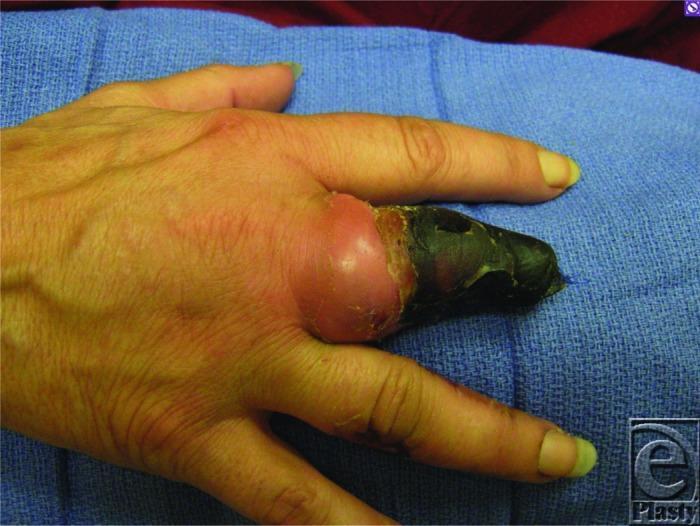
A clinical photograph of a right hand demonstrating advanced digital ischemia and dry gangrene, characteristic of thromboangiitis obliterans (Buerger's disease). The third digit (middle finger) shows a prior partial amputation at the level of the middle phalanx. The residual distal portion of this digit exhibits a dense, black, necrotic eschar consistent with gangrene. There is significant circumferential erythema and swelling proximal to the necrotic tissue, specifically involving the area around the metacarpophalangeal joint, suggesting active inflammation or cellulitis. The surrounding digits show subtle signs of circulatory compromise, including skin pallor and longitudinal nail plate changes. This image serves as an educational example of critical limb-threatening ischemia and the progressive tissue loss associated with small-to-medium vessel vasculitis in tobacco users. The visual findings illustrate the demarcation between necrotic tissue and viable, albeit inflamed, proximal structures.
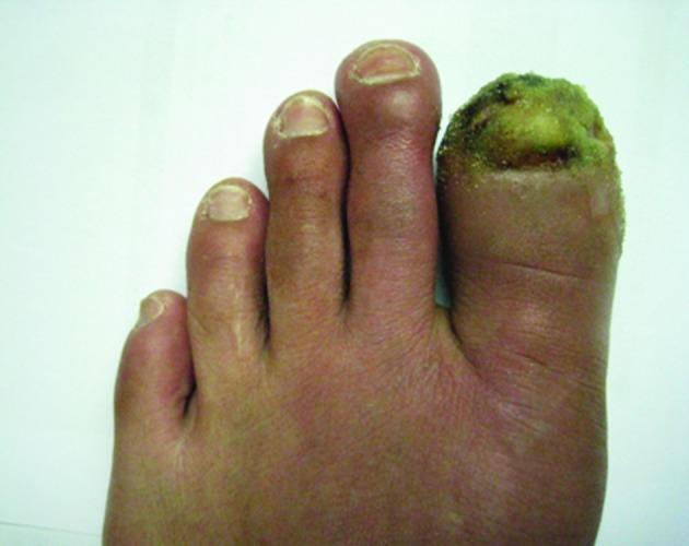
This clinical photograph displays a top-down view of the left foot, specifically highlighting severe pathology of the first toe (hallux). The distal portion of the first toe exhibits total nail plate destruction and a large, infected ulcerated lesion. The ulcer bed presents with a prominent greenish-yellow purulent exudate and areas of necrotic black crusting, suggesting bacterial colonization or secondary infection. The surrounding soft tissue of the first toe is markedly erythematous and edematous. The second, third, and fourth toes show a diffuse reddish-purple discoloration (cyanosis or livedo-like changes) and mild swelling, particularly at the distal phalanges. The nail plates of the remaining toes appear thickened with longitudinal ridges. These visual findings are consistent with thromboangiitis obliterans (Buerger's disease) or severe peripheral vascular disease leading to digital ischemia, gangrene, and secondary paronychia.
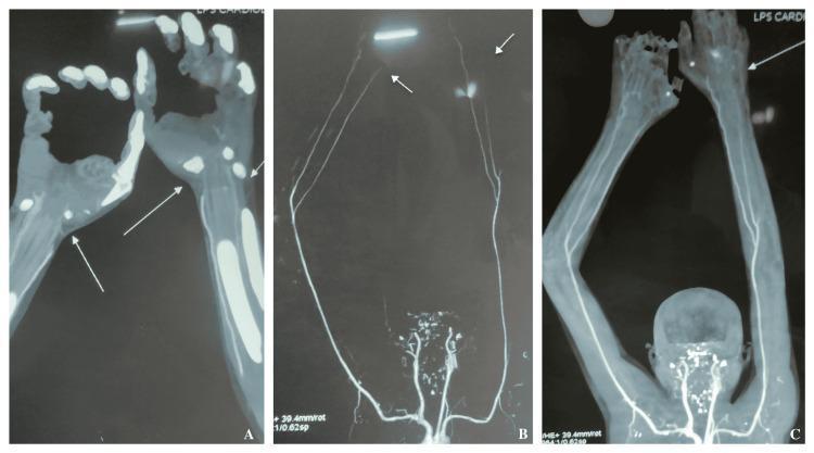
This diagnostic visual consists of three CT angiography (CTA) panels (A, B, and C) depicting the upper extremities of a patient with vascular insufficiency. Panel A focuses on the hands and distal forearms, demonstrating significant bilateral pathology characterized by poor contrast opacification of the palmar arches and digital arteries. The digital vessels appear attenuated, thinned, and interrupted, correlating with clinical findings of distal ischemia or gangrene. Panels B and C provide a more proximal view of the upper limb arterial tree, highlighting the bilateral ulnar and radial arteries. Arrows indicate critical stenoses and areas of diminished flow within the distal ulnar arteries bilaterally. The overall vascular morphology shows threadlike narrowing of distal vessels compared to more proximal arterial segments. These findings are consistent with thromboangiitis obliterans (Buerger's disease) or other severe peripheral arterial diseases, demonstrating a transition from patent proximal vessels to occluded or severely stenotic distal vasculature.
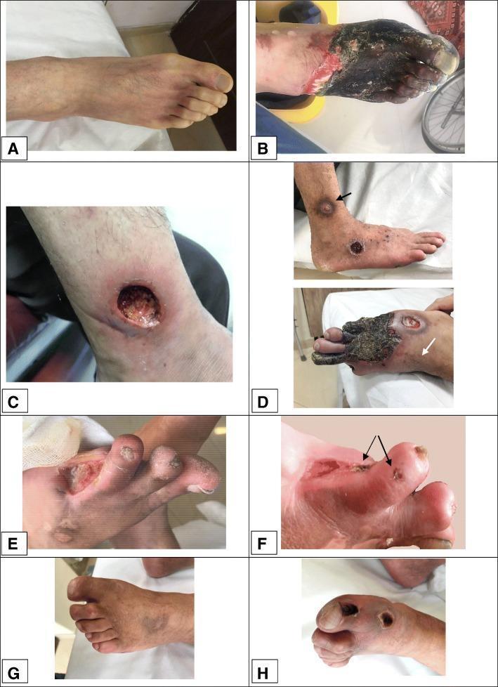
A composite of clinical photographs (panels A-H) demonstrating various stages of dermal gangrene and chronic ulceration in the lower extremities of patients with thromboangiitis obliterans (Buerger’s disease). Panel A shows initial pallor and Raynaud's phenomenon. Panel B illustrates extensive, dark necrotic tissue covering the forefoot and toes with a sharp demarcating border and surrounding erythema. Panel C depicts a deep, circular 'punched-out' ulcer on the lower leg with visible granulation tissue and peripheral edema. Panel D shows multifocal purpuric-like lesions progressing to localized gangrene on the dorsum of the foot and distal digits. Panels E and F provide close-up views of digital ulcers and small gangrenous papules (indicated by arrows) on an amputation stump. Panels G and H show the evolution of localized gangrenous lesions on the toes, transitioning from active necrosis to a dryer, more localized state. This collection highlights the microvascular complications of TAO, including tissue ischemia, necrosis, and subsequent ulceration despite angiogenic medical therapies.
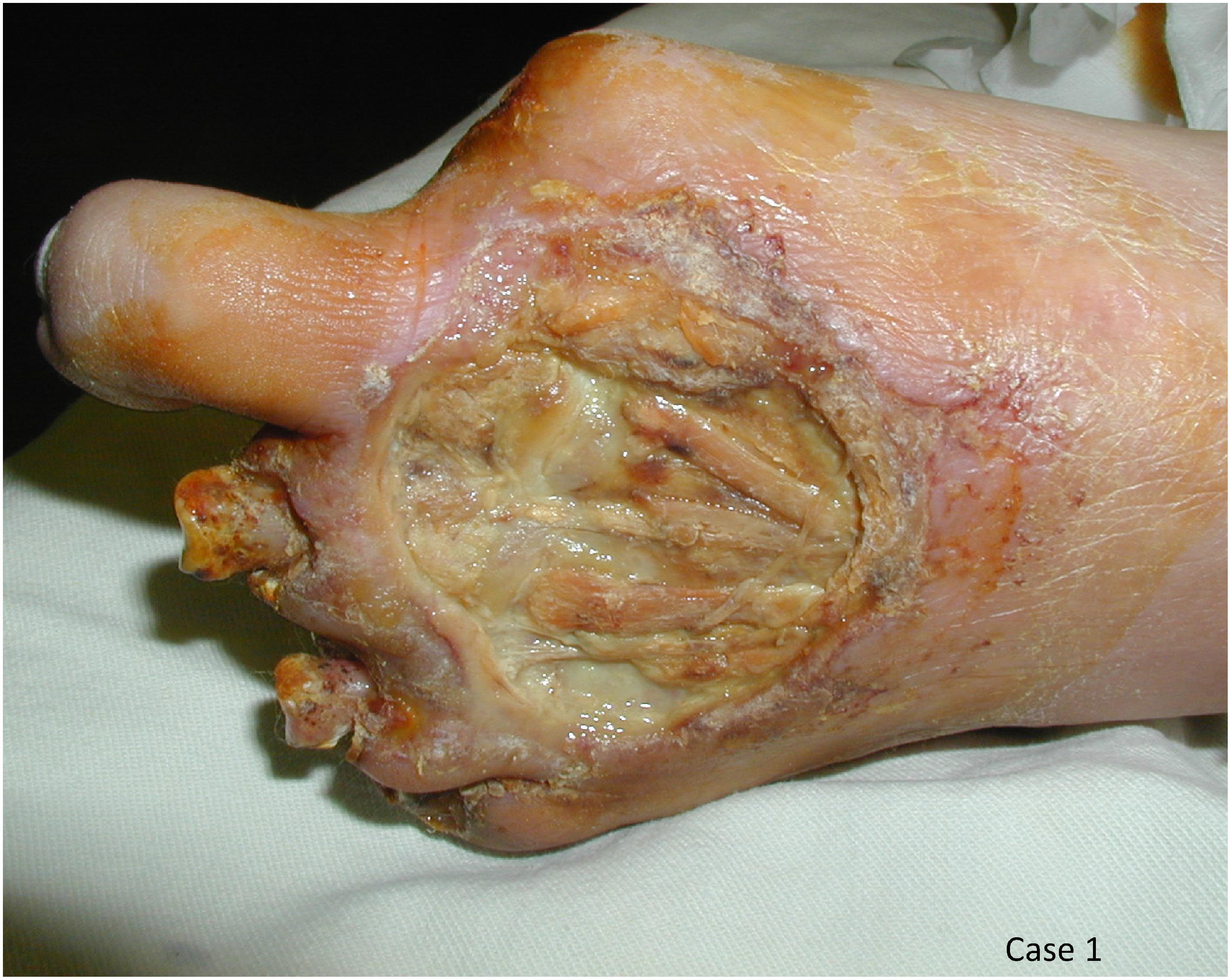
Clinical photograph of a left foot demonstrating severe ischemic complications and tissue loss associated with Buerger's disease (thromboangiitis obliterans) and superimposed atherosclerosis. The image shows a large, deep ulceration on the dorsal and lateral midfoot, exposing underlying extensor tendons, fascia, and subcutaneous tissue. The wound bed appears fibrinous with areas of moist slough. There is evidence of significant structural compromise: the second through fifth digits have undergone partial amputations, leaving irregular stubs with signs of necrosis and chronic scarring. The remaining first toe appears pale with dry, xerotic skin and thin epithelium. Surrounding skin on the midfoot and ankle exhibits varying degrees of erythema, pallor, and hyperkeratosis, indicative of chronic peripheral limb ischemia. This visual serves as a primary example of advanced peripheral arterial disease (PAD) in a patient with multi-decade smoking history and metabolic risk factors, illustrating the progression from digital ulcers to gangrene and auto-amputation.
Thromboangiitis Obliterans (Buerger's Disease)
Overview
Epidemiology
- Typically affects young smokers, usually under 40-45 years of age.
- Historically male-predominant, but the proportion of women has risen to nearly 20%, paralleling increased tobacco use among women (Current Surgical Therapy 14e).
- Worldwide distribution, with highest prevalence in Eastern Europe, the Mediterranean, and Asia. In North America it accounts for less than 1% of severe limb ischemia cases, but was found in 24% of young adults with lower-limb ischemia in one Mayo Clinic series (Schwartz's Principles of Surgery, 11e, p. 1002).
Pathophysiology
Clinical Features
- Early: cold-induced Raynaud phenomenon, instep/arch claudication (often mistaken for orthopedic pathology), and migratory superficial thrombophlebitis (up to 16% of patients, indicating systemic inflammation).
- Progression: calf claudication, ischemic rest pain (the most common presentation, seen in about two-thirds of patients), and ischemic ulcers of fingers/toes progressing to gangrene.
- At least two limbs are almost always involved, and up to 40% of patients have all four limbs affected.
- Rarely, large-vessel or visceral involvement occurs (mesenteric, coronary, cerebral, renal, pulmonary), and mesenteric TAO carries a poor prognosis requiring urgent surgery.
Diagnosis
- Age of onset < 45 years
- History of tobacco use
- Distal extremity ischemia involving two or more limbs
- Angiographic findings suggestive of TAO
- Exclusion of other causes of vasculopathy (diabetes, hyperlipidemia, autoimmune disease, proximal embolic source)
Treatment
- Medical: prostacyclin analogues, aspirin, low-dose intra-arterial streptokinase, bosentan (endothelin-1 receptor antagonist), cyclophosphamide, guanethidine sympathetic blocks, serotonin blockers, sarprogrelate, intramuscular VEGF injections, PDE5 inhibitors.
- Surgical: endovascular angioplasty of tibial/foot artery obstructions; open sympathectomy, arterial reconstruction (limited by lack of suitable target vessels and vein conduits, since migratory phlebitis often damages usable veins), omental transfer, or amputation for non-salvageable limbs.
- Experimental: autologous bone marrow stem cell transplantation, immunoadsorption, gene therapy (beperminogene perplasmid).