

Interpret this image
prostate MRI T2 weighted axial peripheral zone cancer hypointense lesion
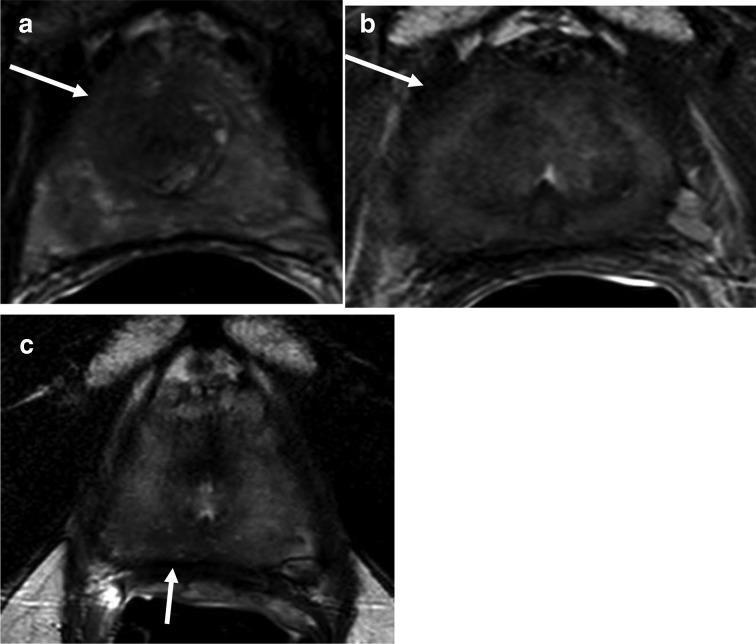
Diagnostic axial T2-weighted (T2W) MRI of the prostate showing a progression from pre-treatment to post-treatment and biochemical recurrence. Image A demonstrates an ill-defined, heterogeneous hypointense lesion in the right apical-mid anterior peripheral and transition zones (indicated by arrow), consistent with primary prostate cancer. Image B shows the prostate after treatment, exhibiting a diffuse iso-to-hypointense signal pattern characteristic of radiation therapy or systemic treatment effects, with the original lesion no longer discernible. Image C illustrates the gland at the time of biochemical recurrence; while it retains the diffuse treatment-related signal changes, a new focal hypointense lesion is identified in the midline to right apical peripheral zone (indicated by arrow). This focal area of signal loss is highly suspicious for recurrent prostate adenocarcinoma. The series serves as an educational example of monitoring treatment response and detecting localized recurrence using MRI in the context of rising PSA levels.
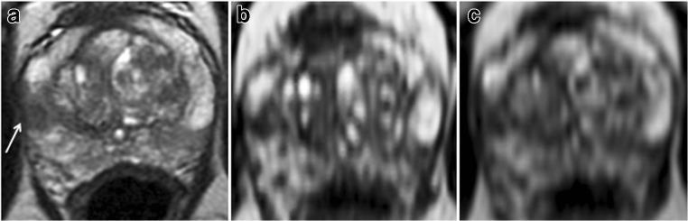
Diagnostic Image Comparison: Comparison of three axial T2-weighted magnetic resonance images (MRI) of the prostate gland at the mid-gland level. Panel (a) shows a conventional 2D axial T2WI demonstrating a hypointense lesion in the right lateral peripheral zone (PZ) marked with a white arrow. In this standard sequence, the lesion appears to have a broad tumor-capsule contact length of 12 mm. Panels (b) and (c) represent 3D thin-sliced cube reformats at two consecutive levels of the same region. These high-resolution reformats clarify the anatomy by reducing partial volume effects, revealing a more minimal capsular contact of approximately 5 mm. The images illustrate the clinical utility of 3D T2W reformatting for more accurate staging of prostate cancer by precisely evaluating the tumor-capsule interface and predicting extra-prostatic extension (ECE). The case relates to a 65-year-old patient with elevated PSA, where histopathology confirmed an organ-confined (pT2) Gleason 4+4=8 lesion.
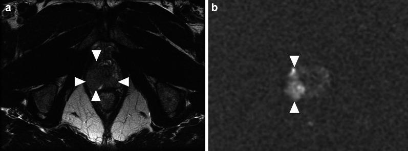
This diagnostic imaging set consists of two axial MRI slices of the prostate apex in a 62-year-old male. Image (a) is an axial T2-weighted (T2W) image showing a large, well-defined, hypointense lesion (arrowheads) located in the right lateral and posterior peripheral zone. The lesion replaces a significant portion of the normal high T2 signal of the peripheral zone tissue. Image (b) is an axial diffusion-weighted image (DWI, b = 2000 s/mm²) at the same level, which demonstrates pronounced high signal intensity (diffusion restriction) corresponding to the lesion seen in the T2W image. While these findings—T2 hypointensity combined with restricted diffusion—are highly characteristic of prostatic adenocarcinoma, the provided clinical context indicates this represents granulomatous (tuberculous) prostatitis following intravesical BCG therapy for bladder cancer. This comparison illustrates a significant pitfall in multiparametric MRI (mpMRI) interpretation, where inflammatory granulomatous processes can closely mimic the imaging phenotype of high-grade prostate cancer.
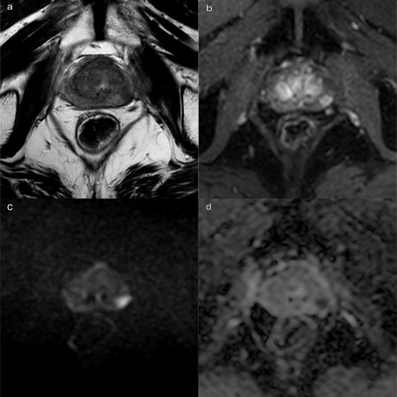
Multiparametric MRI (mpMRI) of the prostate in the axial plane demonstrating a focal recurrence of prostate cancer in the left peripheral zone. Panel (a) is a T2-weighted Turbo Spin Echo (TSE) image showing an 18mm hypointense lesion in the mid-portion of the left peripheral zone with a curvilinear contact surface along the capsule, though no frank extracapsular extension is visible. Panel (b) illustrates Dynamic Contrast-Enhanced (DCE) imaging using a T1-weighted Dixon sequence with fat suppression, revealing early focal arterial enhancement in the same region. Panels (c) and (d) show Diffusion-Weighted Imaging (DWI) at a high b-value of 2500 s/mm² and the corresponding Apparent Diffusion Coefficient (ADC) map, respectively. The lesion exhibits marked hyperintensity on the high b-value DWI and corresponding hypointensity on the ADC map, indicating restricted diffusion and high cellularity characteristic of a PI-RR score of 4. This diagnostic series is used for staging local recurrence and planning targeted biopsy or focal therapy.
MRI Prostate - Interpretation
Modality & Sequence
Normal Anatomy Review
- Peripheral zone (PZ) - normally uniformly bright (hyperintense), forming the outer crescent of the gland
- Transition zone (TZ) / Central gland - normally heterogeneous, with well-demarcated BPH nodules
- Prostatic capsule - a thin hypointense rim around the gland
Findings
PI-RADS Assessment Framework
- T2W (anatomic - seen here)
- DWI/ADC map (diffusion-weighted, primary sequence for peripheral zone lesions)
- DCE (dynamic contrast-enhanced)
| PI-RADS Score | Meaning |
|---|---|
| 1 | Clinically significant cancer highly unlikely |
| 2 | Unlikely |
| 3 | Equivocal |
| 4 | Cancer likely |
| 5 | Cancer highly likely |
Differential Diagnosis for Peripheral Zone Hypointensity
| Diagnosis | Features |
|---|---|
| Prostate adenocarcinoma | Focal, ill-defined, low T2 signal |
| Prostatitis | Can mimic cancer; often more diffuse |
| Post-biopsy hemorrhage | History of recent biopsy; fades over time |
| BPH nodule extending into PZ | More well-circumscribed |
| Granulomatous prostatitis (e.g., post-BCG) | Can closely mimic cancer on mpMRI |
Clinical Correlation
- Full mpMRI (DWI + DCE) for complete PI-RADS scoring
- PSA measurement and clinical context
- If PI-RADS 3-5, MRI-guided or MRI-fusion targeted biopsy of the suspicious lesion
- Concurrent assessment for BPH symptoms (IPSS score, uroflowmetry)
Comparison Image
Important disclaimer: This interpretation is for educational/informational purposes. Formal radiology reporting by a qualified radiologist with access to the full MRI series and clinical history is required for clinical decision-making.