
I have interview tomorrow If CMO asks if a employee collapses at work place what should u do? Tell me sequence of events i should follow in this case Especially mention should i shift patient on strecher
workplace collapse emergency response sequence BLS protocol 2025
https://cprlifeline.net/bls-algorithm
What to Do When an Employee Collapses at the Workplace
Step-by-Step Sequence
STEP 1 - Ensure Scene Safety
STEP 2 - Check Responsiveness
- If no response - proceed immediately.
STEP 3 - Call for Help / Activate Emergency Response
- Shout for help loudly.
- Call the code / emergency number (hospital code blue, or dial 108/112/999 depending on location).
- Send someone to fetch the AED (defibrillator) and emergency crash cart.
- Do NOT leave the patient alone - delegate tasks to bystanders.
STEP 4 - Position the Patient (Critical for your interview)
- Do NOT shift the patient to a stretcher immediately. This is a common mistake.
- Lay the patient flat on their back (supine) on the floor - the floor provides the firm surface needed for effective CPR. A soft stretcher/bed reduces compression effectiveness.
- Only move the patient if they are in immediate danger (fire, unsafe zone).
- Log-roll with neck stabilization if spinal injury is suspected.
STEP 5 - Check Breathing and Pulse Simultaneously (max 10 seconds)
- Look for chest rise, listen and feel for breath.
- Check the carotid pulse with 2 fingers (no more than 10 seconds).
- If no pulse and no breathing - cardiac arrest confirmed.
STEP 6 - Start CPR Immediately (CAB sequence)
| Component | Detail |
|---|---|
| C - Compressions | Hard and fast on the center of the chest; 100-120/min; depth 5-6 cm; allow full recoil |
| A - Airway | Head-tilt chin-lift (or jaw thrust if trauma suspected) |
| B - Breathing | 30:2 ratio (30 compressions : 2 rescue breaths) for single rescuer |
STEP 7 - Attach AED / Defibrillator as Soon as Available
- Apply pads, assess rhythm.
- If shockable rhythm (VF/pulseless VT) - deliver shock, then immediately resume CPR for 2 minutes.
- If non-shockable (PEA/asystole) - continue CPR.
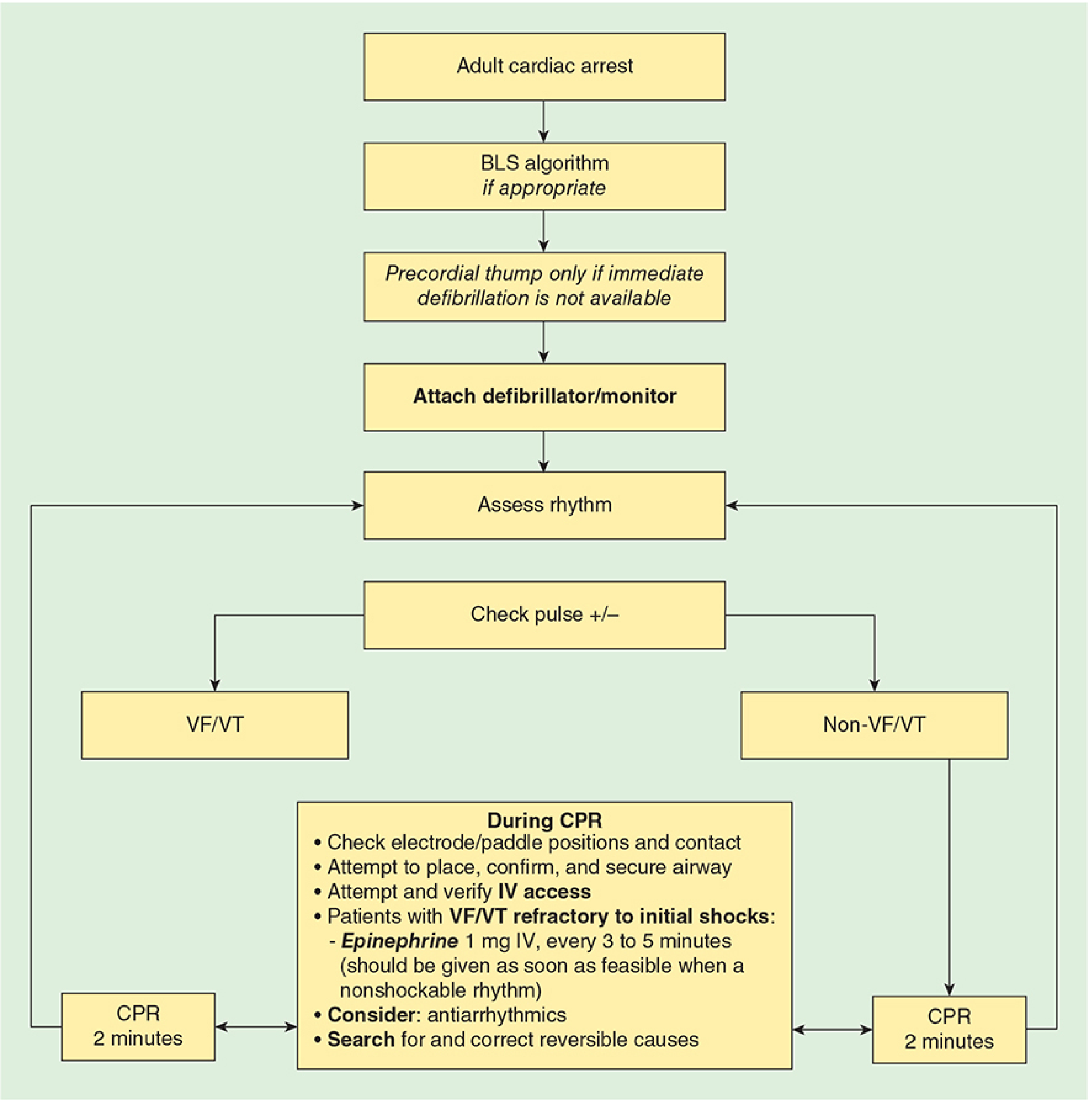
STEP 8 - Advanced Life Support (ACLS)
- IV/IO access.
- Intubation / advanced airway.
- Epinephrine 1 mg IV every 3-5 minutes.
- Identify and treat reversible causes (H's and T's).
STEP 9 - Post-Resuscitation Care (if ROSC achieved)
- NOW transfer to stretcher safely.
- ICU/CCU admission.
- 12-lead ECG, targeted temperature management, treat underlying cause.
STEP 10 - Documentation
Regarding the Stretcher - Direct Answer
Do NOT shift the patient to a stretcher during CPR.
Quick Memory Aid for the Interview
Methods of shifting employee onto stretcher in collapse case,trauma case,neck injury case and any other case
methods of transferring patient onto stretcher trauma spinal neck injury log roll scoop 2024
https://www.hartwellmedical.com/wp-content/uploads/2021/09/S…
scoop stretcher draw sheet transfer conscious unconscious patient methods EMS
Methods of Shifting a Patient onto a Stretcher
Quick Reference Table
| Situation | Preferred Method |
|---|---|
| Collapse / Cardiac arrest (post-ROSC) | Direct lift / Draw sheet transfer |
| Conscious stable patient | Assisted stand-pivot / Walk-on |
| Unconscious, no spinal concern | Scoop stretcher / Direct lift |
| Trauma - suspected spinal injury | Scoop stretcher / 6+ Lift & Slide |
| Neck / cervical spine injury | Log roll WITH inline cervical stabilization |
| Prone patient with spinal injury | Log roll onto spine board (push technique) |
| Bariatric / obese patient | 6+ Lift & Slide with extra rescuers |
| Bed-to-stretcher transfer (ward) | Draw sheet / Slide board |
| Chest / rib injury (no spinal concern) | Semi-recumbent position, assisted transfer |
Detailed Methods
1. Direct Lift (Cradle/Carries Lift)
- 2-3 rescuers stand on the same side of the patient.
- One supports the head and shoulders, one the torso, one the legs.
- On a coordinated count ("1-2-3 lift"), lift simultaneously and lower onto the stretcher.
- Maintain the patient in a flat supine position.
- No spinal precautions needed if trauma is ruled out.
Key point for CMO: After cardiac arrest and ROSC is achieved, use this to transfer from the floor to the stretcher. Do NOT do this during active CPR.
2. Scoop Stretcher (Orthopaedic Stretcher) - PREFERRED for most trauma
- One rescuer maintains manual inline stabilization (MILS) of the head throughout.
- Measure and adjust scoop stretcher length to the patient.
- Separate the stretcher into its two longitudinal halves.
- Carefully wedge one half under one side of the patient.
- Repeat with the other half on the opposite side.
- Lock both ends (head and foot latches) to close the scoop.
- Apply straps, then lift onto the main stretcher/trolley.
- Only 4 rescuers needed.
- Less spinal motion than log roll (supported by biomechanical studies).
- Patient does not need to be rolled.
The scoop stretcher requires only four rescuers, better motion control compared to log roll - Surgical Neurology International, 2012
3. Log Roll - FOR CERVICAL / NECK INJURY & BACK EXAMINATION
| Position | Role |
|---|---|
| Head person (Team Leader) | Maintains MILS - hands on bilateral shoulders/head, forearms stabilize the skull. Gives ALL commands. This is the most senior/tallest person. |
| Chest person | Hands on shoulder and lower chest |
| Hip person | One hand on lower back, one under the thigh |
| Leg person | Supports from below the knee (for tall/heavy patients) |
| 5th person | Slides spine board under patient / performs back examination |
- Apply cervical collar first (appropriate size).
- Team leader at head calls: "On my count - 1, 2, 3, roll."
- Patient rolled as a single rigid unit - no twisting, no independent segment movement.
- Roll to 30-45 degrees only (not full 90).
- 5th person slides the spine board under, or inspects the back.
- On count, roll patient back down onto the board.
- Secure with head blocks and straps.
- The patient is found prone (must log roll onto board using push technique).
- You need to inspect the back for wounds.
- No scoop stretcher is available.
4. Straddle Lift and Slide
- 4 rescuers: one at head (MILS), one straddles the patient at chest level, one at hips, one at legs.
- Patient is lifted slightly and a spine board is slid underneath from the feet upward.
- Patient is lowered onto the board.
- Produces less motion than log roll per biomechanical data.
5. 6+ Lift and Slide
- Requires 6 or more rescuers (one at head for MILS, others evenly distributed along the body).
- All rescuers lift simultaneously on a count.
- A spine board is slid underneath.
- Best spinal motion control of all manual techniques.
- Limitation: needs a large coordinated team.
6. Draw Sheet / Slide Sheet Transfer
- Position the stretcher parallel and adjacent to the bed at the same height.
- Grip the sheet (or slide board placed underneath) firmly.
- 2 rescuers pull the sheet toward the stretcher while a 3rd supports the patient's head.
- Minimal lifting required - reduces rescuer back injury.
- NOT for spinal injury cases as it may cause lateral flexion.
7. Assisted Stand and Walk-on
- Help the patient sit up slowly (watch for postural hypotension).
- Support under the arm and guide them to stand.
- Walk them to the stretcher and assist them to sit/lie down.
- Always keep one hand on the patient.
8. Recovery Position (Before Stretcher Transfer)
- Roll the patient onto their left lateral side to protect the airway.
- Top knee bent forward to stabilize.
- Head tilted slightly back to keep airway open.
- Monitor until stretcher is ready, then transfer using scoop or direct lift.
The Golden Rules - What to Say in the Interview
"The method of transfer depends on three things: consciousness level, presence of trauma, and suspected spinal involvement."
- Never rush a transfer - a wrong move causes secondary injury.
- MILS (Manual Inline Stabilization) must be maintained throughout for any suspected spinal injury - cervical collar alone is not enough.
- Scoop stretcher > Log roll for trauma (produces less spinal motion) - this is current evidence-based practice.
- Log roll is specifically indicated when: patient is prone, or you need to examine the back.
- The head person is always the team leader and gives all movement commands.
- Stretcher height should be at the hip level of the person holding the head before any transfer.
- Minimum rescuers: Scoop = 4, Log roll = 4 minimum (5 preferred), 6+ lift = 6+.
What maximum possible questions can CMO ask me in an interview regarding employee collapses inwork place
CMO interview questions employee collapse workplace emergency occupational health medical officer
causes of collapse at workplace syncope cardiac arrest hypoglycemia differential diagnosis
All Possible CMO Interview Questions - Employee Collapses at Workplace
CATEGORY 1 - First Response & Basic Protocol
- Check scene safety first, then check responsiveness (tap + shout)
- Scene safe → Check response → Call for help / activate emergency → Check pulse + breathing (max 10 sec) → Start CPR → Attach AED → Advanced Life Support → Post-ROSC care → Document
- NO. Keep on firm floor for CPR. Stretcher only after ROSC is achieved. Only exception: immediate danger at scene.
- C = Compressions first, A = Airway, B = Breathing. (Changed from ABC in 2010 AHA guidelines)
- Rate: 100-120 compressions/minute; Depth: 5-6 cm; Allow full chest recoil; Minimize interruptions (< 10 sec pause)
- 30:2 for single rescuer adult CPR
- 15:2 for two-rescuer pediatric CPR
- Compression-only CPR acceptable if rescuer untrained in rescue breaths
- Shout for help, designate someone to call emergency number (108 / 112), send someone to get AED + crash cart, do not leave patient alone
- Automated External Defibrillator. Attach as soon as available. It will analyze rhythm and tell you when to shock. Shock if VF or pulseless VT. Resume CPR immediately after every shock.
CATEGORY 2 - Causes of Collapse
| Category | Examples |
|---|---|
| Cardiac | Cardiac arrest (VF/VT), heart attack (MI), arrhythmia, heart block |
| Vasovagal / Reflex | Vasovagal syncope (commonest cause), situational syncope (coughing, straining) |
| Metabolic | Hypoglycemia (diabetic employee), hyponatremia, hypocalcemia |
| Neurological | Seizure (epilepsy), stroke/TIA, TBI from fall |
| Respiratory | Severe asthma, pulmonary embolism, hypoxia |
| Orthostatic | Postural hypotension (getting up quickly, dehydration, antihypertensive drugs) |
| Toxic / Chemical | Chemical inhalation (factories), CO poisoning, drug overdose |
| Heat-related | Heat stroke, heat exhaustion (outdoor/industrial workers) |
| Psychological | Panic attack, hyperventilation, pseudosyncope |
| Trauma | Fall → head injury, spinal injury |
| Feature | Syncope | Cardiac Arrest |
|---|---|---|
| Pulse | Present | Absent |
| Breathing | Present (normal) | Absent / gasping |
| Recovery | Rapid, spontaneous | No recovery without CPR |
| Skin | Pale, sweaty | Cyanosed, mottled |
| Duration | Brief (seconds) | Sustained |
- Hypoglycemia until proven otherwise. Check blood glucose (glucometer). If conscious: oral glucose. If unconscious: IV dextrose 25-50 mL of 50% dextrose OR 1 mg glucagon IM.
- Panic attack / hyperventilation syndrome causing respiratory alkalosis → syncope. Reassure, calm environment, re-breathe into a bag (controversial now - use with caution).
- Scene safety is paramount - do NOT enter without PPE if toxic fumes suspected. Call hazmat team. Decontaminate before treatment if needed. Consider CO poisoning → 100% high-flow oxygen.
CATEGORY 3 - Patient Transfer & Positioning
- ONLY after ROSC (return of spontaneous circulation) is achieved, OR if the scene is immediately dangerous.
- Direct lift (2-3 person lift) onto stretcher after ROSC. Floor is used during CPR.
- Orthopaedic stretcher that splits into 2 halves, slides under patient without rolling them. Used in trauma, suspected spinal injury. Preferred over log roll as it causes less spinal motion.
- Minimum 4 people. One at head (team leader, maintains cervical spine), others at chest/hip/legs. Roll as single unit on count. Used for neck injury, examining the back, or prone patient.
- Recovery position (left lateral / left-side-down). Prevents airway obstruction and aspiration.
- Supine with legs elevated 15-30 degrees (Trendelenburg-modified / passive leg raise) to improve venous return.
- Semi-recumbent (sitting up at 45 degrees) to reduce cardiac workload and ease breathing.
- Left lateral tilt (15-30 degrees) to relieve aortocaval compression by the gravid uterus. Critical after 20 weeks gestation.
CATEGORY 4 - Clinical Knowledge (Advanced)
| 4 H's | 4 T's |
|---|---|
| Hypoxia | Tension pneumothorax |
| Hypovolemia | Tamponade (cardiac) |
| Hypo/Hyperkalemia (electrolytes) | Toxins (poisoning/drugs) |
| Hypothermia | Thrombosis (PE or coronary) |
- Early recognition and call for help
- Early CPR
- Early defibrillation
- Early advanced life support
- Post-resuscitation care
- ROSC achieved (signs of life - pulse, breathing, movement)
- Declared dead by authorized doctor
- Rescuer is exhausted and no relief available
- Patient has a valid Do-Not-Resuscitate (DNR) order
- Injuries incompatible with life
- Return of Spontaneous Circulation - patient has a pulse again
- Post-ROSC: ABC stabilization, 12-lead ECG, IV access, blood glucose, oxygen titration (SpO2 94-98%), targeted temperature management, transfer to ICU/CCU
- Epinephrine (Adrenaline): 1 mg IV every 3-5 minutes (for any rhythm)
- Amiodarone: 300 mg IV for VF/pulseless VT refractory to 3 shocks (then 150 mg if needed)
- Atropine: No longer recommended in asystole/PEA (removed from guidelines)
- Sodium bicarbonate: Only for specific cases (hyperkalemia, tricyclic overdose)
- Biphasic: 120-200 J (as per manufacturer recommendation), escalate if needed
- Monophasic: 360 J
- NO - a seizure patient is not in cardiac arrest. Protect from injury (padded surroundings), do not restrain, do not put anything in mouth, turn to recovery position after seizure stops, check airway. Call for help. Give benzodiazepine if prolonged (>5 min = status epilepticus).
CATEGORY 5 - Systems, Administration & Occupational Health
- First aid box (stocked)
- AED accessible and maintained
- Trained first aiders (ratio as per local regulations)
- Emergency contact numbers displayed
- Crash cart / emergency trolley in medical room
- Stretcher / scoop stretcher / cervical collars
- Clear evacuation / emergency plan
- Time of collapse
- Time CPR started
- Time and number of shocks given
- Drugs administered (name, dose, time, route)
- Time of ROSC (if achieved)
- Vitals at each stage
- Time of transfer to hospital
- Name of all responders
- Incident report (for occupational health records)
- Conduct mock drills regularly
- Ensure all staff trained in BLS/CPR
- Inspect and replenish first aid kits
- Maintain AED (check battery, pads monthly)
- Identify high-risk employees (cardiac history, diabetes, epilepsy)
- Display emergency contact numbers at all locations
- Have clear escalation protocol in place
- Immediate supervisor of the employee
- HR department
- CMO / Medical department head
- Family/next of kin
- Occupational health department
- If death occurs: police, coroner, regulatory authority (as per local law)
- Duty of care - you MUST respond
- Document everything (protects you legally)
- Do not abandon a patient mid-resuscitation
- Respect DNR if valid and verified
- Incident report is mandatory (workplace injury/illness reporting)
- OSHA / factory act compliance for industrial injuries
- Adequate number of trained first aiders (varies by country/state)
- First aid kit maintained and inspected
- First aid room (for workplaces above a certain size)
- Records of all first aid treatment maintained
- In India: Factories Act 1948 mandates this
- Pre-employment medical examination
- Annual health check-ups
- Identify and monitor employees with chronic diseases
- Ergonomic assessment (prevent heat stroke, exertion in high-risk zones)
- Stress management programs
- Substance abuse policy
- Adequate water, rest breaks, ventilation
CATEGORY 6 - Scenario-Based / Situational Questions
- Shout for help loudly first (delegate AED/crash cart to others), then start CPR yourself. Do not leave patient. Single-rescuer CPR: 30:2, call code between compressions if possible.
- Use START triage (Simple Triage And Rapid Treatment):
- Breathing? No → open airway → if still no breathing → BLACK (deceased in mass casualty)
- Respiratory rate > 30? → RED (immediate)
- Perfusion (pulse) absent? → RED
- Follows commands? No → RED
- Walking? → GREEN (minor)
- Attend RED first.
- Do NOT act on a verbal claim of DNR. Start CPR. DNR must be a verified, written, legally valid document present at scene. If you cannot verify, always err on the side of starting resuscitation.
- Immediately apply manual inline stabilization (MILS) of the cervical spine
- Use jaw thrust (not head-tilt chin-lift) for airway opening
- Apply appropriately sized cervical collar
- Transfer using log roll or scoop stretcher with MILS maintained throughout
- Minimize any neck movement at every step
- Resume CPR immediately for 2 minutes without interruption. Check rhythm every 2 minutes. The rhythm is non-shockable (PEA or asystole) - give epinephrine, search for 4H's and 4T's. AED not shocking does NOT mean stop CPR.
- No fixed time limit in guidelines. Continue until: ROSC, doctor declares death, valid DNR confirmed, physically unable to continue, or clearly futile (prolonged arrest with no reversible cause). Typically > 20-30 min without ROSC in non-hypothermic patients guides the decision.
- Privacy concern - ensure same-gender staff assist if possible, but life over privacy
- Confined space - may need to move patient to open area before starting CPR (only justified move before ROSC)
- Door may be locked - break open if no response
- Floor may be wet - check electrical safety before using AED
Master Tip for the Interview
A good answer to any CMO scenario question follows this structure: "First I ensure safety → Then I assess the patient → Then I act in the correct sequence → Then I document and report."
What maximum possible questions CMO may ask regarding chest pain for an employee
chest pain workplace emergency management MONA protocol ECG ACS 2025 guidelines
MONA BATMAN protocol chest pain ACS pre-hospital initial management aspirin oxygen nitrate morphine 2025
All Possible CMO Interview Questions - Chest Pain in an Employee
CATEGORY 1 - First Response at the Workplace
- Do NOT dismiss it. Take every chest pain seriously until proven otherwise.
- Sit the patient down immediately (semi-recumbent, 45 degrees) - reduces cardiac workload
- Loosen tight clothing, ensure fresh air
- Check: Is he conscious? Breathing? Pulse present?
- Call for help / activate emergency response
- Get the first aid kit, AED, and oxygen to the bedside
- Call ambulance (108/112) if cardiac cause suspected
- Semi-recumbent (45 degrees sitting up) - eases breathing and reduces venous return to an overloaded heart
- NOT flat supine - worsens dyspnea
- NOT standing - risk of collapse
- If he collapses and loses pulse - then flat supine on the floor for CPR
- Pulse rate, rhythm, volume
- Blood pressure (both arms - difference >20 mmHg suggests aortic dissection)
- Respiratory rate
- SpO2 (oxygen saturation)
- Temperature
- Blood glucose (rule out hypoglycemia as cause)
- GCS / conscious level
CATEGORY 2 - History Taking (SOCRATES)
| Letter | Question |
|---|---|
| S - Site | Where exactly is the pain? Central, left side, right side? |
| O - Onset | Sudden or gradual? At rest or exertion? |
| C - Character | Crushing/squeezing (cardiac)? Sharp/stabbing (pleuritic)? Tearing (dissection)? Burning (GERD)? |
| R - Radiation | Jaw, left arm, shoulder (cardiac)? Back between shoulder blades (dissection)? |
| A - Associated symptoms | Sweating, nausea, vomiting, breathlessness, palpitations, syncope? |
| T - Timing | Constant or intermittent? How long? |
| E - Exacerbating/Relieving | Worse with breathing (pleuritic)? Worse with movement (musculoskeletal)? Relieved by antacids (GERD)? Relieved by nitrates (angina)? |
| S - Severity | Pain score 1-10 |
- Past cardiac history (angina, previous MI, angioplasty, bypass)
- Risk factors: hypertension, diabetes, smoking, obesity, high cholesterol, family history
- Current medications (especially antiplatelets, anticoagulants, nitrates, PDE5 inhibitors - contraindicate nitrates)
- Any recent surgery, travel, immobility (raises DVT/PE risk)
- Any trauma to chest (even minor)
- Occupational exposure (chemicals, dust - occupationally relevant)
CATEGORY 3 - Differential Diagnosis
| Condition | Key Feature |
|---|---|
| ACS (MI / Unstable Angina) | Central crushing pain, radiation to jaw/left arm, sweating |
| Aortic Dissection | Sudden TEARING pain radiating to the back, BP difference between arms |
| Pulmonary Embolism (PE) | Pleuritic chest pain + dyspnea + recent immobility/travel/surgery |
| Tension Pneumothorax | Sudden sharp pain + absent breath sounds one side + tracheal deviation |
| Esophageal Rupture (Boerhaave) | Severe pain after vomiting, surgical emphysema |
| Cardiac Tamponade | Beck's triad: hypotension + muffled heart sounds + raised JVP |
- Cardiac: Stable angina, pericarditis, myocarditis
- Respiratory: Pneumonia, pleuritis, simple pneumothorax, asthma
- GI: GERD (acid reflux), esophageal spasm, peptic ulcer, pancreatitis, cholecystitis
- Musculoskeletal: Costochondritis (Tietze syndrome), rib fracture, intercostal muscle strain, precordial catch syndrome
- Neurological: Herpes zoster (shingles - dermatomal distribution before rash appears)
- Psychological: Panic attack, anxiety, hyperventilation
| Feature | Cardiac | Musculoskeletal |
|---|---|---|
| Character | Crushing, pressure, heavy | Sharp, stabbing, localized |
| Radiation | Jaw, arm, shoulder | None |
| Trigger | Exertion, stress | Movement, palpation |
| Tenderness | No chest wall tenderness | Chest wall TENDER on pressing |
| Sweat/nausea | Present | Absent |
| ECG | Abnormal | Normal |
Key: If chest wall is tender on palpation → less likely to be cardiac. But never rule out MI based on this alone.
| Feature | MI | Aortic Dissection |
|---|---|---|
| Pain character | Crushing, builds up | Sudden MAXIMAL at onset - "tearing" |
| Radiation | Left arm, jaw | Back (between scapulae) |
| BP | May be low (shock) | BP DIFFERENCE between arms > 20 mmHg |
| ECG | ST changes | Often normal or non-specific |
| Treatment difference | Give aspirin, anticoagulate | Do NOT anticoagulate - needs urgent CT aorta |
Critical interview point: Aortic dissection mimics MI but anticoagulation can be fatal. This is why BP in BOTH arms must be measured.
- Sudden onset pleuritic chest pain (worsens on breathing)
- Breathlessness out of proportion to clinical findings
- Recent long flight/drive, surgery, or prolonged sitting (office workers)
- Tachycardia + hypoxia (low SpO2)
- ECG: S1Q3T3 pattern (though non-specific)
- Immediate action: oxygen, IV access, call ambulance, anticoagulate only in hospital
CATEGORY 4 - ECG & Investigations
- 12-lead ECG - within 10 minutes of presentation (this is the AHA/ACC standard)
- ST elevation ≥ 1 mm in 2 or more contiguous limb leads
- ST elevation ≥ 2 mm in 2 or more contiguous precordial leads (V1-V6)
- New Left Bundle Branch Block (LBBB) - treat as STEMI equivalent
- Posterior MI: ST depression in V1-V3 (mirror image)
- ST depression (horizontal or downsloping)
- T-wave inversion (especially deep symmetrical - Wellens pattern = critical LAD disease)
- No ST elevation
- Difference: NSTEMI has raised troponin; Unstable Angina has normal troponin
| Leads | Territory | Artery |
|---|---|---|
| II, III, aVF | Inferior wall | Right Coronary Artery (RCA) |
| V1-V4 | Anterior wall | Left Anterior Descending (LAD) |
| I, aVL, V5-V6 | Lateral wall | Left Circumflex (LCx) |
| V1-V2 | Posterior / Right ventricle | RCA |
| Test | Purpose |
|---|---|
| Troponin I or T (high-sensitivity) | Gold standard for MI - rises in 3-6 hrs, peaks 24 hrs |
| CK-MB | Less specific, useful if troponin unavailable |
| ECG | Rhythm + ischemia pattern |
| CBC | Anaemia (can worsen ischemia), infection (PE/pneumonia) |
| BMP / Electrolytes | K+, Na+, renal function |
| D-dimer | If PE suspected (low-risk patient) |
| CXR | Pneumothorax, pulmonary edema, mediastinal widening (dissection) |
| ABG | If hypoxic or acidotic |
| Lipid profile | Risk stratification (not acute) |
| Blood glucose | Diabetic, metabolic cause |
| PT/INR | If anticoagulation planned |
- Most sensitive and specific cardiac biomarker
- High-sensitivity Troponin (hs-cTn) can detect MI within 1-3 hours
- A SINGLE negative troponin does NOT rule out MI - must repeat at 3 hours and 6 hours
- Troponin can also rise in non-cardiac conditions: PE, myocarditis, sepsis, renal failure, stroke
CATEGORY 5 - Treatment & Drug Protocol
Important 2025 update: MONA is largely outdated. Current evidence:
- Aspirin - YES, still FIRST-LINE. 300 mg loading dose chewed immediately. Irreplaceable.
- Oxygen - ONLY if SpO2 < 94%. Routine O2 in normoxic patients may INCREASE infarct size (coronary vasoconstriction).
- Nitrates - For symptom relief only. CONTRAINDICATED if: systolic BP <90 mmHg, RV infarct (inferior STEMI), patient has taken PDE5 inhibitor (Sildenafil/Viagra) within 24-48 hours.
- Morphine - Use with caution. Evidence suggests it may REDUCE absorption of antiplatelet drugs (P2Y12 inhibitors) and is associated with worse outcomes. Reserve for severe unrelieved pain only.
- Sit patient in comfortable position (semi-recumbent)
- Aspirin 300 mg - chewed (not swallowed whole) immediately - if no contraindication
- Oxygen - only if SpO2 <94%
- GTN (Glyceryl Trinitrate / Nitroglycerin) - sublingual 0.4 mg spray or tablet for pain relief - check BP first, no hypotension, no PDE5 inhibitors
- IV access - establish peripheral IV line
- 12-lead ECG - as soon as possible
- Call ambulance for hospital transfer (target: door-to-balloon time <90 min for STEMI)
- Clopidogrel or Ticagrelor (P2Y12 inhibitor) - if available and ACS confirmed (180 mg Ticagrelor or 300-600 mg Clopidogrel loading dose)
- Monitor - continuous ECG, SpO2, BP every 5 min
- Systolic BP < 90 mmHg (hypotension)
- Right ventricular (RV) infarction - inferior STEMI (II, III, aVF) - can cause fatal hypotension
- Patient took Sildenafil (Viagra), Tadalafil (Cialis) within 24-48 hours
- Severe aortic stenosis
- Primary PCI (Percutaneous Coronary Intervention) = preferred - door-to-balloon time target <90 min
- Thrombolysis (fibrinolysis) - if PCI not available within 120 min - use streptokinase or tPA - door-to-needle time <30 min
- Both require hospital - your job at workplace is to stabilize and transfer FAST
- Every minute of STEMI without reperfusion = ~1 million cardiac muscle cells die
- Target: ECG diagnosis within 10 min, reperfusion (PCI) within 90 min of first medical contact
- As a workplace medical officer: your responsibility is FAST recognition, FAST aspirin, FAST transfer
- Thrombolytics (streptokinase, tPA) - risk of major bleeding
- Heparin infusion
- Beta-blockers IV - can worsen cardiogenic shock
- ACE inhibitors (oral, given after stabilization in hospital)
- Statins (loading dose in hospital)
CATEGORY 6 - Occupational / Systems Questions
| Category | Examples |
|---|---|
| Exertion | Heavy physical labor, emergency response drills |
| Heat stress | Boiler rooms, outdoor workers, dehydration |
| Psychological stress | High-pressure jobs, long shifts, deadlines |
| Chemical exposure | CO poisoning (factories), nitrates/solvents (can paradoxically cause angina on withdrawal) |
| Shift work | Night shifts disrupt circadian rhythm, increase cardiac risk |
| Sedentary workers | Office workers - increased VTE/PE risk |
| Dynamic exertion | Unfit employee suddenly doing strenuous activity |
- Pre-employment cardiac screening (ECG, BP, lipid profile, fasting glucose)
- Annual health check-ups for high-risk workers
- Identify employees with known CAD, hypertension, diabetes - place in appropriate job roles
- Stress management programs, EAP (Employee Assistance Programs)
- Ergonomic assessment, rest breaks, hydration
- No smoking policies
- Fitness programs, canteen healthy food options
- Emergency response training for all staff (BLS/CPR)
- AED placement in accessible locations
- Immediate: stabilize and transfer
- Documentation: complete medical incident report
- Inform: HR, next of kin, department head, senior management
- Follow-up: coordinate with treating hospital for updates
- Return to work: fitness-for-duty assessment before employee resumes
- Review: root cause analysis - was this preventable? (heat, overwork, missed diagnosis at screening?)
- Report: occupational injury reporting as per Factories Act / OSHA
- Depends on severity, job type, and treating cardiologist's clearance
- Uncomplicated MI: 2-6 weeks minimum
- After angioplasty/stent: 1-2 weeks for desk jobs, longer for physical labor
- Requires: fitness-for-duty assessment, stress test (if needed), cardiologist's written clearance
- Sedentary jobs: sooner than physically demanding roles
- High-risk jobs (driver, operator of heavy machinery): strict medical fitness standards
CATEGORY 7 - Scenario-Based / Trick Questions
- Do NOT accept this. Central chest pain must be treated as cardiac until proven otherwise.
- Sit him down, check vitals, get ECG
- Reassure him but insist on evaluation
- Remember: "Response to antacid does NOT rule out MI" - Tintinalli's Emergency Medicine
- This is CARDIOGENIC SHOCK or obstructive shock (PE, tamponade)
- DO NOT give nitrates (fatal hypotension)
- DO NOT give morphine
- Lay flat (not semi-recumbent), legs up
- IV access - rapid fluid bolus (cautiously if cardiogenic)
- Oxygen
- Call ambulance IMMEDIATELY - this is a critical emergency
- Prepare for CPR if deteriorates
- This is GTN-induced hypotension (common in RV infarction / hypovolemia)
- Lay patient flat, legs raised
- IV fluids rapidly (250-500 mL normal saline bolus)
- Oxygen
- No more nitrates
- Call for urgent transfer
- Check for right ventricular (RV) involvement - inferior MI commonly involves the right ventricle
- Ask: any hypotension? Check right-sided ECG leads (V4R - ST elevation = RV infarct)
- If RV infarct: GTN is ABSOLUTELY CONTRAINDICATED - these patients depend on high preload and nitrates cause catastrophic BP drop
- Also: avoid diuretics for the same reason
- Give IV fluids instead to maintain RV filling
- Diagnosis: Aortic Dissection
- Do NOT give aspirin
- Do NOT give anticoagulants
- Do NOT give thrombolytics
- Control BP urgently (IV labetalol or esmolol in hospital)
- Urgent CT angiography chest + abdomen in hospital
- Call ambulance - this is a surgical emergency
- SpO2 88% = hypoxemia - give high-flow O2 immediately (this IS indicated, unlike in normoxic MI)
- Consider: PE (physical labor, immobility during breaks), pneumothorax (trauma), MI
- Scene safety first (construction site hazards)
- Full primary survey
- Get ECG, call ambulance
- Atypical MI presentation - women, elderly, and diabetics commonly present atypically
- Women with MI often present with: nausea, fatigue, jaw pain, epigastric pain, breathlessness - WITHOUT classical crushing central chest pain
- Treat as suspected ACS regardless: ECG, aspirin, oxygen if hypoxic, transfer
- Never dismiss female chest pain as "anxiety" or "indigestion" - high-stakes CMO teaching point
- YES - diabetics have "silent ischemia" - impaired pain perception (peripheral and autonomic neuropathy)
- They can have massive MI with minimal or no pain
- Their "mild" chest pain may represent a large infarct
- Treat with same urgency as any chest pain: ECG, troponin, aspirin, transfer
Master Framework for Any Chest Pain Answer
Structure every answer as: Recognize → Assess → Stabilize → Investigate → Diagnose → Transfer
- "I will treat every chest pain as cardiac until proven otherwise"
- "Time is muscle - my goal is ECG within 10 minutes and transfer within 30 minutes"
- "I will check BP in BOTH arms to rule out aortic dissection"
- "Nitrates are contraindicated in inferior STEMI with RV involvement and in patients on PDE5 inhibitors"
- "A single negative troponin does not rule out MI - I will repeat at 3 and 6 hours in hospital"
- "Women and diabetics often present atypically - I will not anchor on the absence of classic symptoms"
What questions CMO may ask in epigastric pain in gastritis
epigastric pain differential diagnosis gastritis peptic ulcer acute management guidelines 2024
All Possible CMO Interview Questions - Epigastric Pain / Gastritis in an Employee
CATEGORY 1 - First Response at the Workplace
- Do NOT assume it is "just gastritis" - epigastric pain has dangerous mimics (MI, aortic dissection, pancreatitis)
- Sit the patient comfortably
- Do a quick primary assessment: is he conscious, what are his vitals (BP, pulse, SpO2)?
- Take a focused history (SOCRATES)
- Rule out cardiac cause first - especially in males > 40, diabetics, hypertensives (inferior MI presents as epigastric pain)
- Inferior MI (RCA territory - leads II, III, aVF) classically presents as epigastric pain, nausea, and vomiting - identical to gastritis
- Diabetics and elderly have "silent" or atypical presentations
- Giving antacids to an MI patient wastes critical time
- Always get an ECG first for any epigastric pain with sweating, breathlessness, or risk factors
CATEGORY 2 - History Taking
| Letter | What to Ask |
|---|---|
| S - Site | Upper abdomen (epigastric)? Central? Radiates to back (pancreatitis/ulcer)? To right shoulder (gallbladder)? |
| O - Onset | Sudden (perforation, MI) or gradual (gastritis, ulcer)? |
| C - Character | Burning/gnawing (ulcer, GERD), colicky (gallstone), constant dull ache (gastritis), boring/band-like radiating to back (pancreatitis) |
| R - Radiation | Back = pancreatitis or posterior ulcer; Right shoulder tip = gallbladder / diaphragm irritation; Jaw/arm = cardiac |
| A - Associated | Nausea/vomiting? Hematemesis (vomiting blood)? Melena (black tarry stool)? Fever? Bloating? Heartburn? |
| T - Timing | Relationship to meals (critical - see Q7 below) |
| E - Exacerbating/Relieving | Food worsens (gastric ulcer), food relieves (duodenal ulcer), antacid relieves (acid-related), worsens lying flat (GERD) |
| S - Severity | 1-10 pain score; has it been progressively worsening? |
- Pain within 30 minutes of eating → Gastric ulcer (food stimulates acid on raw ulcer)
- Pain 2-3 hours after eating (hunger pain, relieved by food) → Duodenal ulcer
- Pain at night waking patient up → Duodenal ulcer (50-88% nocturnal pain)
- No relationship to food → Functional dyspepsia or other cause
- (Source: Yamada's Textbook of Gastroenterology)
- NSAIDs (ibuprofen, diclofenac, aspirin) - most common cause of drug-induced gastritis/ulcer
- Steroids - increase ulcer risk especially combined with NSAIDs
- Alcohol - major cause of acute gastritis
- Anticoagulants (warfarin, clopidogrel) - risk of GI bleeding
- Bisphosphonates (for osteoporosis) - can cause esophagitis/gastritis
- Iron or potassium tablets - can irritate gastric mucosa
- Ask also about smoking (delays ulcer healing, increases recurrence)
CATEGORY 3 - Differential Diagnosis
| Category | Condition | Key Distinguishing Feature |
|---|---|---|
| Common GI | Gastritis | Burning, associated with NSAIDs/alcohol/stress/H. pylori |
| Peptic ulcer (gastric or duodenal) | Meal-related timing; gnawing; relieved by antacids | |
| GERD / Esophagitis | Burning, worse lying flat, acid taste in mouth | |
| Functional dyspepsia | All symptoms, normal endoscopy | |
| Hepatobiliary | Acute cholecystitis | RUQ > epigastric, fever, Murphy's sign positive, fat-rich meal trigger |
| Biliary colic | Episodic, colicky, radiates to right shoulder/back | |
| Hepatitis | Jaundice, dark urine, tenderness over liver | |
| Pancreatic | Acute pancreatitis | Severe epigastric pain radiating to back, worse lying flat, relieved leaning forward, vomiting, raised amylase/lipase |
| Cardiac | Inferior MI | Sweating, nausea, no relief with antacids, ECG changes |
| Surgical emergency | Perforated ulcer | Sudden SEVERE pain, board-like rigid abdomen - peritonitis |
| Ruptured AAA | Pulsatile mass, hypotension, tearing pain | |
| Other | Gastroenteritis | Diarrhea + vomiting, fever |
| Mesenteric ischemia | Severe pain disproportionate to examination |
| Feature | Gastric Ulcer | Duodenal Ulcer |
|---|---|---|
| Pain timing | Within 30 min of food | 2-3 hours after food (hunger pain) |
| Food effect | Often worsens pain | Relieves pain |
| Night pain | Less common (32-43%) | Very common (50-88%) |
| Weight | Weight LOSS (pain with food → avoid eating) | Weight GAIN (eat to relieve pain) |
| Cause | More often H. pylori + NSAIDs | More often H. pylori + stress |
| Cancer risk | Higher (needs biopsy) | Lower |
| Feature | Gastritis/Peptic Ulcer | Acute Pancreatitis |
|---|---|---|
| Pain character | Burning/gnawing | Severe, constant, boring |
| Radiation | Minimal | Straight through to back |
| Posture | No change | Worsens lying flat; relieved leaning forward |
| Vomiting | Mild | Severe, persistent, does NOT relieve pain |
| Trigger | NSAIDs, alcohol (chronic) | Alcohol binge, gallstones |
| Abdomen | Soft or mildly tender epigastrium | Guarding, tenderness |
| Blood tests | Normal amylase/lipase | Elevated amylase/lipase (3x normal) |
- When asked to point to where it hurts, patient points to a single discrete epigastric spot with one finger
- This is moderately predictive of duodenal ulcer (not diagnostic alone)
- (Source: Yamada's Textbook of Gastroenterology)
CATEGORY 4 - Red Flag Symptoms (Must Know for CMO)
| Red Flag | Significance |
|---|---|
| Hematemesis (vomiting blood) | Upper GI bleed - active bleeding |
| Melena (black tarry stool) | Upper GI bleed (digested blood) |
| Unintentional weight loss | Gastric cancer |
| Progressive dysphagia (difficulty swallowing) | Esophageal/gastric cancer |
| Persistent vomiting | Pyloric obstruction, cancer |
| Palpable epigastric mass | Gastric cancer, pancreatic mass |
| Jaundice | Pancreatic cancer, hepatobiliary disease |
| Age > 55 with new onset dyspepsia | Malignancy risk |
| Family history of GI cancer | Increased cancer risk |
| Anaemia (pallor, fatigue) | Chronic GI bleed |
Any red flag = Urgent endoscopy + specialist referral. Do NOT treat empirically.
- Sudden severe epigastric pain with abdomen as hard as wood on palpation
- This means peritonitis - most likely perforated peptic ulcer
- This is a surgical emergency - do NOT give anything orally
- IV access, nil by mouth, call ambulance, urgent surgical referral
- Erect chest X-ray shows free air under the diaphragm (pneumoperitoneum)
CATEGORY 5 - Investigations
- Blood glucose (rule out hypoglycemia)
- ECG (rule out inferior MI - mandatory for any epigastric pain with risk factors)
- SpO2, BP, pulse
- Blood tests: CBC (anaemia, infection), LFTs, amylase/lipase (pancreatitis), renal function, electrolytes, coagulation (if bleeding)
- H. pylori testing: Urea breath test (best non-invasive), stool antigen test, serology (less specific)
- Endoscopy (OGD): Definitive for gastritis, ulcer, cancer - mandatory if red flags present
- Ultrasound abdomen: Gallstones, pancreatitis, liver disease
- Erect CXR: Free air under diaphragm = perforation
- CT abdomen: If pancreatitis, dissection, or AAA suspected
- Test for H. pylori infection
- Patient drinks urea labeled with C-13 isotope; H. pylori breaks it down → labeled CO2 detected in breath
- Non-invasive, accurate (sensitivity 95%, specificity 96%)
- Used for: initial diagnosis, and to CONFIRM eradication 4 weeks after completing treatment
- Must stop PPIs 2 weeks and antibiotics 4 weeks before the test (false negative otherwise)
CATEGORY 6 - Treatment
- Sit comfortably, avoid eating or drinking (especially if vomiting)
- ECG first to rule out MI
- Antacid (liquid or chewable) - immediate symptom relief: Gelusil, Digene, Eno, Mucaine gel
- PPI (Proton Pump Inhibitor): Omeprazole 20 mg or Pantoprazole 40 mg - reduces acid production
- Anti-emetic if vomiting: Ondansetron 4-8 mg or Metoclopramide 10 mg
- Stop causative agent: Withhold NSAIDs, alcohol
- IV fluids if dehydrated or cannot tolerate orally
- Advise rest, soft bland diet (avoid spicy food, coffee, alcohol)
- Refer to hospital if red flags, severe pain, or no response
| Drug Class | Examples | Mechanism | Notes |
|---|---|---|---|
| Antacids | Gelusil, Mucaine, Eno, Milk of Magnesia | Neutralize existing acid chemically | Fast acting, symptom relief only |
| H2 Blockers | Ranitidine, Famotidine | Block H2 receptors on parietal cells → reduce acid | Moderate potency, faster onset than PPI |
| PPIs | Omeprazole, Pantoprazole, Rabeprazole, Esomeprazole | Irreversibly block H+/K+ ATPase (proton pump) | Most potent acid suppression; take 30 min before meals |
| Sucralfate | Sucralfate | Forms protective coating over ulcer | Especially for duodenal ulcer |
| Misoprostol | Cytotec | PGE1 analogue - increases mucus and bicarbonate, reduces acid | Used for NSAID-induced ulcer prevention |
- PPI (Omeprazole 20 mg or Pantoprazole 40 mg) - twice daily
- Clarithromycin 500 mg - twice daily
- Amoxicillin 1 g - twice daily (or Metronidazole 400 mg if penicillin allergy)
- PPI + Bismuth + Tetracycline + Metronidazole (14 days)
- Higher eradication rate but more side effects
- Co-prescribe PPI (Omeprazole/Pantoprazole) with NSAID therapy
- Use Misoprostol (cytoprotection) - but causes diarrhea, not well tolerated
- Use COX-2 selective NSAIDs (Celecoxib) - spares gastric mucosa more than non-selective
- Advise taking NSAIDs WITH food (reduces direct mucosal irritation)
- Use the lowest effective dose for the shortest time
CATEGORY 7 - Complications
| Complication | Presentation | Action |
|---|---|---|
| GI Bleed (Hemorrhage) | Hematemesis, melena, pallor, hypotension | Emergency - IV access, fluids, urgent endoscopy |
| Perforation | Sudden severe pain, rigid abdomen, free air on CXR | Surgical emergency - nil orally, IV fluids, urgent surgery |
| Pyloric Obstruction / Stenosis | Projectile vomiting, succussion splash, weight loss | Endoscopy, surgery |
| Gastric Cancer | Weight loss, dysphagia, mass, anemia | Urgent endoscopy + biopsy |
| Penetration | Ulcer erodes into adjacent organ (pancreas) → pain radiates to back | CT scan, specialist referral |
- Hematemesis (vomiting fresh blood or "coffee ground" material)
- Melena (black, tarry, foul-smelling stool)
- Pallor, cold sweats, dizziness = hypovolemia
- Low BP, high pulse = hemorrhagic shock
- Lay patient flat, legs up (treat shock)
- IV access (2 large-bore cannulas)
- IV fluids (saline/Ringer's)
- Oxygen
- Call ambulance urgently
- Nil by mouth
- Do NOT give NSAIDs or anticoagulants
CATEGORY 8 - Occupational / Systems Angle
| Factor | Mechanism |
|---|---|
| Work stress | Stress → increased corticotropin → increased acid secretion (stress gastritis) |
| Shift work / irregular meals | Disrupts acid buffering by food |
| Skipping meals | Acid acts on empty stomach mucosa |
| Canteen food quality | Spicy, oily food worsens symptoms |
| NSAID use | Occupational pain (back pain, joint pain) treated with NSAIDs |
| Alcohol | Social drinking culture in some workforces |
| H. pylori | Spreads via fecal-oral route - overcrowded work settings, poor hygiene |
| Chemical exposure | Acid fumes, solvents - can cause chemical gastritis |
| Tobacco smoking | Delays ulcer healing, increases recurrence |
- Eat small, frequent meals (every 3-4 hours) - prevents acid accumulating on empty stomach
- Avoid: spicy food, fried food, coffee, tea, carbonated drinks, alcohol, cigarettes
- Do not take NSAIDs or aspirin on empty stomach
- Take PPI 30 minutes BEFORE breakfast for maximum effect
- Avoid lying down immediately after meals (prevents GERD)
- Reduce workplace stress (counseling, workload review)
- Drink adequate water
- Complete the full course of antibiotics if H. pylori positive (do not stop early)
- Simple acute gastritis: Rest for 1-2 days, return when pain-free and tolerating oral diet
- Peptic ulcer: 1-2 weeks depending on severity
- GI bleed or perforation: Return only after hospital discharge and specialist clearance
- H. pylori eradication treatment: Continue working while on treatment (no sick leave needed)
CATEGORY 9 - Scenario-Based / Trick Questions
- YES - do NOT be falsely reassured
- Relief of pain with antacid does NOT rule out MI - nitrates also relieve esophageal spasm
- Response to antacid has poor diagnostic specificity
- Still need: pulse, BP, ECG (especially if any risk factors)
- (Tintinalli's Emergency Medicine: "Response to medications poorly discriminates between cardiac and noncardiac chest/epigastric pain")
- Inferior MI with atypical presentation - diabetics have autonomic neuropathy and silent/atypical ischemia
- Sweating is a red flag - gastritis alone does not cause diaphoresis
- Get ECG immediately
- Do NOT give antacid and send him back to work without ruling out MI
- Lay patient flat, raise legs (treat shock position)
- IV access - two large bore cannulas
- IV fluids - 0.9% normal saline or Ringer's lactate rapidly
- Oxygen by mask
- Keep nil by mouth
- Monitor BP, pulse, SpO2 every 5 minutes
- Call ambulance urgently
- Do NOT give NSAIDs, aspirin, or anticoagulants
- Document everything and notify next of kin
| Feature | Hematemesis (GI bleed) | Hemoptysis (Lung bleed) |
|---|---|---|
| Source | Stomach/esophagus | Lungs/airways |
| Color | Dark red, "coffee ground," mixed with food | Bright red, frothy, mixed with sputum |
| pH | Acidic | Alkaline |
| Preceded by | Nausea, retching | Coughing |
| Associated | Melena, history of ulcer/alcohol | Cough, breathlessness, TB, PE |
- Stop NSAIDs immediately
- Start PPI (Omeprazole 20 mg BD) for 4-8 weeks
- Test for H. pylori (urea breath test or stool antigen)
- Use paracetamol as alternative for back pain (safer on stomach)
- If must use NSAID for back pain: co-prescribe PPI prophylactically, use COX-2 inhibitor
- Refer for endoscopy if red flags or no response in 4 weeks
Master Summary - The 3 Golden Rules for Epigastric Pain
Rule 1: Always rule out MI with an ECG - inferior MI = epigastric pain Rule 2: Look for red flags (hematemesis, melena, weight loss, dysphagia) - these need urgent endoscopy Rule 3: Treat the cause, not just the symptom - find if it is H. pylori, NSAIDs, or stress gastritis and address it