
Why is there hypersensitivity reaction in lungs in parasitic ethology
pulmonary hypersensitivity parasitic infection eosinophilia
tropical pulmonary eosinophilia chest X-ray parasitic lung
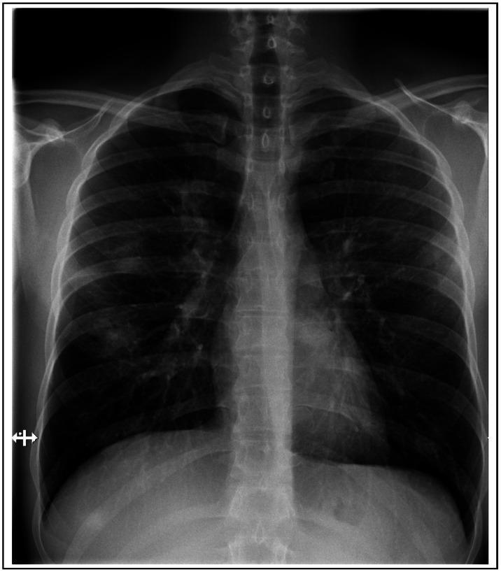
This diagnostic image is a posteroanterior (PA) chest X-ray illustrating the thoracic cavity of a patient with respiratory symptoms. The primary clinical finding is the presence of multifocal, patchy pulmonary infiltrates distributed bilaterally throughout both the left and right lung fields. These areas of increased opacity appear irregular in morphology and are scattered across the lung parenchyma rather than being localized to a single lobe. The cardiac silhouette, mediastinal contours, and diaphragmatic surfaces appear within normal limits. The costophrenic angles are clear, suggesting no significant pleural effusion. This radiographic presentation is consistent with diffuse parenchymal lung involvement, such as that seen in certain infectious processes, tropical eosinophilia, or inflammatory responses. It serves as a classic educational example for identifying non-specific pulmonary infiltrates on plain film radiography, highlighting the importance of correlating imaging findings with clinical history and laboratory data such as peripheral eosinophilia.
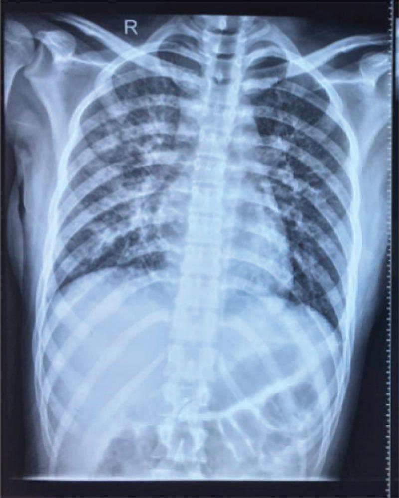
This diagnostic image is a posterior-anterior (PA) chest X-ray of a 22-year-old female patient, demonstrating significant bilateral pulmonary pathology. The lung fields exhibit diffuse, patchy, and reticulonodular infiltrates that are symmetrically distributed throughout the upper, middle, and lower zones. These increased opacities obscure the fine details of the pulmonary vasculature and indicate extensive alveolar or interstitial involvement. The hila appear somewhat prominent, likely due to the surrounding parenchymal density. The mediastinal contour, heart size, and diaphragmatic outlines are visible but partially blurred by the overlying lung disease. There is no clear evidence of significant pleural effusion or pneumothorax on this view. In a clinical context involving rapid onset dyspnea, fever, and peripheral eosinophilia, these radiographic findings are consistent with acute eosinophilic pneumonia (AEP) or severe community-acquired pneumonia. The image serves as an educational example of diffuse lung disease and the characteristic radiographic presentation of inflammatory pulmonary eosinophilic syndromes.
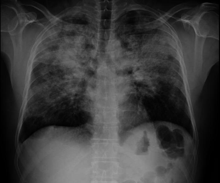
A posterior-anterior (PA) chest x-ray demonstrating extensive, bilateral, multifocal opacities. The primary finding is the presence of several foci of consolidation and ground-glass densities scattered throughout the lung parenchyma, with a notable predominance in the upper and middle lung zones and towards the periphery. These opacities are ill-defined and patchy, characteristic of alveolar filling processes. The cardiac silhouette, mediastinal contours, and hila appear within normal limits. The trachea is midline, and there is no evidence of significant pleural effusion or pneumothorax. The diaphragmatic domes are clear, and the lung volumes are preserved. In the clinical context of asthma and peripheral eosinophilia, these radiographic features are consistent with Chronic Eosinophilic Pneumonia (CEP), highlighting the ‘photonegative’ of pulmonary edema pattern where opacities are peripheral rather than central. This image serves as a key diagnostic example for radiological evaluation of eosinophilic lung diseases.
Pulmonary Hypersensitivity in Parasitic (Helminthic) Etiology
Why the Lung is the Target
The Core Immunological Mechanism
Step 1 - Th2 Polarization
- IL-4 - promotes IgE class switching
- IL-5 - key eosinophil growth and activation factor
- IL-13 - drives mucus production and airway hyperresponsiveness
- IgE - the antibody of Type I hypersensitivity
Step 2 - IgE-Mediated (Type I) Hypersensitivity
- Bronchospasm and wheeze
- Increased vascular permeability
- Mucus hypersecretion
Step 3 - Eosinophilic Tissue Damage
- Major Basic Protein (MBP)
- Eosinophil Cationic Protein (ECP)
- Eosinophil Peroxidase
The Two Main Clinical Syndromes
1. Loeffler Syndrome (Simple Pulmonary Eosinophilia)
- Cause: Transpulmonary migration of helminth larvae - primarily Ascaris lumbricoides, also hookworms (Ancylostoma, Necator), Strongyloides, Toxocara
- Mechanism: Larvae cross from pulmonary capillaries into alveoli, triggering acute Type I hypersensitivity + eosinophilic inflammation
- Features: Low-grade fever, nonproductive cough, dyspnea, wheeze, peripheral eosinophilia, Charcot-Leyden crystals in sputum, migratory "fleeting" pulmonary infiltrates on CXR
- Key point: Self-limited (resolves in 1-2 weeks) because larvae pass through and complete their cycle
- Fishman's Pulmonary Diseases, block 14
2. Tropical Pulmonary Eosinophilia (TPE)
- Cause: Filarial nematodes - Wuchereria bancrofti and Brugia malayi; microfilariae are trapped in the pulmonary vasculature/parenchyma
- Mechanism: Unlike Loeffler syndrome, microfilariae lodge in the lung and trigger a persistent, intense immune hyperreactivity - markedly elevated IgE, massive eosinophilia, eosinophilic granulomas and interstitial infiltrates
- Features: Nocturnal paroxysmal cough and bronchospasm ("nocturnal asthma"), weight loss, lymphadenopathy, marked eosinophilia (often >3000/µL), very high serum IgE, bilateral reticulonodular infiltrates on CXR
- Danger: Unlike Loeffler syndrome, TPE does NOT self-resolve - it can progress to chronic restrictive/fibrotic lung disease if untreated
- Murray & Nadel's Respiratory Medicine, block 14
Summary Table
| Feature | Loeffler Syndrome | Tropical Pulmonary Eosinophilia |
|---|---|---|
| Parasite | Ascaris, hookworms, Strongyloides | Wuchereria bancrofti, Brugia malayi |
| Mechanism | Larval transpulmonary migration | Microfilariae trapped in lungs |
| Hypersensitivity type | Type I (IgE-mediated) | Intense Th2/IgE/eosinophilic |
| Duration | Self-limited (1-2 weeks) | Persistent; can cause fibrosis |
| IgE | Elevated | Markedly elevated |
| Eosinophilia | Moderate | Extreme (>3000/µL) |
| Treatment | Supportive +/- prednisone | Diethylcarbamazine (DEC) |