
Iron deficiency anemia
Reading File
Now let me search the medical library and PubMed simultaneously for comprehensive coverage.
Finding Sources
Searching PubMed
"iron deficiency anemia"[MeSH Terms] AND "diagnosis"[MeSH Terms]
Now let me read the most comprehensive sources - Robbins Pathologic Basis of Disease (latest edition) and Goldman-Cecil for clinical management, in parallel.
Reading File
Reading File
Reading File
Let me get more content from Robbins and also read the clinical/treatment sections.
Reading File
Reading File
Let me get the key clinical/morphology/treatment sections from Robbins and also the Goldman-Cecil treatment section.
Reading File
Finding Sources
Let me get the clinical features/morphology and treatment sections from Robbins and the pharmacology section from Goodman & Gilman / Katzung.
Reading File
Reading File
Let me get the clinical features/morphology from Robbins and a bit more of the treatment info from Katzung.
Reading File
Reading File
I now have all the core content needed. Let me also quickly get the morphology/clinical features section from Robbins and fetch a relevant diagram.
Reading File
Loading Image
I now have comprehensive content from multiple authoritative sources. Let me also fetch the peripheral blood smear image.
Loading Image
I now have comprehensive, authoritative information from multiple medical textbooks. Here is the full, detailed response on Iron Deficiency Anemia:
Iron Deficiency Anemia (IDA)
Iron deficiency is the most common nutritional disorder in the world and the most frequent cause of anemia globally. It affects ~10% of people in high-resource countries and 25-50% in low-resource settings. - Robbins, Cotran & Kumar Pathologic Basis of Disease
Iron Metabolism - Foundation
Understanding IDA requires knowing how iron is handled in the body:
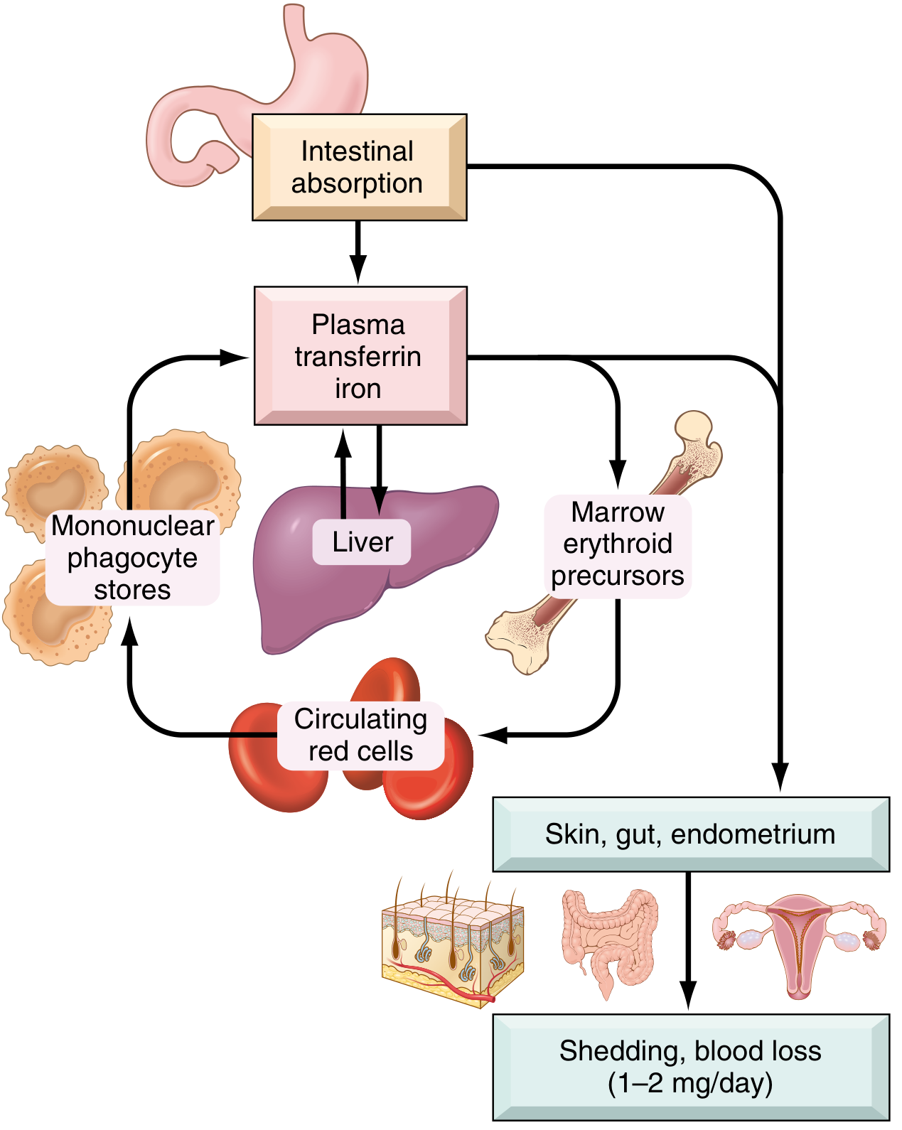
Total body iron distribution (healthy young adults):
| Pool | Males | Females |
|---|---|---|
| Hemoglobin | 2100 mg | 1750 mg |
| Myoglobin | 300 mg | 250 mg |
| Enzymes | 50 mg | 50 mg |
| Storage (ferritin + hemosiderin) | 1000 mg | 400 mg |
| Total | ~3450 mg | ~2450 mg |
- Iron is lost at only 1-2 mg/day via shed epithelial cells. There is no regulated excretory pathway.
- Daily dietary intake in the US: 10-20 mg; only ~20% of heme iron and 1-2% of nonheme iron is absorbed.
- Transferrin is the plasma carrier; normally ~one-third saturated, giving serum iron of ~120 µg/dL (males) and ~100 µg/dL (females). Total iron-binding capacity (TIBC) is normally 300-350 µg/dL.
The key regulator is hepcidin, a liver-derived peptide that inhibits ferroportin (the basolateral iron exporter in enterocytes and macrophages). When iron stores are replete or inflammation is present, hepcidin rises → ferroportin is degraded → iron is trapped in enterocytes and macrophages. When stores are low, hepcidin falls → increased duodenal absorption and iron release from macrophage stores. - Robbins, Cotran & Kumar; Robbins & Kumar Basic Pathology
Etiology and Causes
Iron deficiency arises from four main mechanisms:
1. Dietary Lack
- Rare in high-resource countries where ~2/3 of dietary iron is heme (animal products).
- High-risk groups: infants (breast milk provides only 0.3 mg/L), the impoverished, older adults with restricted diets, and vegans (no heme iron).
- Bioavailability is enhanced by: ascorbic acid, citric acid, amino acids, sugars.
- Bioavailability is reduced by: tannins (tea), carbonates, oxalates, phosphates.
2. Impaired Absorption
- Sprue (celiac disease), fat malabsorption, chronic diarrhea.
- Post-gastrectomy: reduced gastric acidity + rapid transit through duodenum.
3. Increased Requirement
- Infants, children, adolescents (rapid growth).
- Premenopausal women, especially during pregnancy.
- Chronic kidney disease patients on hemodialysis + erythropoietin therapy.
4. Chronic Blood Loss (most common cause in developed countries)
- GI tract (men and postmenopausal women): ulcers, cancers, hookworm infection. In adult males and postmenopausal females, GI blood loss must be presumed until proven otherwise - overlooking this risks missing a GI cancer.
- Uterus: menstrual blood loss (~30 mg iron per cycle); heavy menorrhagia causes significantly more.
- Urinary tract, other sites.
Stages of Progression
Iron deficiency progresses through sequential stages:
- Pre-latent - Storage depletion. Bone marrow iron stores fall; serum ferritin decreases. Hemoglobin and serum iron remain normal.
- Latent - Transport iron depletion. Serum iron falls, TIBC rises, transferrin saturation drops. No anemia yet, but erythroid hyperplasia appears in bone marrow.
- Frank IDA - Iron-deficient erythropoiesis. Anemia appears; microcytic, hypochromic red cells on smear. All iron indices are abnormal. - Robbins, Cotran & Kumar Pathologic Basis of Disease
Morphology
Bone marrow:
- Mild to moderate increase in erythroid progenitors (compensatory erythroid hyperplasia).
- Absence of stainable iron in macrophages - the most diagnostically significant finding; confirmed with Prussian blue stain.
Peripheral blood smear:

- Microcytosis (MCV < 80 fL)
- Hypochromia (MCHC < 30%): zone of central pallor enlarged beyond 1/3 of cell diameter; hemoglobin visible only at the rim.
- Poikilocytosis: characteristic "pencil cells" (small, elongated red cells).
Laboratory Diagnosis
| Test | IDA | Anemia of Chronic Inflammation |
|---|---|---|
| Serum iron | Low (< 30 µg/dL) | Low |
| TIBC | High | Low to normal |
| Transferrin saturation | < 15% (often < 10%) | Low |
| Serum ferritin | Low (< 12 µg/L) | Normal to high |
| Bone marrow iron | Absent | Increased in macrophages |
| Hepcidin | Low | High |
Key distinguishing point: ferritin is low in IDA but high or normal in anemia of chronic inflammation. A low ferritin is essentially diagnostic of IDA; an elevated ferritin does not exclude IDA in the setting of coexisting inflammation (ferritin is an acute-phase reactant).
The transferrin saturation below 15% combined with low serum iron and elevated TIBC is the classic lab triad. - Katzung's Basic and Clinical Pharmacology; Robbins, Cotran & Kumar
Clinical Features
General anemia symptoms: fatigue, pallor, weakness, dyspnea on exertion, palpitations, light-headedness.
Signs specific to severe or long-standing IDA (from iron depletion in non-erythroid tissues):
- Koilonychia - spoon-shaped nails
- Alopecia
- Atrophic glossitis - smooth, painful tongue
- Gastric mucosal atrophy
- Pica - craving for non-food substances (clay) or items like ice (pagophagia) or starch; linked to iron depletion in the CNS
- Intestinal malabsorption
- Plummer-Vinson syndrome (rare triad): microcytic hypochromic anemia + atrophic glossitis + esophageal webs
The dominant symptoms often relate to the underlying cause (GI disease, gynecologic condition, malnutrition, malabsorption). - Robbins, Cotran & Kumar Pathologic Basis of Disease
Workup and Investigation
For adult men and postmenopausal women with confirmed IDA:
- Colonoscopy first, then upper endoscopy + push enteroscopy if colonoscopy negative.
- Duodenal biopsy to screen for celiac disease.
- Test and treat H. pylori if present (reduces iron absorption; can cause microerosions).
- If all three endoscopic procedures are negative: capsule endoscopy.
- Only after negative GI workup: pursue non-GI causes.
For premenopausal women: GI evaluation warranted only after menorrhagia and other obvious gynecologic causes are excluded. - Goldman-Cecil Medicine
Treatment
Oral Iron (first-line for most patients)
Oral iron corrects anemia as rapidly as parenteral iron when GI absorption is intact. Ferrous (Fe²⁺) salts are preferred over ferric forms for better absorption.
| Preparation | Tablet size | Elemental iron/tablet | Usual dose |
|---|---|---|---|
| Ferrous sulfate (hydrated) | 325 mg | 65 mg | 2-4 tabs/day |
| Ferrous sulfate (desiccated) | 200 mg | 65 mg | 2-4 tabs/day |
| Ferrous gluconate | 325 mg | 36 mg | 3-4 tabs/day |
| Ferrous fumarate | 325 mg | 106 mg | 2-3 tabs/day |
- Target: 200-400 mg of elemental iron/day (about 25% is absorbed = 50-100 mg incorporated into Hgb daily).
- Continue treatment for 3-6 months after correcting the underlying cause to replenish stores.
- Response monitoring: reticulocytosis appears in 5-7 days; hemoglobin and red cell indices normalize over weeks.
- GI side effects (nausea, cramps, constipation, diarrhea) are dose-related; can be mitigated by taking with meals or switching to a different ferrous salt. Black stools are expected.
Parenteral Iron (for specific indications)
Reserved for patients who:
- Cannot tolerate or absorb oral iron (malabsorption, post-gastrectomy, IBD involving proximal small bowel)
- Have very high ongoing iron requirements (hemodialysis + erythropoietin)
- Need rapid repletion
Available IV forms:
- Iron dextran (50 mg elemental iron/mL) - can be given as a total-dose infusion; risk of anaphylaxis requires a test dose first.
- Sodium ferric gluconate complex - lower risk of anaphylaxis vs. iron dextran; multiple infusions needed.
- Iron sucrose - low risk; commonly used in CKD patients.
- Ferric carboxymaltose (newer) - high-dose single infusion; favorable safety profile.
Mechanism of safety: ferric iron is stabilized in a carbohydrate shell, allowing slow controlled release and avoiding the toxicity of free ionic iron. - Katzung's Basic and Clinical Pharmacology
Differentiation from Anemia of Chronic Inflammation
This is a common and clinically important distinction:
- Anemia of chronic inflammation (ACI) results from IL-6-driven hepcidin upregulation, which sequesters iron in macrophages and restricts delivery to erythroid precursors.
- Unlike IDA, ACI shows elevated ferritin, elevated storage iron in marrow macrophages, and reduced TIBC.
- Red cells in ACI may be normocytic/normochromic or mildly microcytic/hypochromic.
- Treatment of ACI targets the underlying disease; erythropoietin helps some patients (especially those with cancer). - Robbins, Cotran & Kumar Pathologic Basis of Disease
Sources: Robbins, Cotran & Kumar Pathologic Basis of Disease | Robbins & Kumar Basic Pathology | Goldman-Cecil Medicine | Katzung's Basic and Clinical Pharmacology, 16e | Goodman & Gilman's Pharmacological Basis of Therapeutics
Turners syndrome obg
Reading File
This is a broad OBG (Obstetrics & Gynecology) topic - Turner syndrome with OBG focus. Let me search multiple sources simultaneously.
Finding Sources
Searching PubMed
"Turner syndrome"[MeSH Terms] AND "fertility" OR "pregnancy"
Now let me search for OBG-specific textbooks and read multiple sources simultaneously.
Finding Sources
Reading File
Reading File
Excellent content. Let me read more from Robbins on management and the Maternal-Fetal Medicine and Developing Human sections for OBG-specific content.
Reading File
Reading File
Reading File
Now let me search specifically for OBG-focused content - fertility, HRT, pregnancy management in Turner syndrome.
Finding Sources
Loading Image
Excellent! Now let me read the Berek & Novak's Gynecology section and the Goldman-Cecil treatment section for the OBG management details.
Reading File
Reading File
Reading File
Let me get more Berek & Novak content on Turner syndrome treatment and also look for HRT/management specifics.
Reading File
Finding Sources
Let me read the Goodman & Gilman estrogen treatment section and the Campbell Walsh Urology key points for management details.
Reading File
Reading File
Let me get the key management/treatment section from Campbell Walsh on Turner syndrome and Creasy-Resnik on prenatal.
Reading File
Reading File
I now have comprehensive content from multiple authoritative textbooks. Let me compile the full, detailed OBG-focused response on Turner Syndrome.
Turner Syndrome - OBG Perspective
Quick definition: Turner syndrome is the most common sex chromosome abnormality in phenotypic females - characterized by complete or partial monosomy of the X chromosome, causing hypergonadotropic hypogonadism, primary amenorrhea, streak ovaries, and infertility.
Incidence & Genetics
- Incidence: ~1 in 2000-2500 live female births (1 in 3000 by some sources). However, ~99% of 45,X fetuses abort spontaneously; it is the most common cytogenetic abnormality in spontaneous abortions, accounting for ~18% of all chromosomally abnormal abortions.
- Origin of error: In ~75-80% of cases, the X chromosome retained is maternal in origin - i.e., the paternal X or Y is missing (error in paternal gametogenesis/nondisjunction).
Karyotypes
| Type | Karyotype | Frequency |
|---|---|---|
| Classic monosomy | 45,X | ~57% |
| Isochromosome (loss of short arm) | 46,X,i(X)(q10) | ~14% |
| Ring chromosome | 46,X,r(X) | Structural variant |
| Short/long arm deletion | 46,X,del(Xp) or del(Xq) | Structural variant |
| Mosaic 45,X/46,XX | Most common mosaic | ~10-15% |
| Mosaic 45,X/46,XY | Y-bearing mosaic | ~2-5% |
Mosaicism may be detected in up to 75% of cases with sensitive molecular techniques - conventional cytogenetics detects only ~30%. Mosaicism with a higher proportion of 46,XX cells = milder phenotype; a very small number can even conceive spontaneously. - Robbins, Cotran & Kumar Pathologic Basis of Disease
Pathogenesis of Gonadal Failure
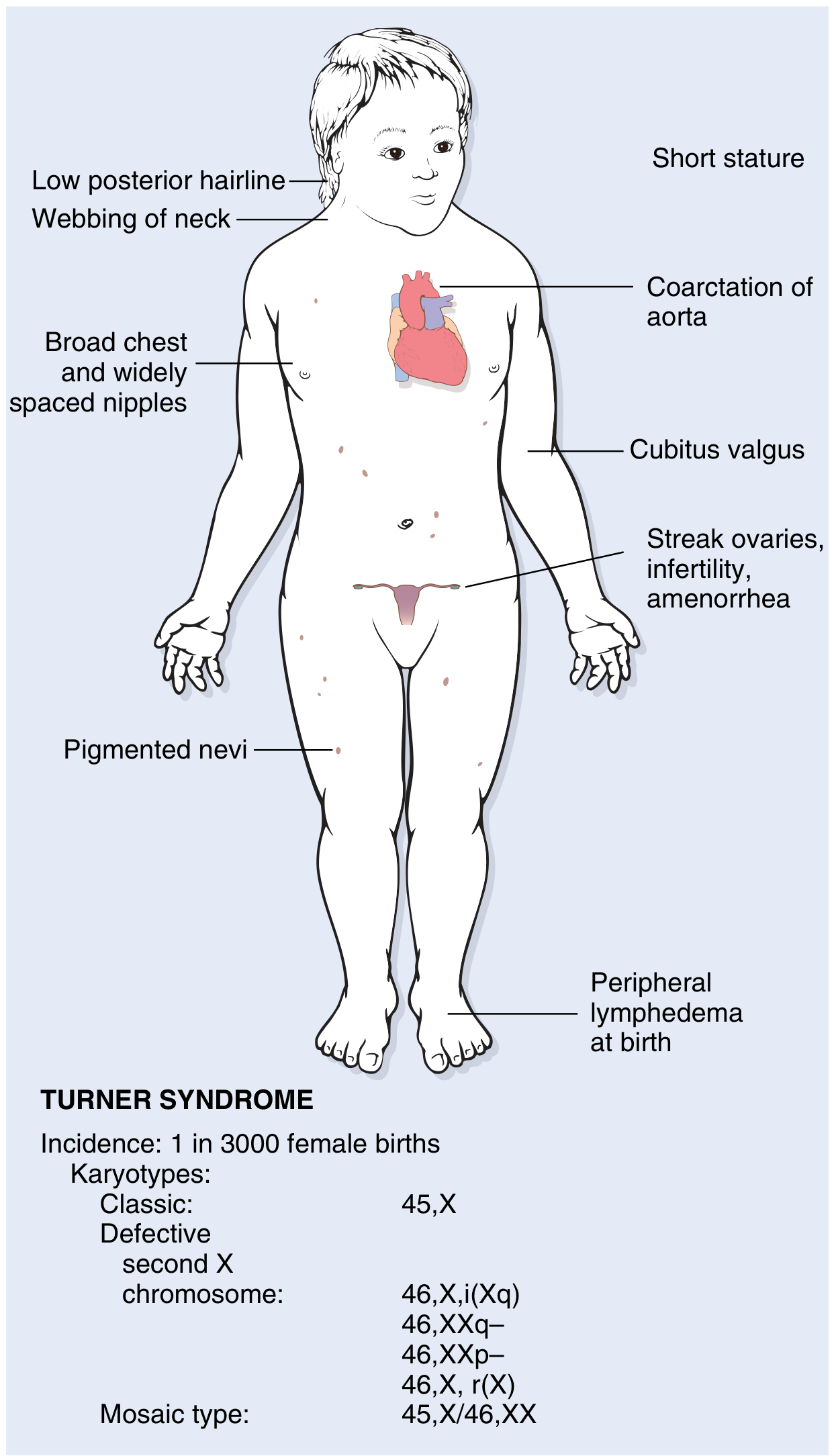
- Fetal ovaries develop normally for the first 18 weeks of gestation.
- The second X chromosome is required to maintain oocytes - without it, accelerated apoptotic loss of oocytes occurs.
- By age 2 years, all oocytes are lost - "menopause occurs before menarche."
- Ovaries are replaced by streak gonads: white, fibrous, 2-3 cm structures in the broad ligament, devoid of follicles and oocytes.
- Result: no estrogen/progesterone production → elevated FSH and LH (hypergonadotropic hypogonadism). - Robbins, Cotran & Kumar; Campbell Walsh Urology
Key gene: SHOX (Xp22.33, pseudoautosomal region, escapes X inactivation) - haploinsufficiency causes short stature. Both males and females normally have 2 copies.
Clinical Features (OBG-Relevant)
In Infancy/Neonatal Period
- Cystic hygroma / nuchal lymphedema (cystic distension of lymphatic channels at the nape)
- Peripheral lymphedema of hands and feet at birth
- Webbed neck (residual from resolved lymphedema)
- Low posterior hairline
In Childhood/Adolescence
- Short stature (below 3rd percentile) - universal in non-mosaic Turner syndrome
- Failure of puberty in ~90% of affected females
- Primary amenorrhea (most common presentation in teens)
- Absent or minimal secondary sexual characteristics (no breast development, minimal pubic hair)
- Infantile genitalia
- Shield chest with widely spaced nipples
- Cubitus valgus (increased carrying angle of arms)
- Short 4th metacarpals; hypoplastic nails
- High-arched palate; multiple pigmented nevi; low-set ears; micrognathia; hypertelorism
Cardiovascular (most common cause of death in childhood)
- Coarctation of aorta (~30%) - 5% of females with coarctation have Turner syndrome
- Bicuspid aortic valve (most common structural defect)
- Aortic root dilation (30% of patients)
- 100-fold increased risk of aortic dissection
Other Systemic Features
- Horseshoe kidney (renal anomaly)
- Hashimoto thyroiditis (autoimmune hypothyroidism in up to 50%, especially isochromosome Xq)
- Glucose intolerance, insulin resistance, obesity, NAFLD - metabolic syndrome in a subset
- Sensorineural hearing loss
- Subtle deficits in visual-spatial processing (intelligence generally normal)
- Osteoporosis (from chronic estrogen deficiency)
OBG-Specific: Amenorrhea Classification
Turner syndrome presents as primary amenorrhea (absence of menstruation by age 13 without secondary sexual development, or by age 15 with breast development).
WHO Classification: Turner falls into WHO Group III - Hypergonadotropic Hypogonadism:
- Elevated FSH and LH (reflecting absent gonadal feedback)
- No endogenous estrogen production
- Streak ovaries on pelvic imaging
Turner is the most common cause of hypergonadotropic hypogonadism in women with primary amenorrhea. - Berek & Novak's Gynecology
Prenatal Diagnosis
Turner syndrome may be identified prenatally by:
- Ultrasound findings: increased nuchal translucency, cystic hygroma, lymphedema, coarctation of aorta, renal anomalies
- Cell-free fetal DNA (cfDNA) screening
- Amniocentesis/CVS karyotyping: definitive
Important counseling point: ~90% of fetuses with incidentally detected 45,X/46,XX or 45,X/46,XY mosaicism prenatally will have a normal phenotype at birth ("ascertainment bias"). This has profound implications for prenatal genetic counseling. - Campbell Walsh Urology
Diagnosis
- Karyotype (gold standard) - at least 25-30 cells analyzed; FISH for Y-material if needed
- Hormonal profile: markedly elevated FSH and LH, low estradiol
- Pelvic ultrasound: streak ovaries, infantile uterus
- Echocardiography: bicuspid aortic valve, coarctation
- Renal ultrasound: horseshoe kidney
- Bone age radiograph
- Thyroid function tests (TSH, anti-TPO antibodies)
- Audiogram (sensorineural hearing loss)
- Fasting glucose / HbA1c
Management
1. Growth Hormone (GH) Therapy
- Recombinant human GH at higher-than-replacement doses (~67 µg/kg/day)
- Often combined with oxandrolone (anabolic androgen) to improve final height
- Estrogen therapy is delayed to allow maximal GH-driven growth
- Mean adult height gain: ~5 cm greater compared to no treatment - Goodman & Gilman's; Creasy & Resnik's
2. Estrogen Replacement Therapy (HRT)
- Started at age ~11-12 years (or bone age ~11) once growth is maximized
- Goal: induce puberty, develop secondary sexual characteristics, protect bone health, prevent cardiovascular metabolic effects of estrogen deficiency
- Regimen:
- Low-dose estradiol started first (to mimic early puberty)
- Gradually increased over 2-3 years
- Cyclic progesterone added once breakthrough bleeding occurs or after 1-2 years of estrogen - to protect endometrium
- Continued until average age of menopause (~50 years)
- Estrogen therapy significantly improves bone mineral density; also associated with neurocognitive and behavioral benefits in girls with Turner syndrome
3. Y Chromosome Material - Gonadectomy
- Mandatory karyotyping to exclude Y-chromosomal material (PCR for SRY if standard karyotyping uncertain)
- 5-10% of Turner patients have Y-cell lines (full 45,X/46,XY or Y-chromosome fragments)
- Risk of gonadoblastoma (in-situ germ cell cancer) is ~12% in those with Y material
- Gonadoblastoma can undergo malignant transformation to dysgerminoma
- Gonadectomy is recommended in all patients with confirmed Y chromosomal material - Berek & Novak's; Campbell Walsh Urology
4. Fertility and Reproductive Options
Spontaneous fertility:
- Extremely rare in classic 45,X (~1-2%)
- More common in mosaic patients (45,X/46,XX) - spontaneous puberty in up to 30%, but premature ovarian failure is common and oocyte reserve must be assessed urgently
- Pregnancy rate with hypergonadotropic amenorrhea approximately 6-8% (Goldman-Cecil)
Fertility preservation:
- Oocyte/embryo cryopreservation is considered experimental but offered to selected mosaic patients with residual follicles
- Must be done before complete follicular depletion
Assisted reproduction:
- Oocyte donation (donor oocytes + partner sperm → IVF → embryo transfer) is the primary option
- The uterus and fallopian tubes are normal - successful pregnancy via donor oocytes is possible
- Endometrium is prepared with exogenous estrogen and progesterone
Critical OBG Warning - Pregnancy Risks in Turner Syndrome:
Women with Turner syndrome contemplating pregnancy have a markedly increased risk of aortic root rupture/aortic dissection during pregnancy (due to pre-existing aortic root dilation and bicuspid aortic valve). This is potentially fatal.
- Baseline aortic imaging (MRI/CT) is mandatory before pregnancy
- Aortic dissection risk is 100-fold higher than the general population
- Goldman-Cecil: "Whether women with gonadal dysgenesis should be offered pregnancy via donor oocytes is now the subject of debate because of markedly increased incidence of aortic rupture secondary to medial necrosis during pregnancy."
- All women with Turner syndrome must be counseled regarding cardiovascular risks before attempting pregnancy. - Goldman-Cecil Medicine; Berek & Novak's Gynecology
Multidisciplinary Surveillance (Lifelong)
| System | Screening |
|---|---|
| Cardiovascular | Echo, aortic MRI every 5-10 years (more frequently if abnormal); BP monitoring |
| Thyroid | TSH, anti-TPO antibodies annually |
| Metabolic | Fasting glucose/HbA1c annually; lipid profile |
| Renal | Ultrasound; BP screening |
| Audiology | Annual audiogram |
| Bone | DEXA scan (osteoporosis prevention with HRT) |
| Eyes | Screen for strabismus, ptosis |
| Neurocognitive | Learning disability screening in childhood |
| Endocrine | Growth monitoring; GH therapy; HRT continuity |
Key OBG Summary Points
| Feature | Detail |
|---|---|
| Karyotype | 45,X (57%), mosaics 43%, structural X variants |
| Incidence | 1 in 2000-2500 live female births |
| Ovaries | Streak gonads (fibrous, devoid of follicles) |
| Hormones | FSH/LH very high; estradiol very low |
| Presentation | Primary amenorrhea, absent puberty, short stature |
| Fertility | Virtually absent in 45,X; occasional in mosaic; donor oocyte IVF possible |
| Y material | 5-10%; gonadectomy mandatory (gonadoblastoma risk 12%) |
| Main pregnancy risk | Aortic dissection (100x increased risk) |
| Treatment | GH + oxandrolone → estrogen replacement → cyclic progesterone |
| Cancer risk | Gonadoblastoma → dysgerminoma (if Y material present) |
Sources: Robbins, Cotran & Kumar Pathologic Basis of Disease | Robbins & Kumar Basic Pathology | Berek & Novak's Gynecology | Goldman-Cecil Medicine | Campbell Walsh Wein Urology | Goodman & Gilman's Pharmacological Basis of Therapeutics | Creasy & Resnik's Maternal-Fetal Medicine | The Developing Human: Clinically Oriented Embryology
This is a shared conversation. Sign in to Orris to start your own chat.