
Transport of blood
Reading File
Now let me search the medical library for comprehensive content on blood transport.
Finding Sources
Finding Sources
Excellent - found key sections. Let me read all the primary content in parallel.
Reading File
Reading File
Reading File
Great content gathered. Let me now read the CO2 transport section and more O2 transport details in parallel.
Reading File
Reading File
Reading File
Excellent content gathered. Let me now fetch the key diagrams and read the O2 dissociation curve and CO2 transport sections.
Reading File
Reading File
Loading Image
I have all the essential content needed. Now I can compile a thorough, well-structured answer on the transport of blood (O2 and CO2 transport), including the key diagrams.
Transport of Blood (O₂ and CO₂)
The "transport of blood" in physiology refers primarily to how blood carries oxygen (O₂) from the lungs to tissues and carbon dioxide (CO₂) from tissues back to the lungs. Both gases are transported by two mechanisms: dissolved in plasma and chemically combined.
I. Transport of Oxygen (O₂)
1. Two Forms of O₂ in Blood
O₂ is carried in blood in two forms:
| Form | Proportion | Mechanism |
|---|---|---|
| Dissolved in plasma | ~2% | Henry's law; proportional to PO₂ |
| Bound to hemoglobin | ~98% | Chemical combination |
Why dissolved O₂ alone is insufficient:
- Solubility of O₂ in blood = 0.003 mL O₂/100 mL blood per mm Hg
- At a normal arterial PO₂ of 100 mm Hg: dissolved O₂ = only 0.3 mL O₂/100 mL blood
- At a resting cardiac output of 5 L/min, this delivers only 15 mL O₂/min
- But the body needs ~250 mL O₂/min at rest - a 17-fold shortfall
- Therefore, hemoglobin binding is essential for adequate O₂ delivery
(Costanzo Physiology 7th Ed; Medical Physiology)
2. Hemoglobin Structure and O₂ Binding
- Normal adult hemoglobin (Hb A) is a tetramer (~68 kDa) with 2α + 2β subunits
- Each subunit contains a heme (iron-porphyrin) group + a globin polypeptide
- Each heme group binds one O₂ molecule → 4 O₂ per Hb molecule
- Iron must be in the ferrous (Fe²⁺) state to bind O₂
- 1 g of hemoglobin A can bind 1.34 mL O₂
- Normal Hb concentration = 15 g/100 mL → O₂-binding capacity = 20.1 mL O₂/100 mL blood
Hemoglobin variants:
| Variant | Feature | Clinical Significance |
|---|---|---|
| HbA (α₂β₂) | Normal adult | - |
| HbF (α₂γ₂) | Fetal; higher O₂ affinity | Facilitates O₂ transfer from mother to fetus |
| HbS (αA₂βS₂) | Deoxygenated form sickles | Sickle cell disease; lower O₂ affinity |
| Methemoglobin | Fe³⁺ (ferric) | Cannot bind O₂; caused by nitrites/sulfonamides |
(Costanzo Physiology 7th Ed)
3. O₂ Content of Blood
The O₂ content (actual O₂ per 100 mL blood) is calculated as:
O₂ content = (O₂-binding capacity × % Saturation) + Dissolved O₂
- Normal arterial blood (98% saturated): O₂ content ≈ 20.0 mL O₂/100 mL
- O₂ delivery = Cardiac output × O₂ content
4. Diffusion of O₂: Alveoli → Blood → Tissues
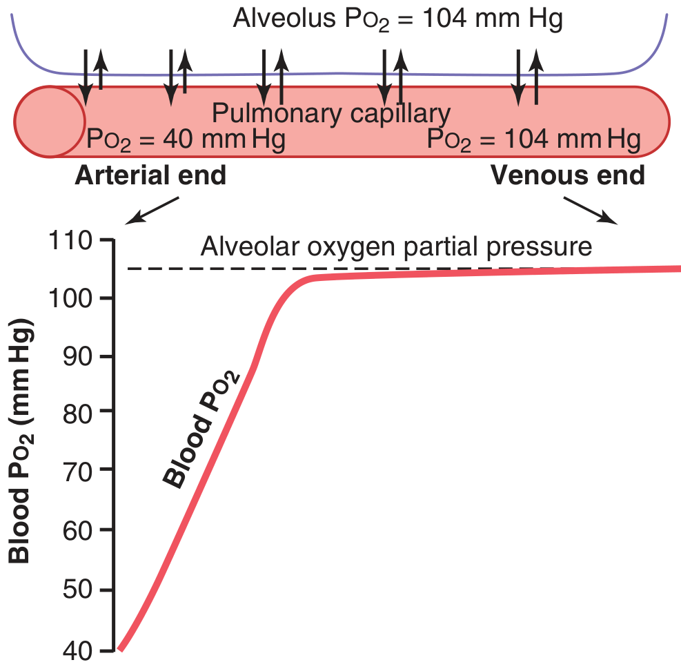
At the lungs (alveolar capillaries):
- Alveolar PO₂ = 104 mm Hg
- Venous blood entering pulmonary capillary PO₂ = 40 mm Hg
- Pressure gradient = 64 mm Hg → drives O₂ into blood rapidly
- Blood reaches near-equilibrium (~104 mm Hg) after only 1/3 of capillary length
Venous admixture ("shunt"):
- ~2% of blood passes through bronchial circulation (unexposed to alveolar air), PO₂ ≈ 40 mm Hg
- This mixes with oxygenated blood → aortic PO₂ drops slightly to ~95 mm Hg
At the tissues (peripheral capillaries):
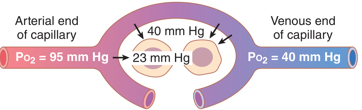
- Arterial blood arrives at PO₂ = 95 mm Hg
- Interstitial fluid PO₂ = 40 mm Hg
- Intracellular PO₂ = ~23 mm Hg (or even lower near mitochondria)
- O₂ diffuses down this gradient into cells
(Guyton & Hall Textbook of Medical Physiology)
5. Factors Affecting O₂-Hemoglobin Dissociation (Bohr Effect)
The oxyhemoglobin dissociation curve describes the sigmoidal relationship between PO₂ and Hb saturation:
Right shift (decreased O₂ affinity, more O₂ released to tissues):
- ↑ PCO₂
- ↑ [H⁺] (acidosis / ↓ pH) - Bohr effect
- ↑ Temperature
- ↑ 2,3-DPG (diphosphoglycerate)
Left shift (increased O₂ affinity, O₂ held tighter):
- ↓ PCO₂
- ↓ [H⁺] (alkalosis / ↑ pH)
- ↓ Temperature
- Fetal hemoglobin (HbF)
- CO poisoning (carboxyhemoglobin)
II. Transport of Carbon Dioxide (CO₂)
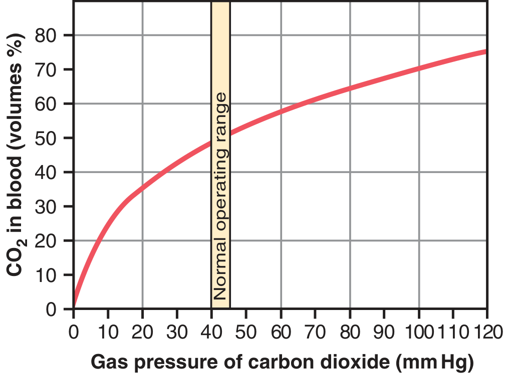
CO₂ is produced by cellular metabolism and must be carried from tissues to lungs. Average transport: 4 mL CO₂ per 100 mL blood. CO₂ is transported in three forms:
1. Dissolved CO₂ (~7%)
- PCO₂ venous = 45 mm Hg; arterial = 40 mm Hg
- Dissolved CO₂ at 45 mm Hg = 2.7 mL/100 mL; at 40 mm Hg = 2.4 mL/100 mL
- Difference = 0.3 mL/100 mL transported this way
2. As Bicarbonate Ion (HCO₃⁻) - the MAJOR form (~70%)
This is the most important mechanism, driven by carbonic anhydrase inside RBCs:
CO₂ + H₂O →[carbonic anhydrase]→ H₂CO₃ → H⁺ + HCO₃⁻
- Carbonic anhydrase accelerates this reaction 5,000-fold
- H⁺ ions are buffered by hemoglobin inside RBCs
- HCO₃⁻ diffuses out of RBCs into plasma in exchange for Cl⁻ (chloride shift / Hamburger phenomenon)
- The venous RBC thus has higher chloride content than arterial RBC
Importance: Blocking carbonic anhydrase (e.g., acetazolamide) raises tissue PCO₂ to ~80 mm Hg (vs. normal 45 mm Hg)
3. Carbaminohemoglobin (~20-30%)
- CO₂ binds directly to amine (-NH₂) groups on hemoglobin → carbaminohemoglobin (CO₂Hb)
- Reaction is reversible; CO₂ released easily in alveoli where PCO₂ is lower
- A small amount also binds to plasma proteins (less significant - only 1/4 the protein mass of Hb)
| Form | % of CO₂ Transported |
|---|---|
| Dissolved CO₂ | ~7% |
| Bicarbonate (HCO₃⁻) | ~70% |
| Carbaminohemoglobin | ~20-23% |
(Guyton & Hall Textbook of Medical Physiology)
The Haldane Effect
Oxygenation of Hb in the lungs displaces CO₂ - this is the reverse of the Bohr effect:
- When O₂ binds to Hb in alveolar capillaries, Hb releases CO₂ more readily
- This increases CO₂ unloading from blood to alveolar air
- The Haldane effect is actually quantitatively more important than the Bohr effect in enhancing gas exchange
III. Summary: The Full Transport Cycle
LUNGS
  Alveolar PO₂ = 104 mm Hg  →  O₂ loads onto Hb
  Alveolar PCO₂ = 40 mm Hg  →  CO₂ unloads from blood
         ↓
ARTERIAL BLOOD
  PO₂ = 95 mm Hg
  PCO₂ = 40 mm Hg
  O₂ content ≈ 20 mL/100 mL
         ↓
TISSUES
  Interstitial PO₂ = 40 mm Hg  →  O₂ unloads from Hb
  Intracellular PCO₂ rises  →  CO₂ loads into blood
         ↓
VENOUS BLOOD
  PO₂ = 40 mm Hg
  PCO₂ = 45 mm Hg
  CO₂ content rises ~4 mL/100 mL
         ↓
Back to LUNGS
Key numbers to remember:
| Parameter | Arterial | Venous |
|---|---|---|
| PO₂ | 95 mm Hg | 40 mm Hg |
| PCO₂ | 40 mm Hg | 45 mm Hg |
| O₂ content | ~20 mL/100 mL | ~15 mL/100 mL |
| Hb O₂ saturation | ~98% | ~75% |
Sources:
- Guyton and Hall Textbook of Medical Physiology (Chapter 41)
- Costanzo Physiology, 7th Edition
- Medical Physiology (Boron & Boulpaep)
This is a shared conversation. Sign in to Orris to start your own chat.