
Frenectomy
Reading File
Now let me search the medical library for frenectomy content in parallel.
Finding Sources
Finding Sources
Searching PubMed
frenectomy frenum
Good - the main content is in Cummings Otolaryngology block 46. Let me also check the Pfenninger book and read the Cummings content.
Reading File
Finding Sources
Let me read more of the Cummings block for complete content on definitions, indications, and technique, plus the Pfenninger primary care content.
Reading File
Reading File
Let me now read the surgical technique and maxillary frenectomy sections.
Reading File
Reading File
Let me read more of the technique and read the Cummings sections on surgical technique for frenectomy.
Reading File
Reading File
I now have comprehensive information. Let me fetch a couple of the key clinical images to include in the response.
Loading Image
Loading Image
I now have comprehensive information to write a thorough response. Here is the full overview of frenectomy:
Frenectomy
Terminology and Definitions
The term frenectomy (also called frenulectomy) refers to complete surgical removal of a frenum (frenulum) along with its attachment to the underlying bone. It is distinct from related procedures:
- Frenotomy - simple incision/release of the frenulum without excision (the most common procedure for neonates with tongue tie)
- Frenuloplasty - release of the frenulum combined with plastic reconstruction, often using a Z-plasty technique
- Frenectomy - complete removal of the frenulum structure
These procedures are most commonly performed for two distinct anatomical sites: the lingual frenulum (tongue tie/ankyloglossia) and the maxillary labial frenulum (lip tie).
Part 1: Lingual Frenulum (Ankyloglossia / Tongue Tie)
Anatomy
The frenum of the tongue is a triangular fold of mucous membrane extending from the lower midline gingival tissue, along the floor of the mouth, arching to the midline of the undersurface of the tongue. In ankyloglossia, the frenulum attaches too far toward the tongue tip, restricting normal elevation or protrusion.
Epidemiology
- Incidence is approximately 4-10% of newborns (varies by definition used)
- More common in males (male:female ratio ~1.5-2.6:1)
- No racial predilection
- In a US database study, affected children were more often male (63.6%), privately insured (60.1%), and from higher-income zip codes (78.1%)
- Incidence appears to decrease in older children, suggesting some cases resolve spontaneously as the frenulum stretches or naturally breaks over time
Classification
The most widely used system classifies ankyloglossia into four types:
| Type | Description |
|---|---|
| Type I | Frenulum inserts at the very tip of the tongue (anterior ankyloglossia) |
| Type II | Frenulum inserts slightly behind the tongue tip |
| Type III | Frenulum inserts at the mid- to posterior undersurface of the tongue |
| Type IV | Submucosal frenulum ("posterior tongue tie") - flat and less obvious |
Types I and II are considered anterior tongue tie, while Types III and IV are called posterior tongue tie - the latter is a more recently recognized and somewhat controversial category.
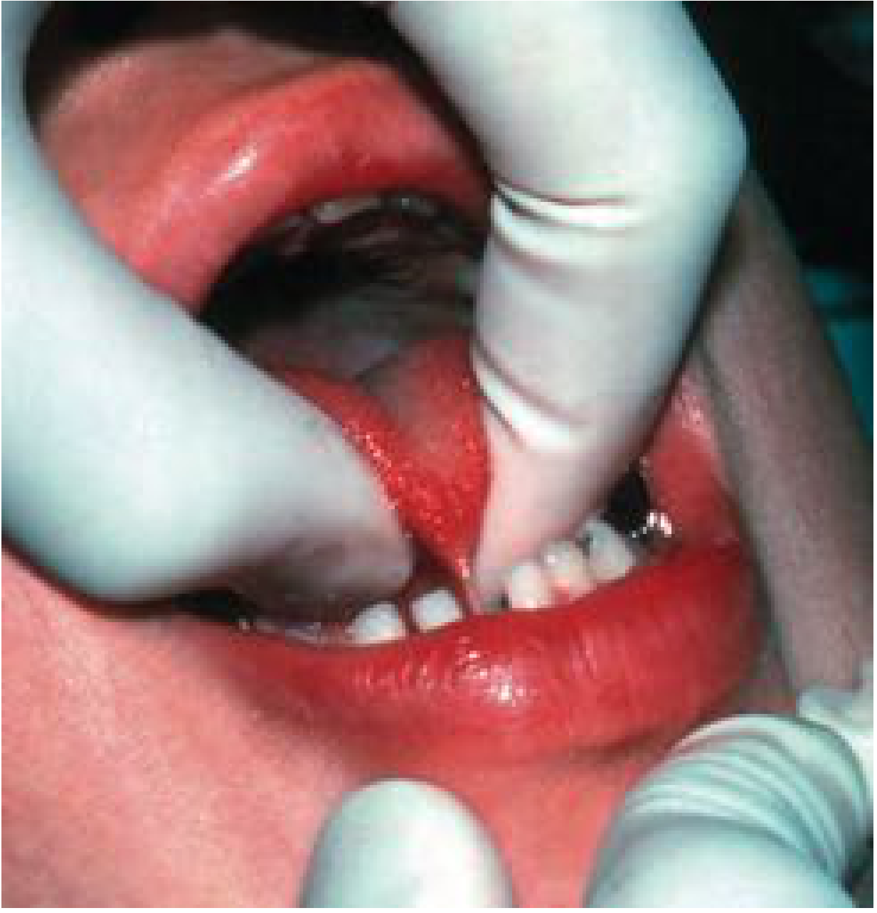
Ankyloglossia with the tongue tethered to the floor of the mouth - Cummings Otolaryngology
Clinical Sequelae
Breastfeeding problems (strongest evidence):
- Difficulty achieving a deep latch and seal
- Maternal nipple pain and soreness
- Reduced milk transfer
- Failure of infant to thrive
- Recurrent mastitis in the mother
Speech concerns (limited evidence):
- Articulation difficulties, especially lingual-dental sounds (t, d) and sibilants (z, s, th)
- Many individuals with ankyloglossia develop entirely normal speech without treatment
Mechanical and social effects:
- Difficulty licking lips or cleaning teeth with tongue
- Diastasis between lower central incisors from frenulum pressure
- Difficulty playing wind instruments
- Social embarrassment/peer ridicule
Indications for Treatment
- Neonates/infants: Poor breastfeeding latch and mechanics, maternal nipple pain, poor weight gain - strongest evidence supports treatment
- Older children: Documented articulation difficulties, particularly after speech therapy has been attempted
- Adolescents/adults: Mechanical concerns, social concerns, dental hygiene issues
- Age under 30 days is associated with higher probability of breastfeeding improvement post-frenotomy
Contraindications (for office-based frenotomy)
- Unstable medical conditions (bleeding disorders)
- Active dental or oral infection
- Orofacial abnormalities (refer to specialist)
- Severe "frozen tongue" ankyloglossia requiring Z-plasty - these require general anesthesia and referral to an experienced surgeon
Surgical Technique: Frenotomy (Office-Based)
Equipment:
- Straight or curved mosquito hemostat
- Surgical scissors (iris or Metzenbaum)
- Tongue retractor (small spoon, wooden tongue blade)
- Optional: topical anesthetic, lidocaine with epinephrine for bleeding control
Steps:
- Immobilize the infant (swaddling, papoose board, parental assistance); crying often improves exposure
- Identify the frenulum and the abnormal portion causing restriction
- For a thin/lucent membranous frenulum (infants under ~4 months): direct snipping without hemostat is acceptable - bleeding is minimal and comparable to biting the lip
- For a thicker, coarser frenulum: apply topical anesthesia (benzocaine syrup on cotton swab - note: FDA has issued a boxed warning against lidocaine/benzocaine in infants for teething due to seizure and methemoglobinemia risk; use only in context of the procedure)
- Optional: Clamp the frenulum with the mosquito hemostat to crush tissue at the snip point - this provides some analgesia to the crushed area; then snip through the crushed tissue only
- A diamond-shaped wound results after frenotomy - this heals by secondary intention
- Control bleeding with a dry or epinephrine-moistened cotton swab; ice may reduce oozing
Post-procedure: Resume normal feeding immediately; no special wound care required.
Frenuloplasty (Z-Plasty)
Used for severe restriction. The diamond-shaped defect created by frenotomy is sutured closed by transposing two triangular tissue flaps - this lengthens the frenulum and prevents re-adhesion. Requires suturing skills and regional or general anesthesia.
Surgical Outcomes
Evidence from multiple cohort studies and RCTs shows that frenotomy for anterior ankyloglossia in newborns:
- Reduces maternal nipple pain during breastfeeding
- Improves breastfeeding mechanics and milk transfer
- Increases probability of resumed breastfeeding if previously stopped
- 73% of mothers who had stopped breastfeeding before frenotomy resumed after the procedure (in one trial with ~4 months follow-up)
- Better outcomes when performed in infants under 30 days vs. over 30 days
A 2017 Cochrane meta-analysis of 5 RCTs found frenotomy consistently reduced short-term maternal nipple pain, though a consistently positive effect on overall breastfeeding duration was not confirmed.
For posterior tongue tie, speech, and social-mechanical concerns - evidence remains limited and controlled trials are lacking.
- Cummings Otolaryngology Head and Neck Surgery, Chapter 207
Part 2: Maxillary Labial Frenulum (Lip Tie)
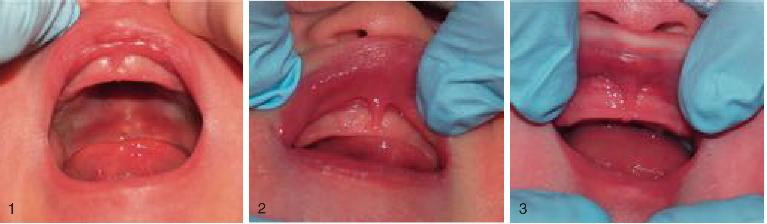
Types 1, 2, and 3 of the maxillary upper lip frenulum - Cummings Otolaryngology
Background
All newborns have some degree of upper labial frenulum. Its evolution over time is poorly understood. Lip tie (tight maxillary frenulum) is increasingly diagnosed and treated, often together with ankyloglossia. It has been described as a condition where the frenulum limits upper lip mobility and function.
Clinical assessment - the "blanching test": Pull the upper lip superiorly and forward to inspect for blanching of the palatine papilla, indicating a tighter attachment.
Classification (4-grade system)
- Grade 1: No tissue attachment of the lip to gingival tissue
- Grade 2: Attachment to free gingival margin
- Grade 3: Attachment between free margin and anterior papilla
- Grade 4: Attachment onto the palate
(There is significant intra- and inter-rater inconsistency in grading, even among experts)
Sequelae
- Breastfeeding difficulties - prevents deep latch and adequate seal; maternal nipple pain
- In older children: dental caries on anterior surfaces of maxillary central incisors (different from baby-bottle caries, which presents lingually); reduced access for tooth brushing and plaque removal
- Midline diastema - a hypertrophic maxillary frenulum may be an etiologic factor (along with genetic factors) for a space between the upper central incisors
Frenectomy Technique (Maxillary)
The surgical approach has changed little since Angle's 1907 description:
- The complete removal of the maxillary frenulum involves excision of its attachment to the underlying periosteum
- The wound may be left open, or primary closure may be attempted
- Timing relative to orthodontics is controversial: if performed before orthodontic treatment (generally after the 6 upper permanent anterior teeth have fully erupted), speed of diastema closure may be affected. Some evidence suggests frenectomy before orthodontics accelerates tooth movement; others find no difference
Laser Frenectomy
Both CO₂ and diode lasers have been used for labial and lingual frenectomy. A 2026 systematic review and meta-analysis (PMID: 41369715) compared diode laser vs. conventional scalpel for labial frenectomy and found evidence in favor of the laser technique for certain outcomes (healing, bleeding, patient comfort).
Summary: Frenectomy vs. Frenotomy vs. Frenuloplasty
| Procedure | Definition | Typical Setting | Anesthesia |
|---|---|---|---|
| Frenotomy | Simple snip/incision of frenulum; no excision | Office/bedside | Topical or none (neonates) |
| Frenectomy | Complete excision of frenulum + periosteal attachment | Office or OR | Local or general |
| Frenuloplasty (Z-plasty) | Release + plastic reconstruction with transposed flaps | OR | General or regional |
Recent Evidence (2025-2026)
Three recent systematic reviews are relevant:
- Frenectomy for ankyloglossia in breastfeeding (PMID: 41204347, 2025): Meta-analysis in children under 5 confirming breastfeeding benefits
- Diode laser vs. scalpel for labial frenectomy (PMID: 41369715, 2026): Meta-analysis of RCTs; laser associated with favorable outcomes vs. conventional scalpel
- Frenotomy for ankyloglossia and feeding in infants (PMID: 42050812, 2026): Part 2 of a comprehensive systematic review on effectiveness, technique, and safety - reinforces evidence for the procedure in feeding-challenged infants
Sources: Cummings Otolaryngology Head and Neck Surgery, Chapter 207 | Pfenninger and Fowler's Procedures for Primary Care, Chapter 82
This is a shared conversation. Sign in to Orris to start your own chat.