

P.D and D.D and management
supraspinatus tendinosis rotator cuff ultrasound shoulder
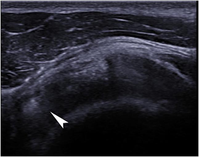
A diagnostic ultrasound image of the right shoulder in a coronal plane, specifically capturing the supraspinatus tendon while the arm is in forced internal rotation. The image reveals a focal hyperechoic formation (indicated by a white arrow) within the tendon substance, characteristic of calcific tendinitis. The lesion is notably brighter than the surrounding tendon tissue and demonstrates posterior acoustic enhancement, which suggests a dense calcification. The adjacent supraspinatus tendon fibers show a heterogeneous echotexture with subtle areas of varied echogenicity, indicative of underlying tendinosis or degenerative changes. The humeral head and overlying deltoid muscle are visible as anatomical landmarks. This imaging is utilized in sports medicine and orthopedics to diagnose rotator cuff pathology, specifically distinguishing between degenerative tears and calcific deposits.
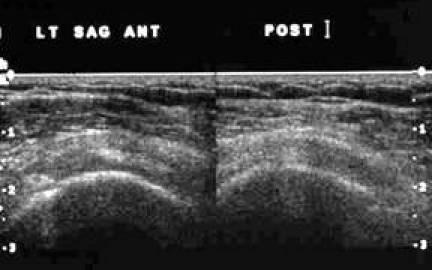
This grayscale diagnostic ultrasound image displays a transverse (short-axis) view of the musculoskeletal structures of the shoulder, specifically identifying the proximal supraspinatus (SST) and infraspinatus (IST) tendons. The image is divided into anterior (ANT) and posterior (POST) segments as indicated by the labels. The superficial layer shows hyperechoic skin and subcutaneous fat. Beneath this, the deltoid muscle appears with characteristic fibrillar echotexture. The primary focus of the image is the deep, rounded, relatively hypoechoic structures representing the SST and IST tendons as they sit adjacent to one another. Below these tendons, the hyperechoic bony cortex of the humerus is visible, characterized by a sharp bright line with posterior acoustic shadowing. This view is clinically significant for evaluating the integrity, thickness, and echogenicity of the rotator cuff tendons in the proximal region to identify pathology such as tendinosis or partial-thickness tears.
Ultrasound Report: Right Shoulder - Ch. Madhavi, 48F
Provisional Diagnosis (P.D.)
- Mildly bulky, heterogeneous supraspinatus tendon
- Fraying of bursal surface fibers
- Minimal subdeltoid bursal effusion (reactive bursitis)
- Mild acromioclavicular (AC) joint arthrosis (co-existing age-related change)
Differential Diagnosis (D.D.)
| Differential | Key Distinguishing Features |
|---|---|
| Partial-thickness rotator cuff tear | Focal hypoechoic/anechoic defect on USG; more sudden onset; arm weakness on empty can test |
| Subacromial bursitis | Predominant bursal fluid; may have calcifications; point tenderness over bursa |
| Calcific tendinopathy | Hyperechoic calcium deposits on USG; acute intense pain; X-ray shows calcification |
| Adhesive capsulitis (frozen shoulder) | Significant restriction of passive range of motion in all planes; typically follows trauma or prolonged immobilization |
| Bicipital tendinopathy | Anterior shoulder pain; tenderness in the bicipital groove; Speed's test positive |
| Acromioclavicular (AC) joint arthritis | Pain localized to the AC joint; positive cross-body adduction test; the mild AC arthrosis on this scan may be contributing |
| Glenohumeral osteoarthritis | X-ray shows joint space narrowing; restricted passive ROM; older age group |
| Full-thickness rotator cuff tear | Complete anechoic defect on USG; positive drop arm test (98% specific); sudden onset usually after trauma |
Management
1. Conservative (First-line - most appropriate at this stage)
- Avoid overhead activities, heavy lifting, and repetitive arm movements
- Relative rest - complete rest is NOT recommended as it leads to loss of muscle power and decreased tendon mechanical properties (Rosen's Emergency Medicine)
- NSAIDs (e.g., ibuprofen 400-600 mg TID or diclofenac 50 mg BD for 1-2 weeks) - short course for pain relief; caution as prolonged use can impair tendon healing
- Paracetamol as adjunct or alternative
- Graduated range-of-motion (ROM) exercises
- Eccentric and concentric strengthening of rotator cuff muscles
- Scapular stabilization exercises
- Refer to physical therapist or sports medicine specialist
- Ice application 20 minutes every few hours for the first 24-48 hours for pain and inflammation
2. Interventional (if conservative fails at 6-8 weeks)
- Peritendinous (NOT intratendinous) injection of corticosteroid + local anesthetic
- Provides short-term pain relief (weeks to months)
- Note: May have detrimental effects on tendon healing at 1 year; use judiciously
- Given the subdeltoid bursal effusion, a subacromial/subdeltoid bursal injection is a reasonable option
- Emerging option for tendinosis; some evidence for rotator cuff tendinopathy
3. Further Investigations to Consider
- Plain X-ray of right shoulder - to assess for calcific deposits, AC joint changes, acromial morphology (hook acromion predisposes to impingement)
- MRI shoulder - if symptoms persist despite conservative treatment, or if partial tear is suspected on clinical exam; MRI gives ~70% accuracy for tendinopathy
- Clinical correlation tests:
- Empty can / Jobe test (supraspinatus)
- Neer test (impingement)
- Hawkins-Kennedy test (impingement)
- Drop arm test (to rule out complete tear)
4. Surgical (Last resort)
- Arthroscopic subacromial decompression (acromioplasty) if conservative management fails over 3-6 months
- Partial debridement for refractory partial-thickness tears
5. General Advice
- Patient education: chronic, degenerative condition - long-term benefit requires adherence to exercises
- Vitamin D supplementation (deficiency impairs tendon healing)
- Avoid sleeping on the affected shoulder
- Ergonomic workplace adjustments if overhead work is involved
- Rosen's Emergency Medicine - Concepts and Clinical Practice, Specific Tendinopathies section, p. 2347-2352
- Rheumatology 2-Volume Set (2022, Elsevier), Shoulder Disorders section