
Lab dx of fungi genaral
fungal laboratory diagnosis KOH mount microscopy hyphae yeast
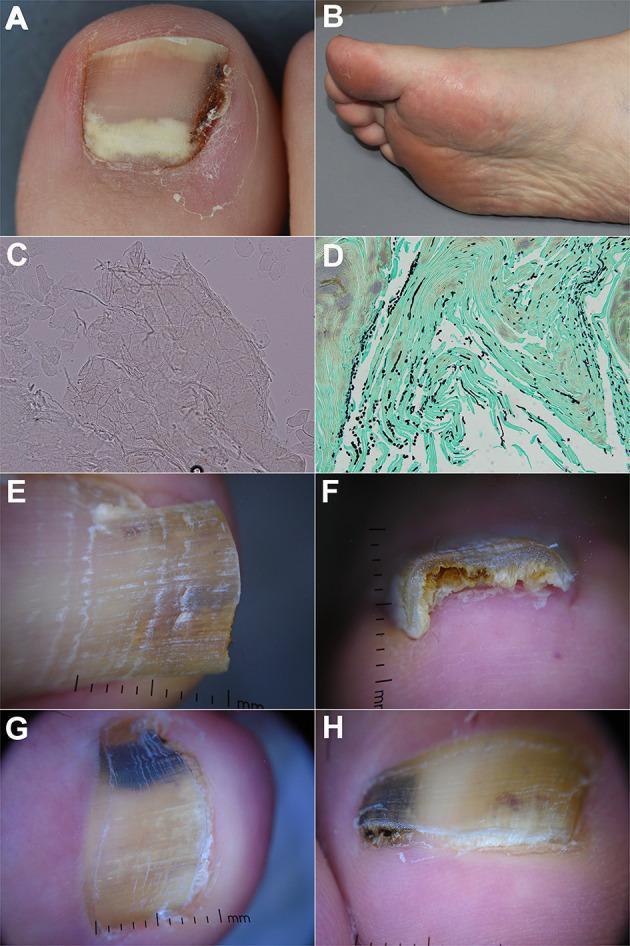
This composite educational image illustrates the clinical, diagnostic, and dermoscopic features of onychomycosis. Panel A shows a clinical photograph of proximal subungual onychomycosis with a characteristic white/yellowish opacity at the lunula. Panel B depicts plantar desquamation (tinea pedis), a common co-infection. Panels C and D show laboratory diagnostics: a potassium hydroxide (KOH) mount with visible septate hyphae (200x) and a Grocott-Gomori methenamine silver (GMS) stained nail clipping (400x) where fungal elements appear black against a green background. Panels E and F provide dermoscopic views of distal lateral subungual onychomycosis, highlighting a spiked pattern, longitudinal striae, and subungual hyperkeratosis at the distal edge. Panels G and H demonstrate fungal melanonychia via dermoscopy, characterized by a multicolor pattern including black and yellow streaks, a reverse triangular pattern (base at the distal edge), and surface scaling. The collection serves to demonstrate the integration of physical examination, microscopy, histopathology, and onychoscopy in the diagnosis of fungal nail infections.
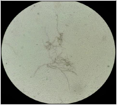
This diagnostic image shows a direct microscopic examination of a clinical specimen using a potassium hydroxide (KOH) mount. The field of view displays clear, translucent fungal elements against a light, granular background characteristic of digested host tissue. The primary features include long, slender, septate hyphae that exhibit regular cross-walls and occasional branching. Associated with these hyphal structures are chains of conidia (spores) in an acropetal arrangement, suggesting fungal proliferation. The morphology is consistent with a phaeohyphomycotic or related filamentous fungal infection, particularly Cladophialophora species in the context of a cerebral abscess. This visual aid is intended for medical education in microbiology and infectious diseases to demonstrate the rapid identification of fungal morphology in clinical exudates. The absence of bacterial structures and the presence of septate hyphae with conidial chains provide a definitive visual diagnostic path for beginning antifungal therapy before culture confirmation.
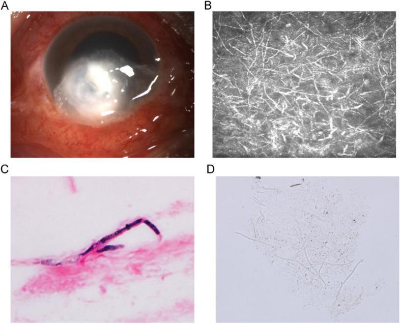
This composite image illustrates the clinical and diagnostic features of fungal keratitis. Panel A is a clinical photograph of the eye showing a significant central and peripheral corneal ulcer characterized by feathery, infiltrating margins, elevated lesions, a dry-appearing surface, and surrounding conjunctival hyperemia. Panel B presents an in vivo confocal microscopy image, revealing a dense network of highly reflective, branching filamentous structures consistent with fungal hyphae within the corneal stroma. Panel C shows a Gram-stained microscopic view of corneal scrapings, where a deep purple, septate fungal hypha is clearly visualized against a pink cellular background. Panel D displays a potassium hydroxide (KOH) wet mount of corneal scrapings under light microscopy, demonstrating translucent, slender, branching fungal hyphae. This series of images demonstrates the progression from clinical presentation to definitive microbiological diagnosis using various imaging and staining modalities, highlighting the morphological characteristics of mycotic corneal infections.
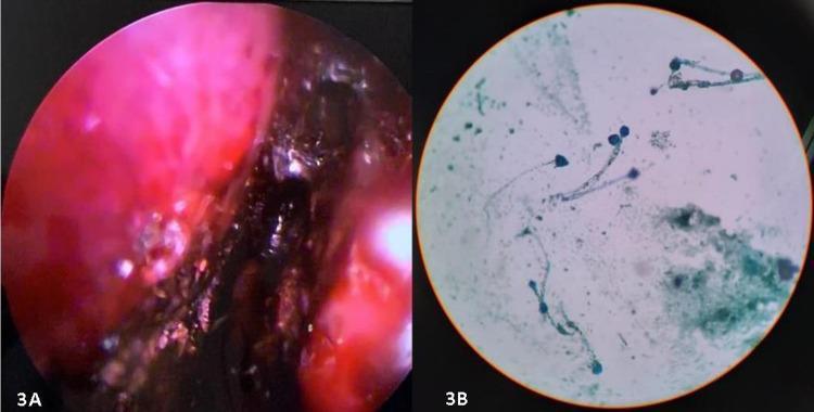
The image consists of two panels demonstrating clinical and laboratory findings associated with invasive fungal sinusitis. Panel A is a diagnostic nasal endoscopy (DNE) photograph showing the right nasal cavity. The mucosa is significantly erythematous and inflamed, featuring extensive dark-brown to black devitalized tissue (eschar) along the turbinates and lateral wall, characteristic of angio-invasive fungal necrosis. Panel B is a bright-field microscopy image of a potassium hydroxide (KOH) mount prepared from a nasal biopsy. It demonstrates fungal elements consistent with Mucorales, characterized by broad, pauciseptate (ribbon-like) hyphae and rounded sporangia/spores. These findings are pedagogically relevant for identifying rhino-orbital-cerebral mucormycosis (ROCM), particularly in immunocompromised or diabetic patients. The combination of visual necrotic tissue (black turbinate sign) and specific fungal morphology provides a comprehensive diagnostic overview of acute invasive fungal infection.
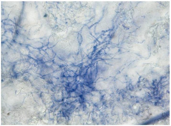
This diagnostic image shows a microscopic direct examination of a skin scrapings sample at 400x magnification. The specimen is processed using a potassium hydroxide (KOH) and Myco-Ink preparation to identify dermatophytes. The visual field demonstrates a network of septate, branching fungal hyphae stained a distinct blue by the Myco-Ink, which binds to chitin in the cell walls. The hyphae appear as elongated, filamentous structures that are intertwined and matted in some regions, typical of Trichophyton quinckeanum morphology. The KOH has cleared the surrounding keratinous epidermal debris, providing high contrast between the darkly stained fungal elements and the lighter, translucent background. This preparation is a critical clinical tool in dermatology for the rapid diagnosis of tinea infections (zoonotic dermatophytosis), allowing for the visualization of hyphal morphology, distribution, and branching patterns necessary for preliminary mycological identification.
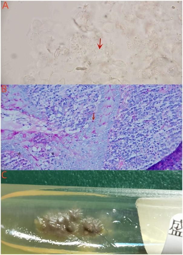
A composite image illustrating the diagnostic mycological and histopathological features of Sporothrix globosa. Panel A (Microscopy): Direct microscopy of an exudate smear using a 10% KOH preparation (400x) reveals slender, translucent, thread-like hyphae (indicated by a red arrow) against a cleared background of squamous cells. Panel B (Pathology): A high-magnification histopathological section (1000x) with Periodic Acid-Schiff (PAS) staining demonstrates intracellular fungal spores (indicated by a red arrow) localized within a multinucleated giant cell, surrounded by a dense inflammatory infiltrate. Panel C (Microbiology): A macroscopic photograph of a fungal culture on a Sabouraud dextrose agar (SDA) slant after 9 days of incubation at 25°C. The culture shows characteristic brownish-beige, moist colonies with a wrinkled, irregular, and slightly raised texture typical of Sporothrix species. This collection of images summarizes the laboratory findings necessary for diagnosing cutaneous disseminated sporotrichosis in a clinical setting.
Laboratory Diagnosis of Fungi - General
Overview of Methods (BOX 60.1)
| Category | Methods |
|---|---|
| Conventional Microbiologic | Direct microscopy, Culture, Identification, Susceptibility testing |
| Histopathologic | H&E, Special stains (GMS, PAS, Mucicarmine), Direct immunofluorescence, In situ hybridization |
| Immunologic | Antibody detection, Antigen detection |
| Molecular | Direct detection (NAA/PCR), Identification, Strain typing |
| Biochemical | Metabolites, Cell wall components, Enzymes |
1. Specimen Collection and Processing
- Selection is based on clinical presentation, radiology, and the most likely fungal pathogen
- Collect aseptically with adequate volume in a sterile, leak-proof container
- Include relevant clinical history - the lab uses this to choose the best processing method
- Sterile body fluids and blood should be cultured when hematogenous spread is suspected
- Skin lesions should be biopsied for both histopathology and culture
- Oral/vaginal mucosal lesions: direct microscopy is preferred over culture (normal flora may confound)
- GI fungal infections: biopsy + histopathology preferred over culture
- Avoid 24-hour sputum/urine collections - they become overgrown with contaminants
- Specimens for fungal culture can be stored at 4°C briefly if processing is delayed (less problematic than bacterial specimens)
2. Direct Microscopy
| Method/Stain | Use | Key Points |
|---|---|---|
| Calcofluor white | Detection of ALL fungi including Pneumocystis | Detects chitin by bright fluorescence; used with KOH; requires fluorescent microscope; 1-2 min |
| KOH (10-20%) | Skin, hair, nail specimens; clearing host material | Dissolves keratin, leaving fungal elements visible; most widely used bedside/clinical method |
| Gram stain | General specimen screen | Most yeasts/hyphae stain gram-positive; Cryptococcus may stain gram-negative or stippled |
| Giemsa stain | Bone marrow, peripheral blood, respiratory specimens | Detects intracellular Histoplasma capsulatum; both intracystic and trophic forms of Pneumocystis |
| India ink | CSF for Cryptococcus neoformans | Negative staining - reveals polysaccharide capsule as a clear halo |
| Fluorescent monoclonal Ab | Pneumocystis jirovecii in respiratory specimens | Sensitive and specific for cysts only (does not stain trophic forms) |
- Septate hyphae - regular cross-walls, branching (e.g., Aspergillus, dermatophytes)
- Pauciseptate/aseptate broad hyphae - ribbon-like, in Mucorales
- Yeast cells with narrow-based budding - Candida, Histoplasma
- Broad-based budding yeast - Blastomyces dermatitidis
- Pseudohyphae - chains of elongated buds (Candida)
- Encapsulated yeast - Cryptococcus neoformans
- Dematiaceous (dark) hyphae - phaeohyphomycosis agents
3. Culture
- Sabouraud Dextrose Agar (SDA) is the standard medium - low pH (5.6) inhibits bacteria; supports most fungi
- Inhibitory Mold Agar (IMA) - with chloramphenicol and cycloheximide for dermatophytes
- Brain Heart Infusion (BHI) agar - for dimorphic fungi at 37°C
- Lysis-centrifugation (Isolator) - best for blood cultures for fungi
- Incubation: most fungi at 25-30°C for mold phase, 35-37°C for yeast/tissue phase
- Can take days to weeks (some dimorphic fungi like Histoplasma may take 4-6 weeks)
- Identify colonies by macroscopic morphology (color, texture, growth rate) then microscopy of sporulation structures
4. Histopathologic Methods
| Stain | What It Does | Key Use |
|---|---|---|
| H&E | General morphology; best for host reaction | Baseline stain; may miss sparse organisms |
| GMS (Grocott-Gomori Methenamine Silver) | Stains fungi black against green background | BEST stain for detecting all fungi in tissue; stains hyphae and yeast |
| PAS (Periodic Acid-Schiff) | Stains yeasts and hyphae pink/magenta | Good all-purpose fungal stain in tissue |
| Mucicarmine | Stains mucin red/pink | Highlights polysaccharide capsule of Cryptococcus neoformans; also stains Blastomyces walls |
| Direct immunofluorescence | Species-specific antibody labeling | High specificity |
| In situ hybridization | Nucleic acid probes in tissue sections | Research and advanced labs |
5. Immunologic (Serologic) Methods
- Useful mainly for histoplasmosis (complement fixation, immunodiffusion) and coccidioidomycosis
- Most other antibody tests lack sensitivity/specificity for invasive infections
- Serial titers monitor disease progression and response to therapy
- More direct and clinically useful for invasive disease
- Cryptococcal polysaccharide antigen (latex agglutination/EIA) - rapid diagnosis of cryptococcal meningitis; very sensitive and specific
- Histoplasma urinary antigen (EIA) - best test for disseminated histoplasmosis
- Aspergillus galactomannan (EIA on serum or BAL) - surrogate marker for invasive aspergillosis
- Candida mannan/anti-mannan - commercially available EIA
- 1,3-β-glucan (Limulus lysate assay) - pan-fungal cell wall marker; detects Candida, Aspergillus, Pneumocystis; does NOT identify the genus
- D-arabinitol in serum - marker of hematogenous candidiasis
- D-mannitol in BAL - possible marker of pulmonary aspergillosis
- (Limited by lack of commercial availability and variable sensitivity)
6. Molecular Methods
| Method | Use |
|---|---|
| PCR (conventional and real-time) | Direct detection of fungal DNA in blood, BAL, tissue |
| Panfungal PCR | Broad-range primers targeting rRNA genes (18S, ITS region) |
| Organism-specific PCR | Targeted detection of Candida, Aspergillus, Pneumocystis, Mucorales |
| Gene chip / microarray | Multiplex identification |
| ITS sequencing | Gold standard for species identification of cultured isolates |
| MALDI-TOF MS | Rapid species identification from colonies - now widely used |
- Meta-analysis of PCR for invasive candidiasis: best results with whole blood, panfungal targets (rRNA, P450 genes), and in vitro detection limit ≤10 CFU/mL
- Strain typing is used for outbreak investigation
7. Biochemical Methods
- Cell wall components: β-glucan, chitin (spectrophotometry)
- Enzymes: urease (Cryptococcus), phenoloxidase (melanin), germ tube test (Candida albicans)
- Assimilation/fermentation profiles: API 20C, Vitek 2 yeast cards
- Chromogenic media: CHROMagar Candida - different Candida spp. produce different colony colors
8. Susceptibility Testing
- Broth microdilution (CLSI M27 for yeasts, M38 for molds) - gold standard
- Disk diffusion for some Candida species
- Commercial systems: Sensititre, Etest
- Important for guiding therapy in resistant/refractory infections (e.g., fluconazole-resistant Candida auris)
Quick Summary Table
| Method | Speed | Sensitivity | Notes |
|---|---|---|---|
| KOH microscopy | Minutes | Moderate | Cheap, rapid; operator-dependent |
| Calcofluor white | Minutes | High | Needs fluorescence microscope |
| Culture (SDA) | Days-weeks | High | Gold standard; slow |
| GMS/PAS histopath | Hours-days | High | Shows tissue invasion |
| Cryptococcal Ag | 1-2 hrs | Very high | Best for crypto meningitis |
| Galactomannan | Hours | Moderate-high | Invasive aspergillosis |
| β-glucan | Hours | Moderate | Pan-fungal; not genus-specific |
| PCR | Hours | High | Not widely standardized |
| MALDI-TOF | Minutes | Very high | Requires culture first |
- Medical Microbiology 9e (Murray), Ch. 60, pp. 661-669
- Sherris & Ryan's Medical Microbiology, 8e, Block 6