
Approach to vesicles bulbous lesions/ immunoflourescent patterns in skin disorders 10 marks question md pathology exam easy to remember simpliform yet from standard reference
direct immunofluorescence pemphigus vulgaris intercellular IgG deposition net-like pattern
direct immunofluorescence bullous pemphigoid linear IgG C3 basement membrane zone
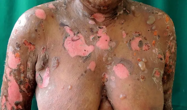
Imaging modality: Clinical photography (dermatology). Anterior view of the upper body demonstrates widespread, pruritic, erythematous plaques with evolution to tense bullae on the arms, trunk, and back. The blisters are dome-shaped and intact in many areas, containing clear serous fluid; numerous lesions have ruptured, leaving bright pink-red, oozing erosions with peripheral crusting. No mucosal involvement is evident. The distribution is symmetric and predominantly involves extensor surfaces and the trunk, consistent with a bullous drug eruption. The clinical morphology may mimic idiopathic bullous pemphigoid, but drug-induced forms are well described and often lack mucosal lesions. Direct immunofluorescence, as reported in this case, shows linear deposition of IgG and C3 along the basement membrane zone, supporting subepidermal blistering at the dermal-epidermal junction. The patient’s history includes recent antibiotic or antihypertensive exposure and fever; these factors raise suspicion for drug-induced bullous pemphigoid rather than idiopathic disease. The images and accompanying information are valuable for dermatology education, pharmacovigilance, and differential diagnosis training. Potential clinical use cases include dermatology reference, medical student teaching, and AI-assisted image retrieval for blistering diseases. Clinically important terms include bullous pemphigoid, blistering eruption, drug-induced bullous eruption, IgG and C3 linear deposition, and basement membrane.
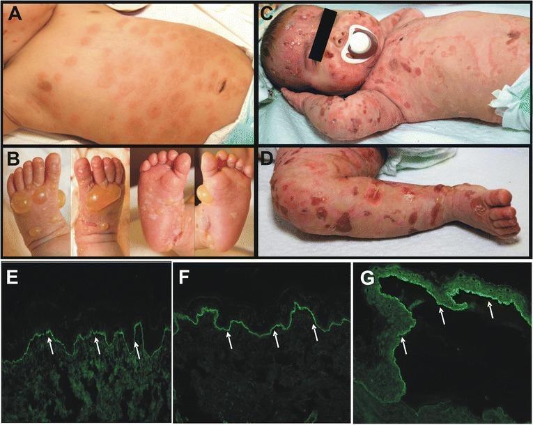
A multi-panel figure illustrating the clinical and diagnostic hallmarks of infantile bullous pemphigoid (BP). Panels A-D display clinical photographs of an infant. Panel A shows erythematous, urticarial plaques on the trunk with characteristic raised rims and dusky centers. Panel B depicts multiple tense, fluid-filled bullae and firm blisters on the hands and feet. Panels C and D illustrate a severe disease relapse with widespread blistering, erosions, and denuded skin across the face, trunk, and extremities. Panels E-G present immunofluorescence microscopy (200x magnification). Direct immunofluorescence (E and F) demonstrates continuous linear deposits of IgG and C3 along the epidermal basement membrane zone (indicated by white arrows). Indirect immunofluorescence on salt-split skin (G) shows circulating IgG antibodies binding specifically to the epidermal roof of the blister, a diagnostic finding for BP. The image serves as an educational resource for recognizing pediatric autoimmune blistering diseases and understanding the correlation between clinical morphology and immunopathological findings.
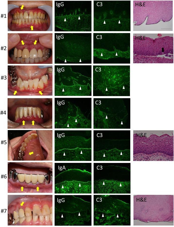
This composite educational graphic displays clinical, histological, and immunofluorescence findings from seven cases (labeled #1-#7) of mucous membrane pemphigoid presenting with oral manifestations. Column 1 features clinical photographs of the oral cavity, highlighting desquamative gingivitis and erythematous lesions (yellow arrows) involving the marginal and attached gingiva. Columns 2 and 3 present direct immunofluorescence (DIF) micrographs of buccal mucosa biopsies, demonstrating characteristic linear deposition of IgG (or IgA in case #6) and C3 (white arrowheads) along the basement membrane zone (BMZ). Column 4 provides histopathological images (H&E stain), showing subepithelial junctional separation at the BMZ (black arrow in case #2) and extensive tissue destruction in more severe cases. This visualization illustrates the diagnostic triad for autoimmune bullous diseases: clinical presentation of chronic gingival inflammation, histological evidence of subepithelial clefting, and the definitive DIF pattern of linear immunoreactant deposition at the dermal-epidermal junction. The content is suitable for dermatology, pathology, and dentistry education regarding immunobullous disorders.
dermatitis herpetiformis direct immunofluorescence granular IgA dermal papillae
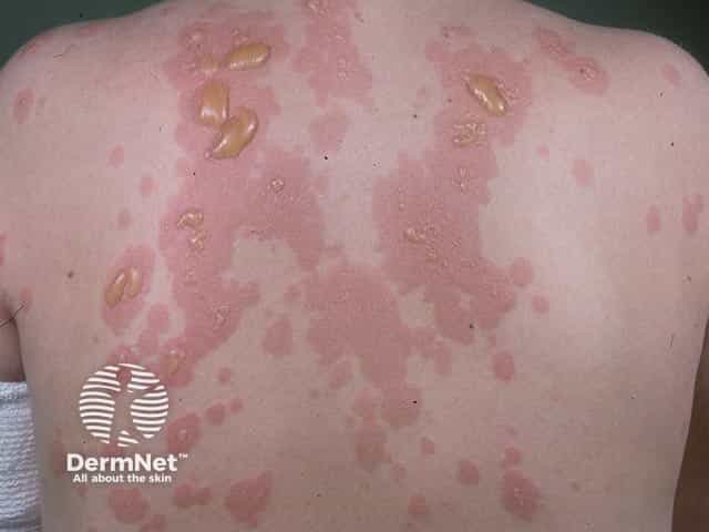
Clinical photography of the patient's back illustrating classic dermatitis herpetiformis manifestations. Modality: in vivo dermatologic imaging using standard color photography under ambient lighting; posterior trunk view highlighting a symmetric eruption. Anatomical region includes the dorsal trunk with involvement of the back's extensor surfaces. Visual features include clustered, erythematous urticarial plaques overlying tense vesicles and bullae, with gold-yellow crusts in places and superficial erosions. The lesions are discrete to coalescent, often arranged in grouped vesiculopapular configurations, with intense pruritus suggested by confluent erythema. The dermal involvement shows papillary microabscesses clinically represented by small vesicles with clear fluid; in Dermatitis Herpetiformis, histopathology reveals subepidermal vesicles with neutrophilic microabscesses in the dermal papillae; direct immunofluorescence demonstrates granular IgA deposition at the tips of dermal papillae. Clinically, dermatitis herpetiformis has a strong association with gluten-sensitive enteropathy (celiac disease) and responds to dapsone therapy and strict gluten-free diet. This image is relevant for educational assessment, differential diagnosis training (urticarial vasculitis, bullous pemphigoid, linear IgA disease), and database indexing for dermatology, medical education, and skin pathology research. Note the mirrored labeling of body surface and detailed morphology to improve search recall for dermatitis herpetiformis presentations. documentation.
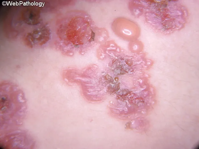
Clinical photography of dermatitis herpetiformis illustrating a typical cutaneous eruption associated with gluten sensitive enteropathy. This in vivo macroscopic dermatology photograph shows clustered intensely pruritic lesions on extensor surfaces. The distribution is symmetrical, involving elbows and knees with additional involvement of buttocks, scapular and sacral regions; face and scalp may be affected in some cases. Lesions comprise erythematous papules with small firm topped vesicles atop plaques, often with urticarial morphology and surrounding excoriations from scratching, leading to crusted erosions. Morphologic features include grouped vesiculopapular lesions in a pruritic distribution. Clinically this dermatitis herpetiformis is strongly associated with gluten sensitive enteropathy and antibodies to transglutaminases. Pathophysiology involves IgA autoantibodies depositing in the tips of dermal papillae; biopsy when performed can reveal neutrophilic microabscesses at papillary tips and dermal neutrophilic eosinophilic infiltrates, while direct immunofluorescence shows granular IgA deposits at dermal papillae tips with C3. The condition often follows a prolonged course but may remit; treatment includes dapsone or sulfapyridine and a strict gluten free diet. This image supports educational emphasis on clinical diagnosis, differential considerations bullous diseases and dermatitis herpetiformis mimics, and management implications. Clinical correlation with serology and biopsy confirms diagnosis and guides therapy.
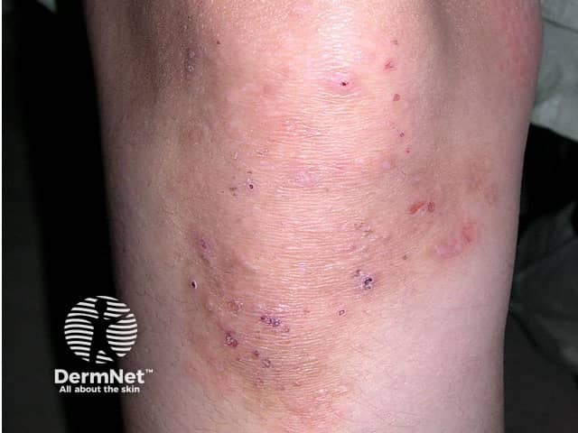
Imaging modality: Clinical photography, high-resolution close-up dermal photograph of the left/right elbow region captured with standard color balance to document cutaneous vesiculopapular eruption. Anatomical site: Antecubital fossa/extensor surface of the elbow, with overlying erythema and excoriations; similar lesions may appear on knees, buttocks, and back. Visual features: grouped vesicles and papules on an erythematous base, often coalescing into urticarial plaques; surrounding scaling and crusts; itching leads to scratch marks; lesions are intensely pruritic, arranged symmetrically on extensor surfaces, and may show crusted erosions. Pathology/diagnostic significance: While the image shows surface morphology compatible with dermatitis herpetiformis, definitive diagnosis relies on direct immunofluorescence demonstrating granular IgA deposition at dermal papillae, and histology showing subepidermal vesicles with neutrophils clustering at the dermal papillae. Clinically, dermatitis herpetiformis is strongly associated with celiac disease and gluten sensitivity; management includes dapsone for symptom control and a strict gluten-free diet. Differential considerations include linear IgA disease, bullous pemphigoid, and eczematous dermatitis. This image is useful for dermatology education, tele-dermatology triage, and comparative pathology during clinical examination and teaching archives; it supports recognition of extensor-predominant vesiculopapular itch, aids patient counseling, and guides biopsy or immunofluorescence testing. This image also supports clinical correlation, patient education, biopsy planning, and care.
Approach to Vesiculobullous Lesions & Immunofluorescence Patterns in Skin Disorders
1. Definition
2. Stepwise Clinical Approach (Algorithm)
- Age of onset, mucosal involvement, drug history, pruritus vs pain, flaccid vs tense bullae.
- Nikolsky sign (lateral traction on perilesional skin causes epidermal separation) - positive in intraepidermal disease (pemphigus group), negative in subepidermal disease (pemphigoid group) - Henry's Clinical Diagnosis, Tintinalli's Emergency Medicine.
- Cytology of blister floor - shows rounded acantholytic cells in pemphigus (rapid bedside test).
| Level | Disease |
|---|---|
| Subcorneal | Pemphigus foliaceus, Staphylococcal scalded skin syndrome |
| Suprabasal | Pemphigus vulgaris |
| Subepidermal (at dermo-epidermal junction) | Bullous pemphigoid, Dermatitis herpetiformis, Linear IgA disease, EBA, Pemphigoid gestationis |
3. Master Table - Immunofluorescence Patterns (the "10-mark" core)
| Disease | DIF Pattern | Antibody/Deposit | Target Antigen | Salt-split localization |
|---|---|---|---|---|
| Pemphigus vulgaris | Intercellular, "fishnet"/"chicken-wire", throughout epidermis with basal predominance | IgG (+C3) | Desmoglein 3 (± Dsg1) | - |
| Pemphigus foliaceus | Intercellular, same fishnet pattern but confined to upper/superficial epidermis | IgG (+C3) | Desmoglein 1 | - |
| IgA pemphigus | Intercellular, upper epidermis | IgA | Desmocollin 1 | - |
| Bullous pemphigoid | Linear, smooth band along basement membrane zone (BMZ) | IgG + C3 | BP180 (collagen XVII), BP230 | Epidermal (roof) |
| Epidermolysis bullosa acquisita (EBA) | Linear at BMZ (looks identical to BP on routine DIF) | IgG | Type VII collagen | Dermal (floor) |
| Pemphigoid gestationis | Linear C3 (± IgG) at BMZ | C3 predominant | BP180 | Epidermal (roof) |
| Dermatitis herpetiformis | Granular deposits at the tips of dermal papillae | IgA (+C3) | Epidermal transglutaminase (TG3) | - |
| Linear IgA bullous dermatosis | Linear IgA at BMZ | IgA | LAD-1 (BP180 fragment) | Variable (roof or floor) |
| Bullous SLE / "Lupus band test" | Granular, "full house" (IgG+IgM+IgA+C3+C1q) at BMZ | Multiple immunoreactants | dsDNA/BMZ antigens | - |
| Lichen planus (non-bullous but IF-taught) | "Shaggy" fibrin deposits at BMZ | Fibrin | - | - |
4. Simple Mnemonics to Remember
- "Pemphigus = cells fall apart, so antibody sits BETWEEN cells" → intercellular fishnet/chicken-wire pattern (like a net holding fish - when torn, cells separate → flaccid blister, +Nikolsky).
- "Pemphigoid = antibody sits at the FLOOR (basement), so the roof stays intact" → linear band at BMZ → tense blister, -Nikolsky.
- "D for Dermatitis herpetiformis = Dots/Granules at Dermal papillae tips" - granular, not linear.
- Roof vs floor trick for subepidermal diseases: "Bullous Pemphigoid Binds the Roof" vs "EBA Exits through the floor" (roof = epidermal side, floor = dermal side on salt-split skin).
- "Full house = Lupus" - when DIF shows all immunoglobulins + complement together, think SLE.
5. Representative Images
6. Exam Writing Tip
Include Pathogenesis and morphology too
Approach to Vesiculobullous Lesions & Immunofluorescence Patterns - With Pathogenesis & Morphology
1. Clinical Approach (Recap)
FIG 22.8 Levels of blister formation (Robbins):
(A) Subcorneal      → Pemphigus foliaceus
(B) Suprabasal       → Pemphigus vulgaris
(C) Subepidermal     → Bullous pemphigoid, Dermatitis herpetiformis
2. Disease-wise Pathogenesis, Morphology & IF Pattern
A. Pemphigus Vulgaris (PV) & Pemphigus Foliaceus (PF)
- IgG autoantibodies bind desmoglein 1 (Dsg1) and desmoglein 3 (Dsg3) - transmembrane desmosomal cadherins that hold keratinocytes together.
- PV → antibodies against Dsg3 (± Dsg1) → deep, suprabasal split (Dsg3 is dominant in deeper epidermis and mucosa).
- PF → antibodies against Dsg1 only → superficial, subcorneal split (Dsg1 dominant in superficial epidermis).
- Antibody binding disrupts desmosomal adhesion directly and may also trigger intercellular proteases → acantholysis (loss of cell-cell cohesion).
- Associated with specific HLA alleles (autoimmune predisposition).
- Gross: Flaccid, easily ruptured bullae on skin/mucosa (PV) → painful oral erosions often the first sign; PF blisters are more superficial, scaly/crusted, mucosa usually spared.
- Microscopy: Rounded, separated acantholytic keratinocytes floating within an intraepidermal blister cavity; minimal inflammation.
- DIF: Intercellular "fishnet"/"chicken-wire" pattern of IgG (± C3) outlining every keratinocyte - basal predominance in PV, superficial layers only in PF.
- IIF: Circulating IgG titer correlates with disease activity - useful for monitoring treatment response.
B. Bullous Pemphigoid (BP)
- IgG autoantibodies (and complement) target hemidesmosomal proteins - BPAG1 (BP230) and BPAG2/BP180 (type XVII collagen) - which anchor basal keratinocytes to the basement membrane.
- Anti-BP180 antibodies are directly pathogenic; antibody binding activates complement and recruits eosinophils and neutrophils, whose enzymes degrade the dermo-epidermal junction → subepidermal split.
- Key contrast with pemphigus: target is in hemidesmosomes (BP) vs desmosomes (pemphigus) - same general mechanism, different location, hence different clinical severity/course.
- Gross: Tense, fluid-filled bullae on erythematous/urticarial base (do not rupture easily, unlike pemphigus); heals without scarring if uninfected; intense pruritus.
- Microscopy: Subepidermal blister with an intact, full-thickness epidermal roof (no acantholysis - key distinguishing point from pemphigus); early lesions show basal cell vacuolization, dermal edema, and an eosinophil-rich (± neutrophil) infiltrate at the dermo-epidermal junction.
- DIF: Linear band of IgG + C3 along the basement membrane zone.
- Salt-split skin: Antibody binds the epidermal (roof) side.
- Special variant: Pemphigoid gestationis (herpes gestationis) - same mechanism, occurs in 2nd-3rd trimester, resolves post-partum, may recur in future pregnancies.
C. Dermatitis Herpetiformis (DH)
- Strongly linked to gluten sensitivity/celiac disease (up to 80% have celiac disease, though few celiac patients develop DH).
- Genetically predisposed individuals form IgA antibodies against dietary gliadin, which cross-react with endomysium and tissue transglutaminase, including epidermal transglutaminase expressed by keratinocytes.
- IgA deposits bind anchoring fibrils connecting hemidesmosomes to the dermis at the papillary tips → recruits neutrophils → papillary tip microabscesses → basal cell vacuolization → subepidermal blister.
- Gross: Intensely pruritic, grouped (herpetiform) vesicles/papules, bilateral and symmetric, over extensor surfaces - elbows, knees, upper back, buttocks.
- Microscopy: Neutrophilic microabscesses at the tips of dermal papillae, basal vacuolization, and subepidermal vesicle formation.
- DIF: Discontinuous, granular IgA deposits selectively at the tips of dermal papillae (± C3) - hallmark finding.
- Responds to dapsone and a gluten-free diet.
D. Other Subepidermal Look-Alikes (for completeness)
| Disease | Pathogenesis (brief) | Morphology highlight | DIF pattern |
|---|---|---|---|
| Epidermolysis bullosa acquisita (EBA) | IgG autoantibodies against type VII collagen (anchoring fibrils below lamina densa) | Fragile skin, trauma-induced blisters, scarring | Linear IgG at BMZ; salt-split → dermal (floor) side |
| Linear IgA bullous dermatosis | IgA antibodies against LAD-1 (a BP180 fragment) | Tense bullae, often annular ("cluster of jewels") | Linear IgA at BMZ |
| Bullous SLE | Antibodies against type VII collagen/BMZ antigens in a lupus background | Vesiculobullous lesions in photo-distributed areas | Granular, "full-house" pattern (IgG+IgM+IgA+C3+C1q) |
3. Quick Memory Anchors
- "Pemphigus = between the bricks (desmosomes), Pemphigoid = below the wall (basement membrane)."
- PV vs PF: "Vulgaris goes Very deep (suprabasal, Dsg3); Foliaceus stays Flat/superficial (subcorneal, Dsg1)."
- BP morphology clincher: tense bulla + eosinophils + NO acantholysis = Bullous Pemphigoid.
- DH clincher: gluten + granular IgA at papillary TIPS + neutrophilic microabscess = Dermatitis Herpetiformis.
- Roof vs floor: "BP Binds the roof, EBA Exits through the floor" (salt-split skin).
4. Image Reference
5. Exam Write-up Suggestion (10 marks)
- Definition + classification by level of split (1-2 marks)
- Approach algorithm - Nikolsky, Tzanck, biopsy, DIF/IIF, salt-split (2-3 marks)
- Pathogenesis + morphology for PV, BP, DH (4-5 marks - these three are asked most often)
- IF pattern summary table + salt-split trick for BP vs EBA (1-2 marks)
Combine them
Approach to Vesiculobullous Lesions & Immunofluorescence Patterns in Skin Disorders
1. Definition
2. Stepwise Clinical Approach (Algorithm)
FIG: Levels of blister formation
(A) Subcorneal    → Pemphigus foliaceus
(B) Suprabasal    → Pemphigus vulgaris
(C) Subepidermal  → Bullous pemphigoid, Dermatitis herpetiformis, LABD, EBA
3. Disease-wise Pathogenesis, Morphology & Immunofluorescence
A. Pemphigus Vulgaris (PV) & Pemphigus Foliaceus (PF)
- IgG autoantibodies bind desmoglein 3 (Dsg3) and/or desmoglein 1 (Dsg1) - transmembrane desmosomal cadherins holding keratinocytes together.
- PV → antibodies against Dsg3 (± Dsg1) → deep, suprabasal split (Dsg3 dominant in deeper epidermis and mucosa).
- PF → antibodies against Dsg1 only → superficial, subcorneal split (Dsg1 dominant superficially).
- Antibody binding directly disrupts desmosomal adhesion and may activate intercellular proteases → acantholysis.
- Associated with specific HLA alleles.
- Gross: Flaccid, easily ruptured bullae on skin/mucosa (PV) - painful oral erosions often the first sign; PF blisters more superficial, scaly/crusted, mucosa usually spared.
- Microscopy: Rounded, separated acantholytic keratinocytes floating within an intraepidermal blister cavity; minimal inflammation.
- DIF: Intercellular "fishnet"/"chicken-wire" pattern of IgG (± C3) outlining every keratinocyte - basal predominance in PV, superficial layers only in PF.
- IIF: Circulating IgG titer correlates with disease activity - useful for monitoring treatment.
B. Bullous Pemphigoid (BP)
- IgG autoantibodies (+ complement) target hemidesmosomal proteins - BPAG1 (BP230) and BPAG2/BP180 (type XVII collagen) - which anchor basal keratinocytes to the basement membrane.
- Anti-BP180 antibodies are directly pathogenic; binding activates complement and recruits eosinophils and neutrophils, whose enzymes degrade the dermo-epidermal junction → subepidermal split.
- Contrast with pemphigus: target is in hemidesmosomes (BP) vs desmosomes (pemphigus) - similar mechanism, different location and severity.
- Gross: Tense, fluid-filled bullae on an erythematous/urticarial base (do not rupture easily); heals without scarring if uninfected; prominent pruritus.
- Microscopy: Subepidermal blister with an intact, full-thickness epidermal roof (no acantholysis - key distinction from pemphigus); early lesions show basal cell vacuolization, dermal edema, and an eosinophil-rich (± neutrophil) infiltrate at the dermo-epidermal junction.
- DIF: Linear band of IgG + C3 along the basement membrane zone.
- Salt-split skin: antibody binds the epidermal (roof) side.
- Variant: Pemphigoid gestationis (herpes gestationis) - same mechanism, 2nd-3rd trimester onset, resolves post-partum, may recur in future pregnancies.
C. Dermatitis Herpetiformis (DH)
- Strongly linked to gluten sensitivity/celiac disease (up to 80% have celiac disease, though only a small fraction of celiac patients develop DH).
- Genetically predisposed individuals form IgA antibodies against dietary gliadin, which cross-react with endomysium and tissue transglutaminase, including epidermal transglutaminase on keratinocytes.
- IgA deposits bind anchoring fibrils connecting hemidesmosomes to the dermis at papillary tips → recruits neutrophils → papillary tip microabscesses → basal vacuolization → subepidermal blister.
- Gross: Intensely pruritic, grouped (herpetiform) vesicles/papules, bilateral and symmetric, over extensor surfaces - elbows, knees, upper back, buttocks.
- Microscopy: Neutrophilic microabscesses at the tips of dermal papillae, basal vacuolization, subepidermal vesicle formation.
- DIF: Discontinuous, granular IgA deposits selectively at the tips of dermal papillae (± C3) - hallmark finding.
- Responds to dapsone and a gluten-free diet.
D. Other Subepidermal Look-Alikes (for completeness)
| Disease | Pathogenesis (brief) | Morphology highlight | DIF pattern |
|---|---|---|---|
| Epidermolysis bullosa acquisita (EBA) | IgG against type VII collagen (anchoring fibrils below lamina densa) | Fragile skin, trauma-induced blisters, scarring | Linear IgG at BMZ; salt-split → dermal (floor) side |
| Linear IgA bullous dermatosis (LABD) | IgA against LAD-1 (a BP180 fragment) | Tense bullae, often annular ("cluster of jewels") | Linear IgA at BMZ |
| Bullous SLE | Antibodies against type VII collagen/BMZ antigens on a lupus background | Vesiculobullous lesions in photo-distributed areas | Granular, "full-house" pattern (IgG+IgM+IgA+C3+C1q) |
| IgA pemphigus | IgA against desmocollin 1 | Intraepidermal, superficial pustules/vesicles | Intercellular IgA, upper epidermis |
4. Master IF Summary Table
| Disease | DIF Pattern | Antibody | Target Antigen | Salt-split localization |
|---|---|---|---|---|
| Pemphigus vulgaris | Intercellular "fishnet", basal predominance | IgG (+C3) | Desmoglein 3 (±1) | - |
| Pemphigus foliaceus | Intercellular fishnet, superficial only | IgG (+C3) | Desmoglein 1 | - |
| Bullous pemphigoid | Linear at BMZ | IgG + C3 | BP180, BP230 | Epidermal (roof) |
| EBA | Linear at BMZ | IgG | Type VII collagen | Dermal (floor) |
| Pemphigoid gestationis | Linear (C3 ± IgG) at BMZ | C3 predominant | BP180 | Epidermal (roof) |
| Dermatitis herpetiformis | Granular at dermal papillae tips | IgA (+C3) | Epidermal transglutaminase | - |
| Linear IgA disease | Linear IgA at BMZ | IgA | LAD-1 | Variable |
| Bullous SLE | Granular "full house" | IgG+IgM+IgA+C3+C1q | dsDNA/BMZ antigens | - |
5. Quick Memory Anchors
- "Pemphigus = between the bricks (desmosomes); Pemphigoid = below the wall (basement membrane)."
- PV vs PF: "Vulgaris goes Very deep (suprabasal, Dsg3); Foliaceus stays Flat/superficial (subcorneal, Dsg1)."
- BP clincher: tense bulla + eosinophils + no acantholysis = Bullous Pemphigoid.
- DH clincher: gluten + granular IgA at papillary tips + neutrophilic microabscess = Dermatitis Herpetiformis.
- Roof vs floor: "BP Binds the roof, EBA Exits through the floor" (salt-split skin).
- "Full house = Lupus" - all immunoglobulins + complement together on DIF = think SLE.
6. Images
7. Exam Write-up Plan (10 marks)
- Definition + classification by level of split (1 mark)
- Approach algorithm - Nikolsky, Tzanck, biopsy, DIF/IIF, salt-split (2-3 marks)
- Pathogenesis + morphology for PV, BP, DH (4-5 marks - the three most commonly asked)
- IF pattern summary table + salt-split trick for BP vs EBA (1-2 marks)