
acute aortic syndrom teach me at the level of mch cardiac surgery level from standard textbooks and literature with pictures and all
Reading File
Now I'll launch parallel searches across the library for AAS at MCh cardiac surgery level.
Finding Sources
Finding Sources
Finding Sources
Finding Sources
Excellent finds. Now let me read the most relevant blocks in parallel - Braunwald's (gold standard for cardiac surgery level), Grainger & Allison's (imaging), Robbins (pathology), and Echocardiography text.
Reading File
Reading File
Reading File
Finding Sources
Excellent. Now let me read the most detailed cardiac surgery-level sections from Braunwald's (the dedicated AAS chapter), Fuster's Heart (IMH/PAU variants), Sabiston (surgical management), and Grainger & Allison (imaging), plus fetch the Robbins pathology image.
Reading File
Reading File
Reading File
Loading Image
Now let me fetch the key images and read the Braunwald's main AAS chapter (block dedicated to aortic dissection) plus the Grainger imaging section and echocardiography text.
Finding Sources
Reading File
Loading Image
Searching PubMed
"acute aortic syndrome" management guidelines
Now let me get the Braunwald's main dedicated AAS chapter and fetch more key images including the CT imaging and classification diagrams.
Finding Sources
Reading File
Loading Image
Loading Image
Now let me get the critical Braunwald's dedicated AAS chapter (block with actual aortic dissection content) and the echocardiography section on dissection, plus one more imaging search.
Reading File
Reading File
Finding Sources
Excellent. Let me now get the Miller's Anesthesia section (which has great surgical/anesthesia detail) and the Current Surgical Therapy section on Type B management, plus fetch the key TEE image.
Reading File
Reading File
Loading Image
Now fetch the DeBakey classification diagram and get more surgical detail from the Current Surgical Therapy on type A management.
Loading Image
Reading File
Finding Sources
Let me get the Grainger imaging protocol section and Schwartz surgical clinical manifestations, plus fetch the TEVAR procedural diagram.
Reading File
Reading File
Loading Image
Excellent - I have comprehensive data from 8 textbooks plus images. Let me also fetch the CT scan image showing TEVAR coverage and get the ESC 2024 guidelines detail.
Loading Image
Reading Article
PMID: 39210722
I now have comprehensive material from 8 major textbooks plus imaging. Let me compose the full MCh-level teaching document.---
Acute Aortic Syndrome (AAS) - MCh Cardiac Surgery Level
Sources: Braunwald's Heart Disease | Fuster & Hurst's The Heart (15e) | Robbins & Kumar Basic Pathology | Grainger & Allison's Diagnostic Radiology | Textbook of Clinical Echocardiography | Miller's Anesthesia (10e) | Current Surgical Therapy (14e) | Schwartz's Principles of Surgery (11e) | Sabiston Textbook of Surgery | 2024 ESC Guidelines [PMID: 39210722]
1. DEFINITION AND SPECTRUM
Acute Aortic Syndrome (AAS) is a unifying term for three distinct but interrelated aortic emergencies that share a common presentation of acute severe chest/back pain and carry high mortality if unrecognized:
| Entity | Core Pathology | Relative Frequency |
|---|---|---|
| Aortic Dissection (AD) | Intimal tear + medial hematoma tracking along dissection plane | ~75% |
| Intramural Hematoma (IMH) | Vasa vasorum rupture / microscopic tear without visible flap | 10-20% |
| Penetrating Aortic Ulcer (PAU) | Atherosclerotic plaque ulcerating through internal elastic lamina | 2-7% |
The pathological relationship between these entities is dynamic: IMH can arise from thrombosis of a false lumen; a PAU can precipitate IMH or dissection; dissection and IMH can be indistinguishable radiologically.
- Grainger & Allison's Diagnostic Radiology, p. 2061
2. THE THREE ENTITIES - ANATOMY AND PATHOPHYSIOLOGY
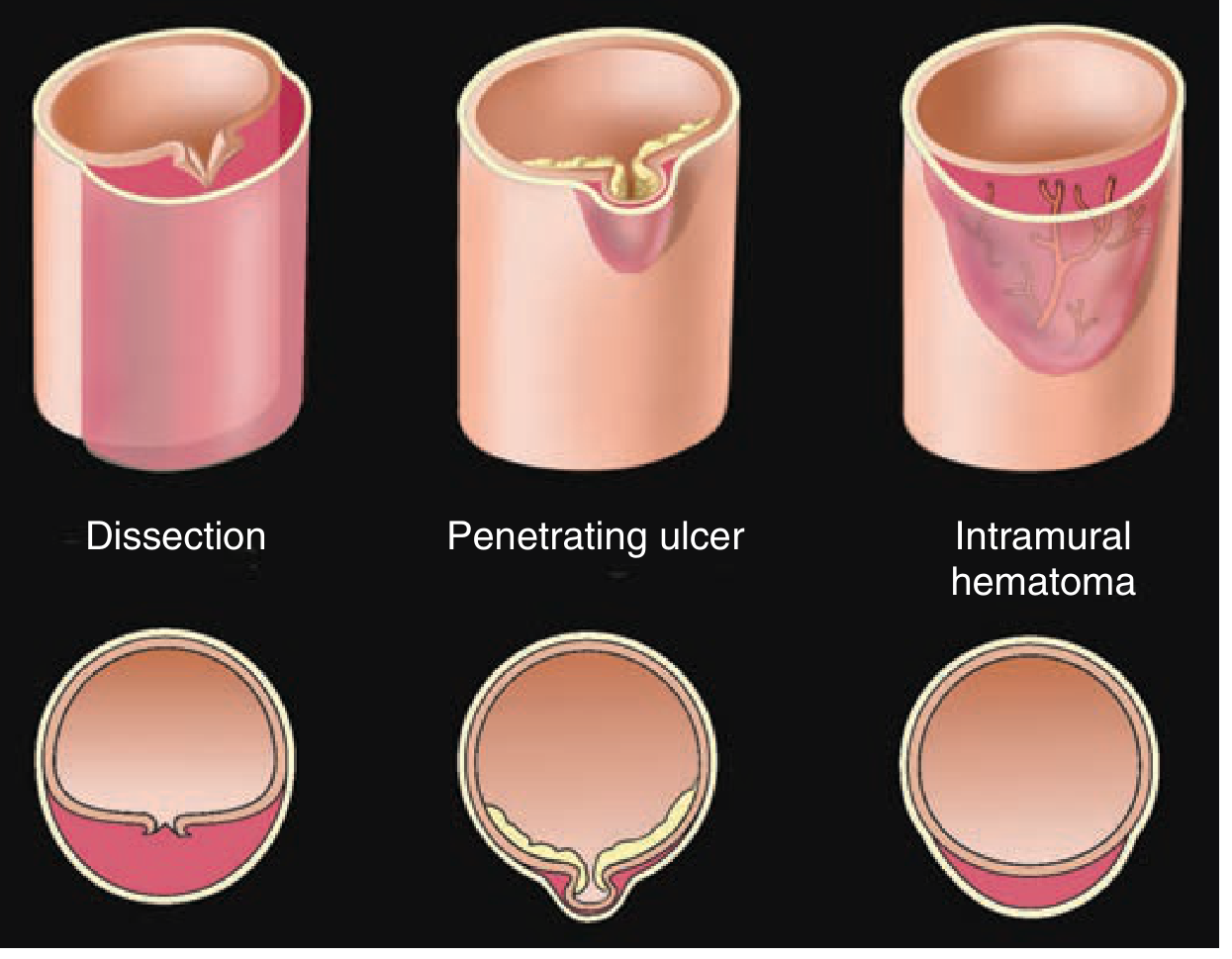
Figure: The three AAS variants - Dissection, Penetrating Ulcer, and Intramural Hematoma. Note the cross-sectional and longitudinal differences in medial involvement. (Fuster & Hurst's The Heart, 15e, Fig. 23-47)
2.1 Aortic Dissection
Mechanism: Blood-filled channel forms within the aortic media when laminar planes split apart. The initiating event is either:
- Primary intimal tear (caused by shear stresses on aortic wall) - most common. Tear is usually transverse/oblique, 1-5 cm long, with jagged edges, situated within 10 cm of the aortic valve in ascending dissections.
- Intramural hemorrhage from vasa vasorum rupturing and weakening the intima at the tear site.
Once established, blood under systemic pressure propagates the dissection plane - usually between the middle and outer thirds of the media - either retrograde toward the heart or distally to iliac/femoral arteries.
Gross Pathology:
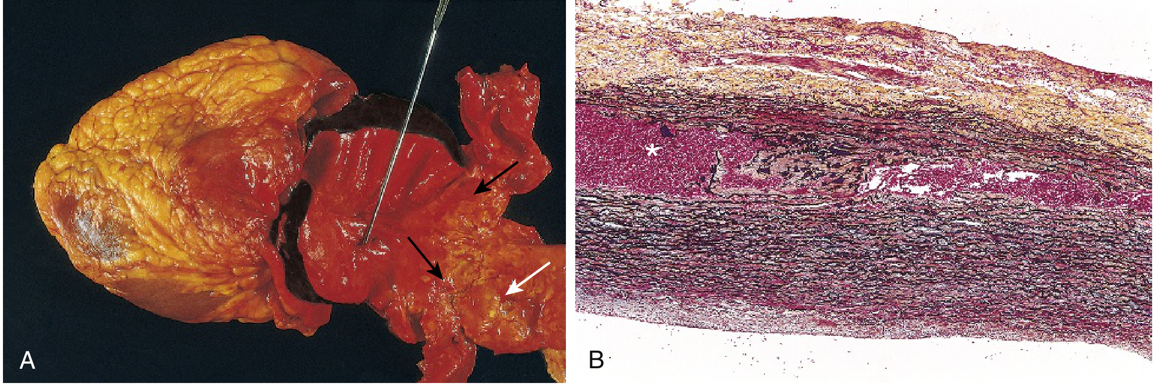
Figure: (A) Opened aorta with proximal dissection from a small oblique intimal tear. Note the intimal tear occurred in a region largely free of atherosclerotic plaque; the distal edge of the IMH (black arrows) lies at the edge of an atherosclerotic zone (white arrow) which arrested propagation. (B) Histology showing dissection plane and IMH (asterisk), Movat stain. (Robbins & Kumar Basic Pathology, Fig. 8.18)
Key observation: Dissection is unusual in the presence of substantial atherosclerosis because medial fibrosis inhibits hematoma propagation. The intimal tear and atherosclerosis are biologically distinct events.
2.2 Intramural Hematoma (IMH)
IMH differs from dissection in that:
- No visible intimal flap delineating true and false lumens
- Hematoma is located circumferentially around the aortic lumen (not obliquely)
- No identifiable entry/exit tear on imaging
Pathogenesis controversy: Arises from either rupture of vasa vasorum within the aortic wall, OR from a microscopic intimal tear not visible radiographically.
Clinical course is variable: The hematoma may:
- Persist unchanged
- Resorb (aorta returns to normal appearance)
- Leave an aneurysm (risk of rupture)
- Transform into frank dissection (the most dangerous evolution)
IMH involves the descending thoracic aorta more frequently than the ascending, tends to occur in older patients, and is more likely to be managed medically than frank dissection.
- Fuster & Hurst's The Heart, p. 769
2.3 Penetrating Aortic Ulcer (PAU)
Mechanism: An atherosclerotic plaque ulcerates through the internal elastic lamina and erodes into the media. This creates:
- Local medial penetration
- Can mimic or result in aortic dissection
- Can cause pseudoaneurysm formation, IMH, or rupture
Epidemiology:
- Accounts for 2-7% of AAS
- Descending aorta in 90% of cases (below the left subclavian artery)
- Often asymptomatic - detected incidentally (87% in one Mayo Clinic series)
- Defined on contrast CT as a focal outpouching through intimal calcification in a region of diffuse atherosclerosis
CT criteria for intervention: Greatest diameter >20 mm or neck >10 mm warrants early intervention even if asymptomatic.
- Sabiston Textbook of Surgery
3. EPIDEMIOLOGY AND RISK FACTORS
| Risk Factor | Mechanism |
|---|---|
| Hypertension (>90% of dissections) | Vasa vasorum narrowing → ECM degeneration + medial SMC loss; abrupt BP surges shear intima |
| Marfan syndrome | FBN1 mutation → fibrillin-1 deficiency → medial cystic necrosis |
| Ehlers-Danlos syndrome type IV | COL3A1 mutation → type III collagen deficiency |
| Bicuspid aortic valve | Associated ascending aortopathy (shared developmental origin) |
| Cocaine use | Acute sympathomimetic BP surge - implicated in 0.5-37% of cases; mean interval 12 hours from use to dissection |
| Pregnancy | Third trimester hormonal vascular remodeling + haemodynamic stress (~10-20/million births) |
| Iatrogenic | Arterial cannulation, catheterization, cardiopulmonary bypass |
| Turner syndrome | Aortic coarctation + bicuspid AV phenotype |
| Vasculitides | Inflammatory weakening of media |
Demographics (IRAD data): 65% male, mean age 63 years (men) / 67 years (women). 32% of patients are >70 years. Younger patients more commonly have genetic disorders.
- Braunwald's Heart Disease, Chapter 42
4. CLASSIFICATION
4.1 Anatomical Classification
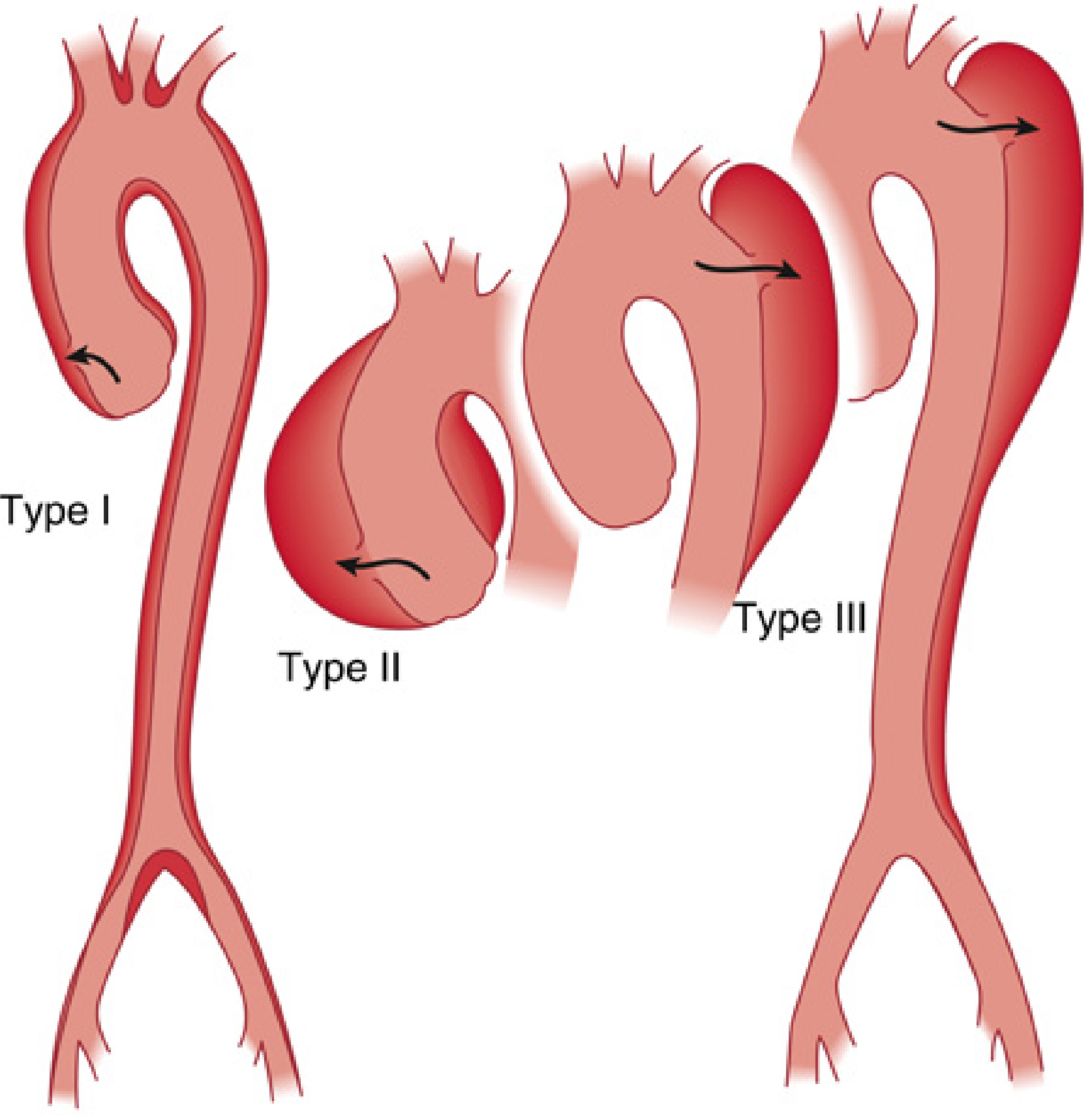
Figure: DeBakey Classification of aortic dissection. Type I, II (ascending - both surgical emergencies), Type III (descending - usually medical/endovascular). (Miller's Anesthesia 10e, Fig. 52.7)
| Classification | Definition | Surgical Implication |
|---|---|---|
| DeBakey I | Ascending tear + dissects entire aorta | Emergency surgery |
| DeBakey II | Ascending tear, limited to ascending aorta | Emergency surgery |
| DeBakey III | Tear distal to L. subclavian; IIIa = thoracic only; IIIb = to iliacs | Usually medical/endovascular |
| Stanford A | Any dissection involving the ascending aorta (= DeBakey I + II) | Emergency surgery |
| Stanford B | Dissection not involving ascending aorta (= DeBakey III) | Usually medical; TEVAR if complicated |
Stanford classification is preferred clinically because the key management decision is whether the ascending aorta is involved. Stanford A accounts for ~75% of cases.
Temporal Classification:
- Acute: <14 days (much higher mortality)
- Subacute: 14 days - 2 months
- Chronic: >2 months (in 20-40%, significant aneurysmal dilatation of the descending/thoracoabdominal aorta develops)
5. CLINICAL PRESENTATION
5.1 Cardinal Symptoms
- Pain is the most common presenting symptom:
- Abrupt onset, maximum intensity at outset (distinguishes from MI where pain builds)
- "Tearing," "ripping," or "knife-like" quality
- Anterior chest pain - suggests ascending aorta involvement (Type A)
- Interscapular/back pain - suggests descending aorta (Type B)
- Abdominal or migratory pain - dissection extending distally
- Silent dissection exists in a small proportion
5.2 Anatomical Complications and Their Presentation
(Critical to understand for MCh level - malperfusion drives decisions)
| Anatomical Level | Structure Compromised | Clinical Manifestation |
|---|---|---|
| Aortic root (retrograde) | Aortic valve commissures disrupted | Acute AR - dyspnoea, pulmonary oedema, diastolic murmur, shock |
| Aortic root (retrograde) | Coronary ostia (RCA most common) | Acute MI - chest pain, ECG changes, elevated troponin; masked dissection if thrombolysis given |
| Pericardial space | Serosanguineous effusion / rupture | Tamponade - JVD, muffled tones, pulsus paradoxus, low voltage ECG, shock |
| Innominate / carotid | Carotid malperfusion | Syncope, TIA, stroke, coma, carotid pulse deficit |
| Subclavian | Brachial malperfusion | Cold arm, pulse deficit, right-left BP differential >20 mmHg |
| Intercostal arteries | Spinal cord (Adamkiewicz artery) | Paraplegia / paraparesis, incontinence |
| Superior mesenteric | Gut ischemia | Nausea, vomiting, abdominal pain, lactic acidosis |
| Renal arteries | Renal malperfusion | Oliguria, anuria, hematuria, hypertension |
| Iliac / femoral | Limb ischemia | Cold, painful limb, Doppler loss, motor deficits |
Older patients (>70 yrs) are less likely to have chest pain and more commonly present with syncope, CVA, or heart failure. Hypotension carries ominous prognosis.
6. DIAGNOSTIC WORKUP
6.1 Risk Stratification - "High Suspicion" Features
The likelihood of dissection is highest with:
- Predisposing condition (Marfan, bicuspid AV, prior aortic surgery, hypertension)
- Abrupt-onset severe pain
- "Tearing" chest pain descriptor
- Evidence of distal compromise - pulse deficit, neurologic symptoms, R-L arm BP differential
6.2 Chest X-Ray
- Widened mediastinum (>8 cm) - classic but non-specific
- Pleural effusion (left hemothorax - blood tracking into pleural space)
- A normal CXR does NOT exclude AAS - 12-15% of dissections have a normal CXR
6.3 Cross-Sectional Imaging - The Diagnostic Standard
CTA is the initial modality of choice:
- Available 24/7, rapid acquisition, full aortic visualization
- Sensitivity and specificity >95%
- Identifies: entry tear, flap extent, true/false lumen, branch vessel involvement, pericardial effusion
- Limitations: contrast load (renal impairment), radiation, no real-time assessment of AR or coronary involvement
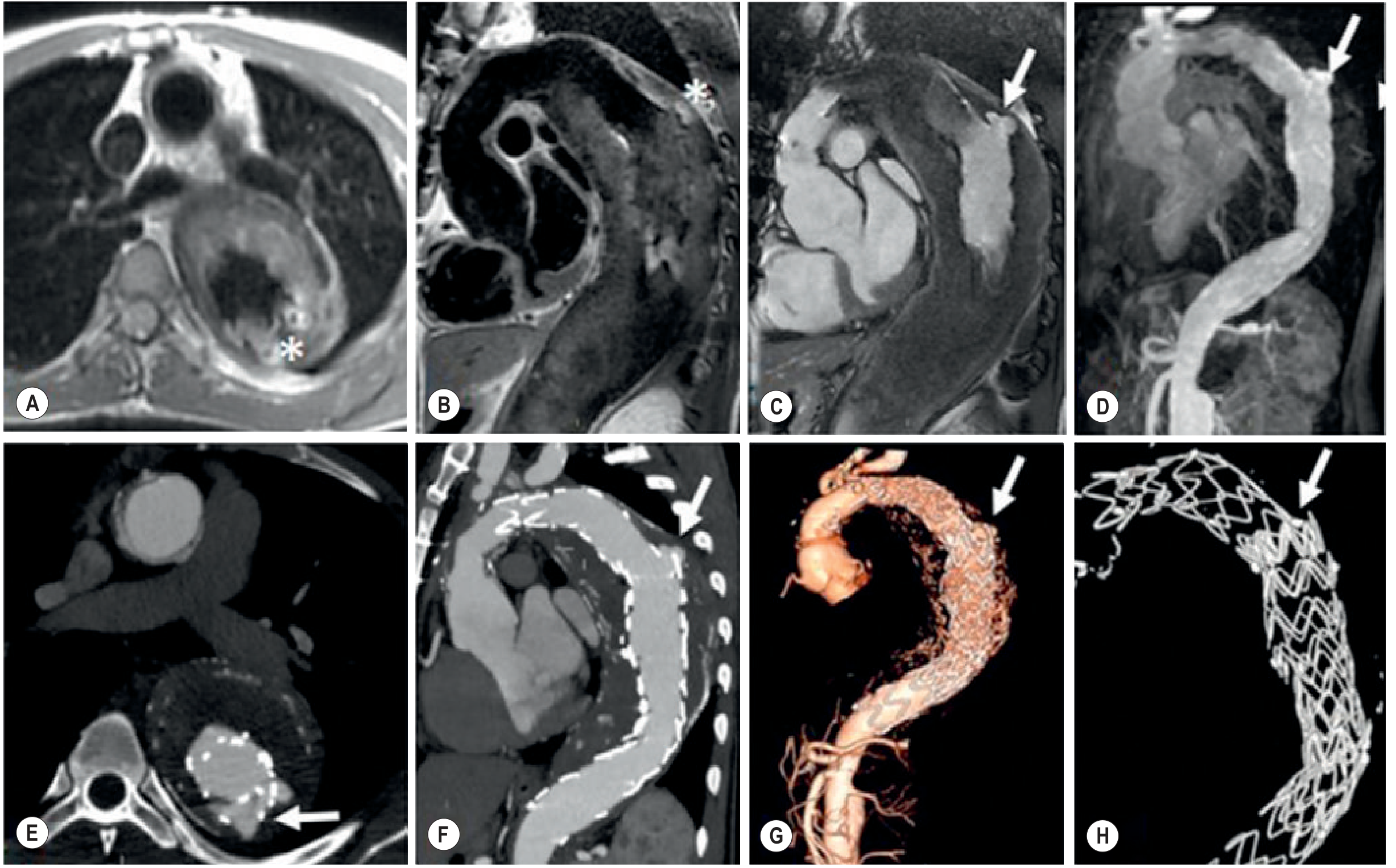
Figure: MR and CTA follow-up after TEVAR for Type B dissection. Note the dissection flap (A), post-stent appearance (E-H), and endoleak detection requiring multimodal imaging. (Grainger & Allison's Diagnostic Radiology, Fig. 17.38)
TEVAR Post-treatment CT - Recognizing False Lumen Behavior:
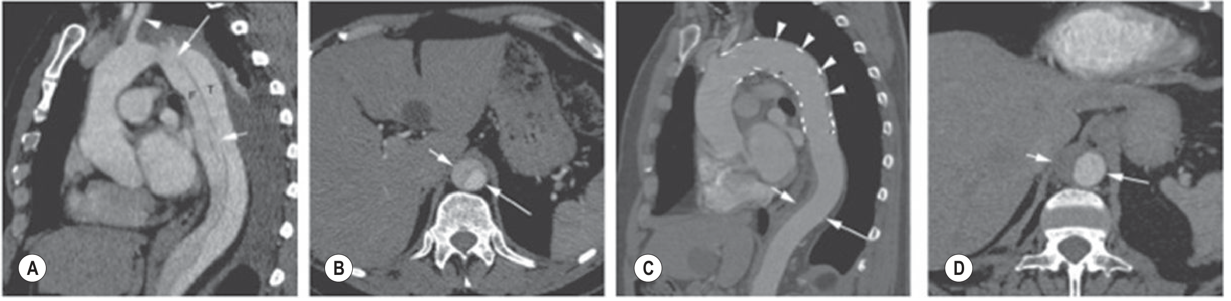
Figure: CT after TEVAR coverage of proximal entry tear showing false lumen thrombosis (ideal outcome). Ongoing false lumen perfusion after TEVAR = risk factor for aneurysmal dilatation. (Grainger & Allison's Diagnostic Radiology, Fig. 79.18)
Echocardiography (TTE/TEE):
- TTE: Limited sensitivity for dissection (poor windows for ascending aorta); if definite undulating intimal flap seen, specificity is high. A negative TTE does NOT exclude dissection.
- TEE: High sensitivity and specificity; particularly useful:
- Intraoperatively to confirm dissection and assess AR before/after repair
- Identifies fenestrated dissection flap, AR severity, pericardial effusion, wall motion abnormalities (RCA involvement)
- Cannot image the distal ascending aorta ("blind spot" between left main bronchus and aorta)
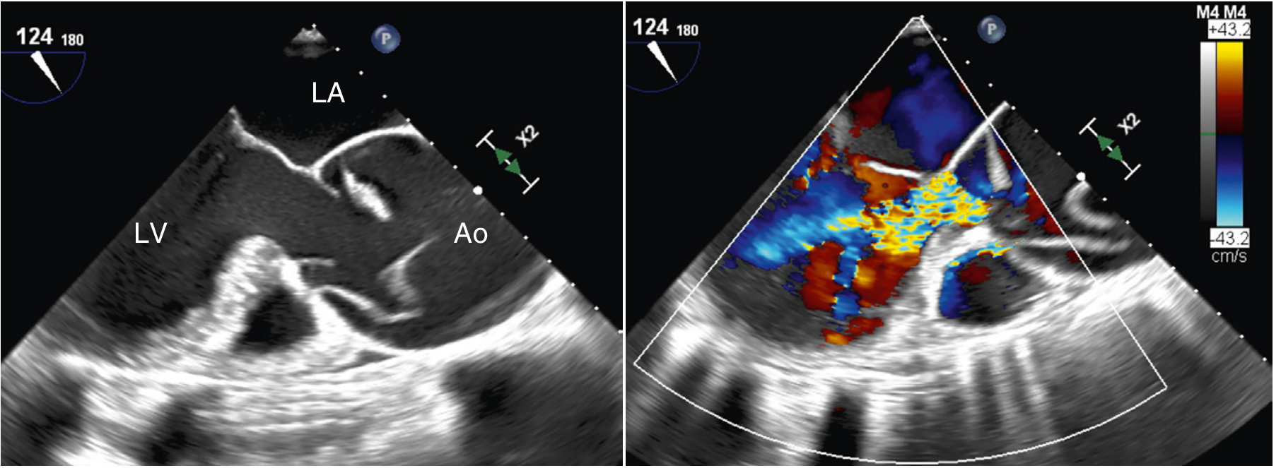
Figure: Intraoperative TEE in a young woman with Type A dissection presenting in cardiogenic shock. (Left) Fenestrated dissection flap adjacent to aortic valve extending into ascending aorta. (Right) Color Doppler showing severe AR filling the entire LVOT. (Textbook of Clinical Echocardiography, Fig. 18.20)
Key TEE findings in AAS:
- Dilated aortic lumen
- Linear, mobile echogenic intimal flap with motion different from aortic wall
- Differential color Doppler flow patterns in true vs false lumen (true lumen: pulsatile flow; false lumen: sluggish or absent flow)
- Pericardial effusion / tamponade
- Regional wall motion abnormalities (coronary involvement)
MRI:
- Highest accuracy; ideal for chronic dissections and follow-up
- No radiation, no contrast required for basic sequences
- Limitations: time-consuming, limited access in emergencies, contraindicated with many pacemakers
7. INITIAL MEDICAL MANAGEMENT (ALL AAS)
Immediate goals - "Anti-impulse therapy":
- Eliminate pain - IV opioids (morphine 2-4 mg IV)
- Reduce systolic BP - target SBP 100-120 mmHg
- Reduce dP/dt (rate of pressure rise) - target HR <60 bpm
First-line agents:
- IV beta-blocker: Labetalol (20 mg IV bolus, then 2 mg/min infusion) or esmolol (500 mcg/kg bolus, then 50-200 mcg/kg/min infusion) - reduces both HR and BP
- If beta-blocker insufficient: Add IV vasodilator (sodium nitroprusside 0.3-10 mcg/kg/min)
- Do NOT use vasodilators alone without beta-blockade - reflex tachycardia increases dP/dt and worsens dissection
Monitoring: Arterial line (use right radial for Type A - left may be compromised), central venous access, urinary catheter, continuous ECG, pulse oximetry bilaterally.
8. SURGICAL MANAGEMENT - TYPE A DISSECTION
8.1 Indications
Stanford Type A dissection = surgical emergency. Untreated mortality: >1% per hour in the first 24 hours; >50% within 48 hours. Fatal complications include aortic rupture, cardiac tamponade, acute AR, acute MI.
Transfer immediately to cardiac surgery. Even octogenarians benefit: 63% 1-year survival with surgery vs 38% with medical management for uncomplicated Type A in this age group.
8.2 Operative Strategy
Exposure: Median sternotomy.
Cannulation: The key challenge is safe cannulation in a friable, dissected aorta. Options:
- Femoral artery - traditional; risk of retrograde malperfusion if cannulating false lumen
- Right axillary/subclavian artery - preferred when femoral access is compromised or in Marfan syndrome; provides antegrade flow and safe cerebral perfusion
- Direct aortic cannulation - only when clearly in true lumen
CPB strategy:
- Venous cannulation via right atrium or bicaval
- Cooling to moderate hypothermia (24-27°C nasopharyngeal) before circulatory arrest
- Hypothermic Circulatory Arrest (HCA) is essential for open distal anastomosis without aortic cross-clamp
Cerebral protection during HCA:
- Antegrade Cerebral Perfusion (ACP) via right axillary/direct carotid cannulation - 10 mL/kg/min to right carotid, maintain R cerebral O2 sat ≥60%
- Retrograde Cerebral Perfusion (RCP) via superior vena cava - simpler but less effective
- ACP is associated with improved neurological outcomes and allows longer safe arrest times
The Distal Anastomosis:
- Resect entire ascending aorta from sinotubular junction to innominate artery with a bevel at the lesser curve of the arch
- Inspect arch for complex intimal disruptions under direct vision
- Confirm brachiocephalic branches arise from true lumen
- Obliterate the false lumen: Two main techniques:
- Teflon felt sandwich: Two strips of felt sandwiching the dissected aortic wall → anastomose Dacron graft to felt-reinforced aorta
- Felt plug into false lumen: Author-preferred technique to avoid late pseudoaneurysm from glue (biological glues are avoided as they can be destructive and promote pseudoaneurysm)
The Proximal Reconstruction:
During rewarming, assess aortic root, aortic valve, and coronary ostia:
- Aortic valve preservation (commissure resuspension) - when no root aneurysm (<5 cm) and valve is structurally sound
- Composite aortic root replacement (Bentall procedure) - when:
- Root aneurysm >5 cm
- Complex aortic valve pathology
- Dissection extending to coronary ostia
- Genetically triggered disease (Marfan, Loeys-Dietz)
- Valve-sparing root replacement (David / Yacoub procedure) - only in experienced centers with appropriate anatomy
Arch Involvement:
- Routine arch replacement is not necessary for most Type A dissections (increases complexity and risk)
- Arch replacement is indicated when:
- Complex intimal disruptions in the arch
- Arch aneurysm >5 cm
- Genetically triggered thoracic aortic disease
Frozen Elephant Trunk (FET):
- For large intimal disruptions in the distal arch or proximal descending aorta
- Antegrade stent-graft deployment under direct vision through the open arch
- Stabilizes the distal aorta, reduces need for early reintervention, addresses distal malperfusion
- Bridges toward second-stage descending aorta treatment
Myocardial Protection:
- In cases with AI + coronary malperfusion: LV venting on bypass before cross-clamp
- Retrograde coronary sinus cardioplegia + direct antegrade coronary ostial cardioplegia
8.3 Surgical Results (IRAD Data)
| Patient Category | Hospital Mortality |
|---|---|
| Overall surgical mortality | 18% |
| Stable patients | ~10-15% |
| Unstable patients (tamponade, shock, stroke, coma, mesenteric ischemia) | >30% |
Postoperative complications to manage in ICU:
- Coagulopathic bleeding (massive transfusion - use thromboelastography-guided protocol)
- Transient neurologic dysfunction / stroke
- Renal insufficiency
- Acute lung injury
- New malperfusion syndromes - persistent false lumen flow
- Refractory hypertension
- Multiorgan failure
9. MANAGEMENT OF TYPE B DISSECTION
9.1 Uncomplicated Type B - Medical Management
Standard of care: Anti-impulse therapy (IV beta-blocker). Target:
- HR <60 bpm
- SBP 100-120 mmHg
Transition to oral agents when stable: beta-blockers + ACE inhibitors + calcium channel blockers as needed.
Early mortality with medical management is ~10% but is similar to emergency surgical repair (which historically had >50% mortality for Type B).
9.2 Complicated Type B - Indications for Intervention
| Indication | Status |
|---|---|
| Rupture with blood outside vessel wall | Absolute |
| Major vessel occlusion with malperfusion syndrome | Absolute |
| Rapid expansion to total aortic diameter ≥4.5 cm | Absolute |
| Uncontrolled pain despite medical therapy | Relative |
| Worsening radiological findings | Relative |
Approximately 20% of acute Type B dissections are complicated by rupture or malperfusion.
9.3 TEVAR for Type B - Technique
TEVAR has largely replaced open surgery for complicated Type B with improved early outcomes.
Principle: Cover the primary entry tear (usually in the proximal descending aorta, zone 3) to redirect flow into the true lumen, promote false lumen thrombosis, and relieve malperfusion.
Coverage zones:
- Zone 3 to Zone 5 (caudal left subclavian to just proximal to celiac axis) is standard
- Zone 2 coverage may be needed if entry tear involves the left subclavian artery
- If Zone 2 coverage is planned non-emergently: left subclavian revascularization first (carotid-subclavian bypass or subclavian-carotid transposition) to reduce risk of vertebrobasilar stroke, left arm ischemia, and paraplegia
Technical steps:
- Iliofemoral access (open or percutaneous under US/fluoroscopy guidance)
- Systemic heparin (ACT >250 sec)
- J-wire advanced into ascending aorta
- IVUS confirmation of true lumen access along entire wire length - critical step
- IVUS delineates LSA origin, celiac axis, compressed true lumen, and branch vessel involvement
- Exchange for stiff Lunderquist wire
- Sequential stent-graft deployment with controlled BP lowering (SBP ~80 mmHg during deployment)
- Post-deployment IVUS + fluoroscopy to confirm coverage and true lumen expansion
Goal: Complete false lumen thrombosis - protects against rupture, aneurysmal dilatation, and improves long-term survival.
Residual false lumen perfusion can occur via:
- Uncovered fenestrations
- Retrograde flow from abdominal aortic fenestrations
- Back-bleeding from branch vessels
9.4 Prognostic Factors Favoring Intervention in "Uncomplicated" Type B
(From Grainger & Allison - these patients have high-risk features)
| Factor | Threshold |
|---|---|
| False lumen expansion rate | >1 cm/year |
| False lumen diameter | >2.2 cm |
| Combined early diameter | >4 cm |
| Entry tear size | >1 cm diameter |
| Partial false lumen thrombosis | (paradoxically increases rupture risk vs complete thrombosis) |
Whether pre-emptive TEVAR in high-risk uncomplicated Type B improves long-term outcomes remains to be proven (INSTEAD-XL trial showed benefit at 5 years; ADSORB trial ongoing).
9.5 Open Surgery for Type B
Reserved for:
- Unsuitable anatomy for TEVAR (extensive branch vessel involvement, aortic tortuosity, inadequate landing zones)
- Genetically triggered aortic disease (Marfan - risk of late stent-graft failure)
- Failed TEVAR
10. INTRAMURAL HEMATOMA - SPECIFIC MANAGEMENT
| Type | Management |
|---|---|
| Type A IMH | Immediate surgical intervention (majority opinion); some Japanese centers challenge this with medical management for uncomplicated cases |
| Type B IMH | Medical management + serial imaging (Class I ESC recommendation); TEVAR (Class IIa) or open surgery (Class IIb) for: expansion, periaortic hematoma, recurrent pain, conversion to frank dissection |
TEVAR for Type B IMH challenge: No primary intimal tear to identify; coverage territory is either presumptive (site of initiation) or targeted at an associated PAU or periaortic hematoma.
Natural history: 60% stabilize; 40% progress (to frank dissection, rupture, or aneurysm). Serial imaging at 1 week, 1 month, 3 months, 6 months, then annually.
11. PENETRATING AORTIC ULCER - SPECIFIC MANAGEMENT
| Situation | Management |
|---|---|
| Asymptomatic PAU | Medical (antiimpulse therapy) + routine surveillance imaging |
| PAU ≥20 mm diameter or neck ≥10 mm | Early intervention regardless of symptoms |
| Symptomatic PAU (pain, expansion) | Urgent intervention |
| Pseudoaneurysm formation or rupture | Emergency TEVAR (preferred) or open surgery |
TEVAR for PAU: Good limited-data results; challenges include severe aortic atherosclerosis extending into access vessels, and laminated thrombus making safe landing zone definition difficult.
Open surgery for PAU: Involved aortic segment resected and replaced with prosthetic graft or homograft; finding normal aorta proximally/distally for anastomosis is technically difficult due to extensive calcification.
12. CHRONIC DISSECTION AND ANEURYSMAL DEVELOPMENT
- Late aneurysm formation occurs in up to 50% of patients with chronic Type B dissection
- Management requires exclusion of false lumen flow by closing all fenestrations between true and false lumens
- Complex (fenestrated or branched) endografts or surgical bypass of major aortic branches may be needed
- Surveillance: CT/MRI at 1, 3, 6, 12 months then annually for all AAS survivors
- Strict BP control (SBP <130 mmHg) is essential for all chronic dissection patients
13. SPECIAL SITUATIONS AT MCh LEVEL
13.1 Coronary Malperfusion with Type A Dissection
- Dissection can shear coronary ostia (usually RCA) causing acute MI presentation
- Do not give thrombolytics if there is any possibility of aortic dissection - can be fatal
- In the OR: direct coronary ostial cardioplegia; if RCA involvement recognized pre-bypass, prepare for coronary artery bypass or ostial reconstruction
13.2 Tamponade with Type A Dissection
- Do NOT drain pericardial tamponade percutaneously pre-operatively (raises BP → worsens dissection; can cause sudden decompression → death)
- Go directly to OR; open pericardium on CPB under controlled conditions
13.3 Marfan Syndrome
- Valve-sparing root replacement preferred when possible (David procedure)
- TEVAR is relatively contraindicated for Type B - late stent-graft failure and aortic dilation distal to the graft
- Open surgery preferred for Type B in young Marfan patients
13.4 Iatrogenic Dissection During Catheterization/CPB
- Type A iatrogenic dissections often have better outcomes than spontaneous (less medial fragility)
- Same surgical principles apply
13.5 Malperfusion Syndrome Strategy
- True lumen collapse (dynamic obstruction) - TEVAR or fenestration restores flow
- Static obstruction from dissection flap occluding branch vessel orifice - may need stenting of the branch vessel in addition to TEVAR
- Visceral malperfusion with established bowel ischemia (rising lactate, peritonism) - consider laparotomy + TEVAR hybrid approach
14. PARAPLEGIA PREVENTION IN AORTIC SURGERY
The most feared complication of descending/thoracoabdominal aortic operations
Risk factors: Extent II (Crawford), cross-clamp time >30 min, emergent repair, renal failure, female sex, advanced age.
Protective strategies:
- Distal aortic perfusion (partial CPB or centrifugal pump) during thoracoabdominal repair
- CSF drainage (lumbar drain) - target CSF pressure <10 mmHg
- Motor evoked potential (MEP) / somatosensory evoked potential (SSEP) monitoring
- Moderate hypothermia (32-34°C)
- Steroid administration
- Re-implantation of intercostal arteries (T8-L2 segment)
- Maintain MAP >90 mmHg post-operatively
15. KEY NUMBERS TO KNOW (MCh Examinations)
| Parameter | Value |
|---|---|
| Incidence of aortic dissection | 5-10 per 100,000/year (increasing with ageing population) |
| Stanford A as proportion of all dissections | ~75% |
| Untreated Type A mortality | >50% at 48 hours; ~1%/hour early |
| Overall surgical mortality (IRAD) | 18% |
| Unstable Type A surgical mortality | >30% |
| Type B complicated | ~20% of all Type B |
| Aneurysm formation in chronic Type B | Up to 50% |
| IMH proportion of AAS | 10-20% |
| PAU proportion of AAS | 2-7% |
| HCA temperature (moderate) | 24-27°C nasopharyngeal |
| Target SBP (medical therapy) | 100-120 mmHg |
| Target HR (anti-impulse) | <60 bpm |
| PAU size threshold for intervention | Diameter >20 mm or neck >10 mm |
16. CURRENT GUIDELINES (2024 ESC)
The 2024 ESC Guidelines for peripheral arterial and aortic diseases (Mazzolai et al., European Heart Journal, 2024; PMID: 39210722) consolidate current evidence:
- Type A AAS: Emergency surgery remains standard of care
- Type B complicated AAS: TEVAR preferred over open surgery
- Type B uncomplicated AAS: Medical management as first-line; TEVAR may be considered in high-risk uncomplicated cases
- IMH Type B: Initial medical management + surveillance (Class I); TEVAR for progression (Class IIa)
- PAU symptomatic: Intervention recommended; TEVAR preferred for descending lesions
SUMMARY FRAMEWORK FOR MCh LEVEL
AAS
├── Type A (ascending involved)
│ ├── ALL → Emergency Surgery
│ ├── Root: resuspend valve vs Bentall vs David
│ ├── Arch: routine replacement only if disrupted/aneurysmal/genetic
│ ├── Distal: HCA + ACP, felt obliteration of FL, Dacron graft
│ └── FET if distal arch/proximal descending involvement
│
├── Type B (descending only)
│ ├── Uncomplicated → Medical (anti-impulse) + surveillance
│ ├── Complicated (rupture/malperfusion/expansion) → TEVAR
│ └── TEVAR unsuitable/genetic → Open surgery
│
├── IMH
│ ├── Type A → Surgical (same as Type A AD)
│ └── Type B → Medical; TEVAR if progressing
│
└── PAU
├── Asymptomatic, small → Medical + surveillance
├── Large (>20mm / neck >10mm) or symptomatic → Intervention
└── TEVAR preferred for descending PAU
Sources consulted for this response:
- Braunwald's Heart Disease (2 Vol Set), Chapters 42 & 84
- Fuster & Hurst's The Heart, 15e, Chapter 23
- Robbins & Kumar Basic Pathology, Chapter 8
- Grainger & Allison's Diagnostic Radiology, Chapters 17 & 79
- Textbook of Clinical Echocardiography, Chapters 16 & 18
- Miller's Anesthesia 10e, Chapter 52
- Current Surgical Therapy 14e, Chapter on Aortic Dissection
- Schwartz's Principles of Surgery 11e, Chapter 22
- Sabiston Textbook of Surgery
- 2024 ESC Guidelines [PMID: 39210722]That is a complete MCh Cardiac Surgery-level teaching document on Acute Aortic Syndrome. Here is a summary of what was covered and the sources used:
What was covered (16 sections):
- Definition and spectrum - AAS as a triad: Dissection (75%), IMH (10-20%), PAU (2-7%)
- Pathophysiology of all three entities with gross pathology and histology images from Robbins
- Epidemiology and risk factors including IRAD demographics, Marfan, cocaine, pregnancy
- DeBakey + Stanford classification with the classic diagram from Miller's Anesthesia
- Clinical presentation - pain characteristics, the full malperfusion syndrome table (Schwartz)
- Diagnostic workup - CXR limitations, CTA protocol, TEE features (intraoperative image included), MRI
- Medical management / anti-impulse therapy - targets, drug choices, monitoring
- Type A surgical strategy - cannulation options, HCA + cerebral perfusion, felt technique, root management decision-making, arch/FET indications, IRAD mortality data
- Type B management - uncomplicated vs complicated, malperfusion indications, TEVAR technique step-by-step including IVUS
- IMH management - Japanese vs Western controversy for Type A; Type B algorithm
- PAU management - size thresholds, TEVAR challenges
- Chronic dissection - surveillance, aneurysmal evolution in 50%
- Special situations - coronary malperfusion (no thrombolytics!), tamponade (no percutaneous drainage!), Marfan, malperfusion strategies
- Paraplegia prevention - CSF drainage, MEP monitoring, intercostal reimplantation
- Key numbers for MCh examinations
- 2024 ESC Guidelines summary
make detailed mch cardiac surgery level note standard sources with pictures and everything about blunt traima aorta injury
Finding Sources
Finding Sources
Finding Sources
Finding Sources
Finding Sources
Finding Sources
Reading File
Reading File
Excellent finds. Now let me read the Grainger imaging section in detail, the Grainger block4 TAI section (TEVAR), Echocardiography section, and Miller's anesthesia operative management - all in parallel.
Reading File
Reading File
Reading File
Reading File
Excellent - I found the Presley CT grading table. Let me now read the rest of the Grainger CT grading table, search for AAST classification, and fetch key images all in parallel.
Reading File
Finding Sources
Loading Image
Searching PubMed
"blunt traumatic aortic injury" management TEVAR
Excellent - found 2022 ACC/AHA guidelines AND a 2026 SVS systematic review. Now read the Fischer's Mastery of Surgery BTAI section and Sabiston + the rest of the Grainger CTA description, and fetch more images in parallel.
Reading File
Reading File
Reading File
Loading Image
Now read the Fischer's section on AAST grading and TEVAR technique, plus the Sabiston open surgical section, and fetch the CXR image and the CT axial image.
Reading File
Reading File
Loading Image
Loading Image
Now let me read the Fischer's TEVAR technique and open surgery section, fetch the anatomical isthmus diagram, and get the 2022 ACC/AHA guideline abstract.
Reading File
Loading Image
Reading Article
PMID: 36322642
Now let me read the Fischer's TEVAR technical steps and open surgical repair sections, and fetch the classification diagram.
Reading File
Loading Image
I now have comprehensive data from 6 textbooks plus 6 images and 2 guidelines. Let me now synthesize the complete MCh-level teaching note.---
Blunt Traumatic Aortic Injury (BTAI) - MCh Cardiac Surgery Level
Sources: Fischer's Mastery of Surgery (8e) | Grainger & Allison's Diagnostic Radiology | Miller's Anesthesia (10e) | Sabiston Textbook of Surgery | Schwartz's Principles of Surgery (11e) | Textbook of Clinical Echocardiography | 2022 ACC/AHA Guideline for Aortic Disease [PMID: 36322642] | SVS Systematic Review 2026 [PMID: 40482894]
1. EPIDEMIOLOGY AND MORTALITY PROFILE
BTAI is the second leading cause of death from blunt trauma, exceeded only by traumatic brain injury.
| Parameter | Data |
|---|---|
| Prehospital mortality | ~80% (die before reaching ER) |
| Of those reaching hospital - die within 24 hrs | 50% |
| Untreated - die within 4 months | 90% |
| US motor vehicle deaths yearly | ~40,000; 20% caused by aortic rupture |
| Median age of BTAI | 41 years (predominantly young males) |
The high prehospital mortality means that only the subset with adventitially contained injuries survive to hospital presentation - these are the patients a cardiac surgeon will encounter. Their continued survival depends on the integrity of the adventitia as the only remaining intact layer.
- Fischer's Mastery of Surgery, p. 7029; Miller's Anesthesia, p. 7775
2. MECHANISMS OF INJURY
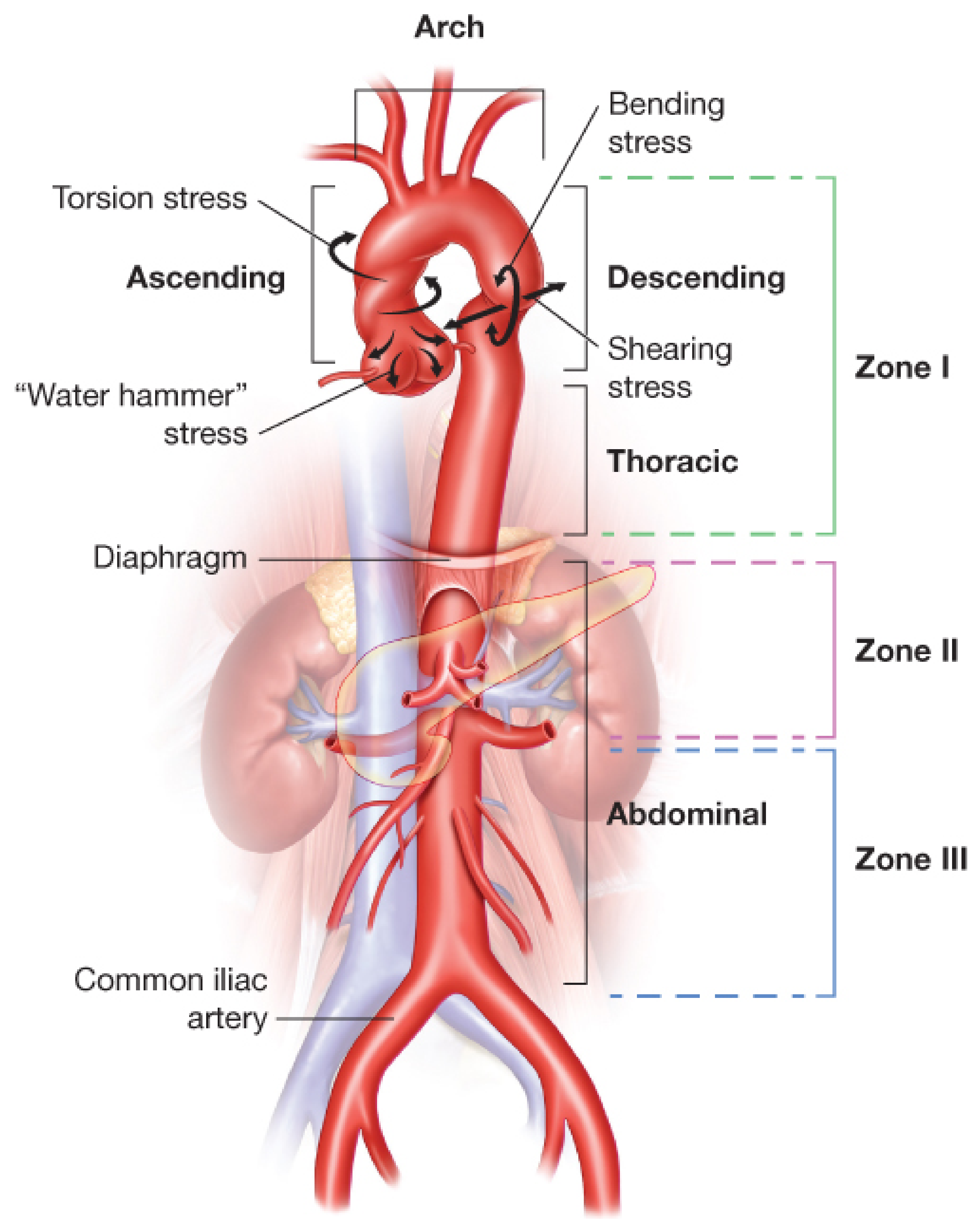
Figure: Biomechanical forces acting on the aorta during blunt deceleration trauma. The isthmus (transition from mobile arch to fixed descending aorta) receives the greatest shear stress. (Fischer's Mastery of Surgery, 8e, Fig. 261.1)
2.1 Three Biomechanical Mechanisms
1. Rapid Deceleration - Shearing (most common)
- The mobile aortic arch continues forward while the relatively fixed descending thoracic aorta (fixed by ligamentum arteriosum, intercostal arteries, and paravertebral tissues) is anchored
- The transition zone - the aortic isthmus (just distal to the left subclavian artery) - receives the maximal shear force
- This accounts for 50-70% of all BTAI
- Mechanism: high-speed MVA (especially >30 mph / 48 km/h), motorcycle crashes, pedestrian vs automobile
2. Osseous Pinch
- Anteroposterior compression of the chest causes the heart and aorta to be crushed between the sternum and vertebral column
- Results in direct full-thickness compression injury
- Involves the ascending aorta close to the innominate artery or just above the aortic valve
- Mechanism: falls from large heights (>10 ft / LD50 = 4 stories), crush injuries
3. Torsional / Hydraulic Mechanism ("Water Hammer")
- Sudden displacement of the heart to the left during AP compression creates torque at the arch-isthmus junction
- Rapid increase in intraluminal aortic pressure (transient hypertension exceeding tensile strength of aortic wall)
- Can cause injury at any aortic segment
Sites of injury by frequency:
-
Aortic isthmus (distal to L. subclavian artery): 50-90%
-
Ascending aorta / aortic arch: 18%
-
Distal thoracic / diaphragmatic aorta: 14%
-
Abdominal aorta: rare (infrarenal; usually "osseous pinch" mechanism)
-
Grainger & Allison's, p. 430; Miller's Anesthesia, p. 7775
3. SPECTRUM OF INJURY (PATHOLOGICAL)
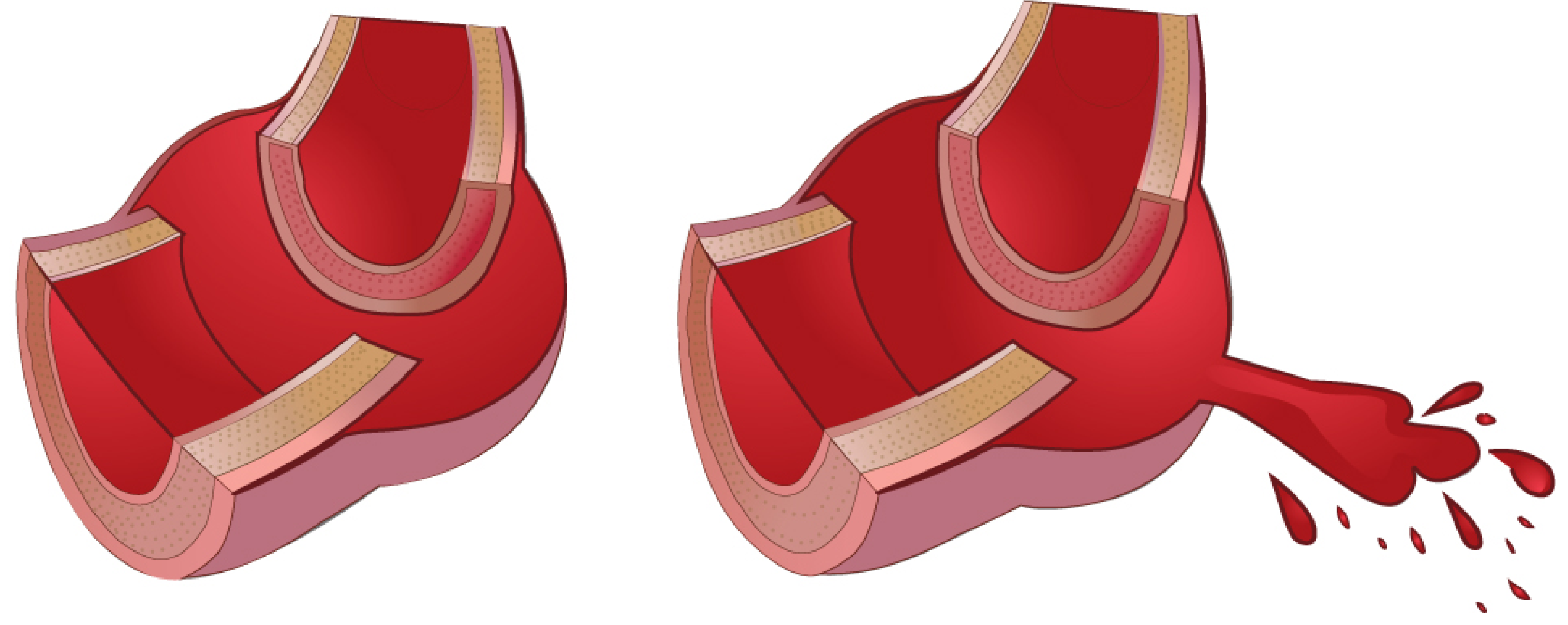
Figure: The two ends of the BTAI spectrum - contained adventitial rupture (survivors) vs complete transmural rupture (prehospital fatalities). (Fischer's Mastery of Surgery, Fig. 261.2 partial)
The spectrum from the least to the most severe:
- Simple subintimal hemorrhage - intimal disruption only, no hemodynamic consequence
- Intimal flap / tear - intima disrupted, media intact
- Intramural hematoma - media involved, adventitia intact
- Pseudoaneurysm - all layers except adventitia disrupted; adventitia + periaortic tissues provide containment (the "false aneurysm")
- Frank transection/rupture - complete transmural disruption → exsanguination (80-90% die at scene)
In 80-90% of patients who die at the scene, there is complete full-thickness aortic rupture. Survivors reaching hospital invariably have the adventitia and/or periaortic mediastinal tissues still intact, maintaining precarious containment.
4. CLASSIFICATION - SVS GRADING SYSTEM (Azizzadeh/ATF)
The Society for Vascular Surgery (SVS) grading system (Azizzadeh et al., J Vasc Surg 2009) is the current standard. It uses CTA findings to classify the extent of aortic wall injury:
| Grade | Injury Description | CTA Findings | Relative Frequency (ATF Registry) |
|---|---|---|---|
| Grade 1 | Intimal tear | Intimal flap, intimal irregularity | ~25% |
| Grade 2 | Intramural hematoma | Medial hematoma, no pseudoaneurysm | ~15% |
| Grade 3 | Pseudoaneurysm | Focal outpouching, periaortic hematoma | ~50% |
| Grade 4 | Free rupture | Contrast extravasation, hemothorax | ~10% |
Grades 1 + 2 = Minimal Aortic Injury (MAI) - emerging evidence supports definitive medical management with serial imaging for MAI.
Grades 3 + 4 require urgent intervention (<24 hours).
Presley Trauma Center CT Grading System (Gavant, 1999) is an alternative radiological grading system:
| Grade | Subgrade | CT Findings |
|---|---|---|
| I (Normal aorta) | Ia | Normal aorta, no mediastinal hematoma |
| Ib | Normal aorta, para-aortic mediastinal hematoma | |
| II (Minimal aortic injury) | IIa | Small (<1 cm) pseudoaneurysm / intimal flap, no mediastinal hematoma |
| IIb | Small pseudoaneurysm / intimal flap with mediastinal hematoma | |
| III (Confined injury) | IIIa | >1 cm well-defined pseudoaneurysm, intimal flap, no arch/great vessel involvement |
| IIIb | >1 cm pseudoaneurysm with arch/great vessel involvement | |
| IV (Total disruption) | IV | Irregular poorly-defined pseudoaneurysm, mediastinal hematoma, total disruption |
(Modified from Gavant ML, Radiographics 1999)
5. CLINICAL PRESENTATION AND INITIAL ASSESSMENT
5.1 The "High Index of Suspicion" Principle
The majority of patients with BTAI do not have specific symptoms or signs referable to the aortic injury on arrival. Clinical diagnosis is impossible; imaging is mandatory in high-energy mechanisms.
Key clinical features that mandate CTA:
- High-speed MVA (>30 mph / 48 km/h), especially frontal or lateral impact
- Motorcycle collision
- Pedestrian struck by vehicle
- Fall from >10 feet
- Airplane crash
- Crush injury
Physical signs that increase suspicion:
- Chest wall ecchymosis / "seat belt sign"
- Sternal fracture (associated with ascending aortic injury)
- Crepitus, flail chest
- Unequal bilateral blood pressure (subclavian involvement or pseudocoarctation)
- Absent or reduced distal pulses
- Hoarseness (recurrent laryngeal nerve compression by periaortic hematoma)
- Paraplegia or paraparesis (spinal cord ischemia)
- Harsh systolic murmur (turbulent flow across pseudoaneurysm)
Critical point: Normal blood pressure on arrival does NOT rule out BTAI.
5.2 ATLS Primary Survey in BTAI Context
| ATLS Element | BTAI Relevance |
|---|---|
| Airway | Often intubated in polytrauma; cervical spine immobilization limits TEE |
| Breathing | Massive hemothorax (blood from periaortic rupture into pleural space) |
| Circulation | Hemorrhagic shock (BTAI + other injuries); tamponade from ascending aortic injury |
| Disability | TBI concurrent in 40-60% of BTAI cases - impacts BP management targets |
| Exposure | Seat belt marks, chest wall ecchymosis, deformity |
FAST exam: Cannot diagnose BTAI directly; screens for pericardial blood (ascending injury), hemothorax.
6. DIAGNOSTIC IMAGING
6.1 Plain Chest X-Ray - Screening Tool, Not Diagnostic
Sensitivity 80-90%, specificity poor (many findings are from venous mediastinal bleeding unrelated to aortic injury)
CXR features suggestive of BTAI:
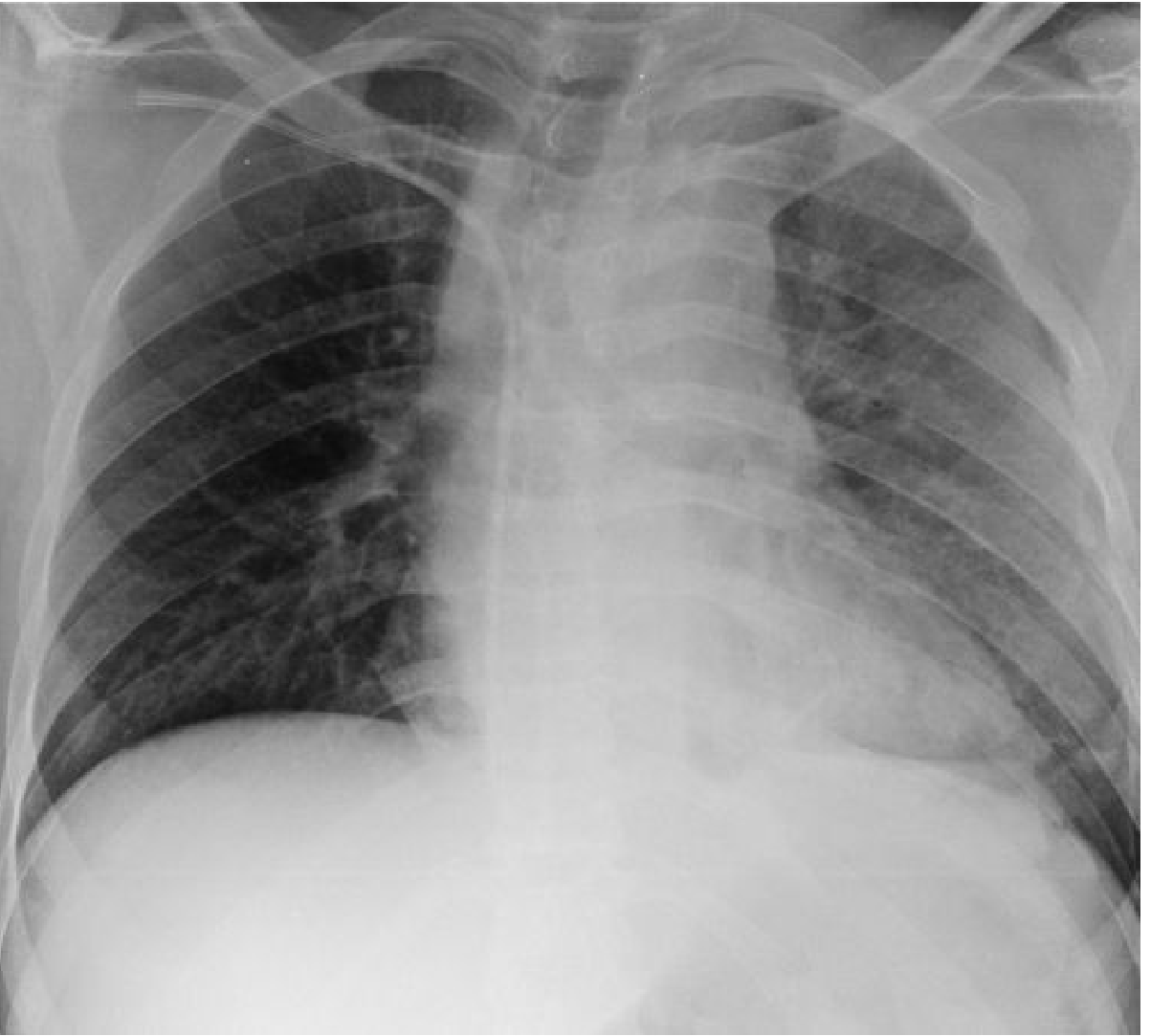
Figure: Supine CXR after road traffic accident demonstrating mediastinal widening (M/C ratio ~0.3), widened right paratracheal stripe, rightward tracheal deviation, and obscured aortic knob - all signs of traumatic aortic injury. (Grainger & Allison's Diagnostic Radiology, Fig. 17.20)
| CXR Finding | Comment |
|---|---|
| Widened mediastinum >8 cm (or M/C ratio >25%) | Most sensitive single sign; often non-specific (venous bleeding) |
| Loss of normal aortic contour (aortic knob obscured) | Periaortic hematoma effacing the aortic knob contour |
| Tracheal deviation to the right | Mediastinal hematoma displacing the trachea |
| Depression of the left mainstem bronchus (>40°) | Hematoma between arch and left main bronchus |
| Nasogastric tube deviation to the right | Classic sign of mediastinal hematoma displacing the esophagus |
| Widened right paratracheal stripe | Hematoma tracking right paratracheal |
| Left apical cap | Extrapleural hematoma tracking to the apex |
| Left hemothorax | Blood from periaortic rupture or hemothorax from rib fractures |
Important: A normal CXR does not exclude BTAI. The decision to perform CTA should be based on mechanism of injury, not CXR appearance.
6.2 CT Angiography (CTA) - Gold Standard
CTA has replaced conventional angiography as the standard of care. Sensitivity and specificity approaching 100% with modern multidetector CT.
Advantages:
- Rapid acquisition (seconds for entire thorax + abdomen)
- Available 24/7 adjacent to ICU/trauma bay
- Simultaneously evaluates brain, thorax, abdomen, and pelvis (mandatory in polytrauma)
- Provides preoperative planning data: aortic diameters, landing zone lengths, access vessel anatomy, relationship to arch vessels
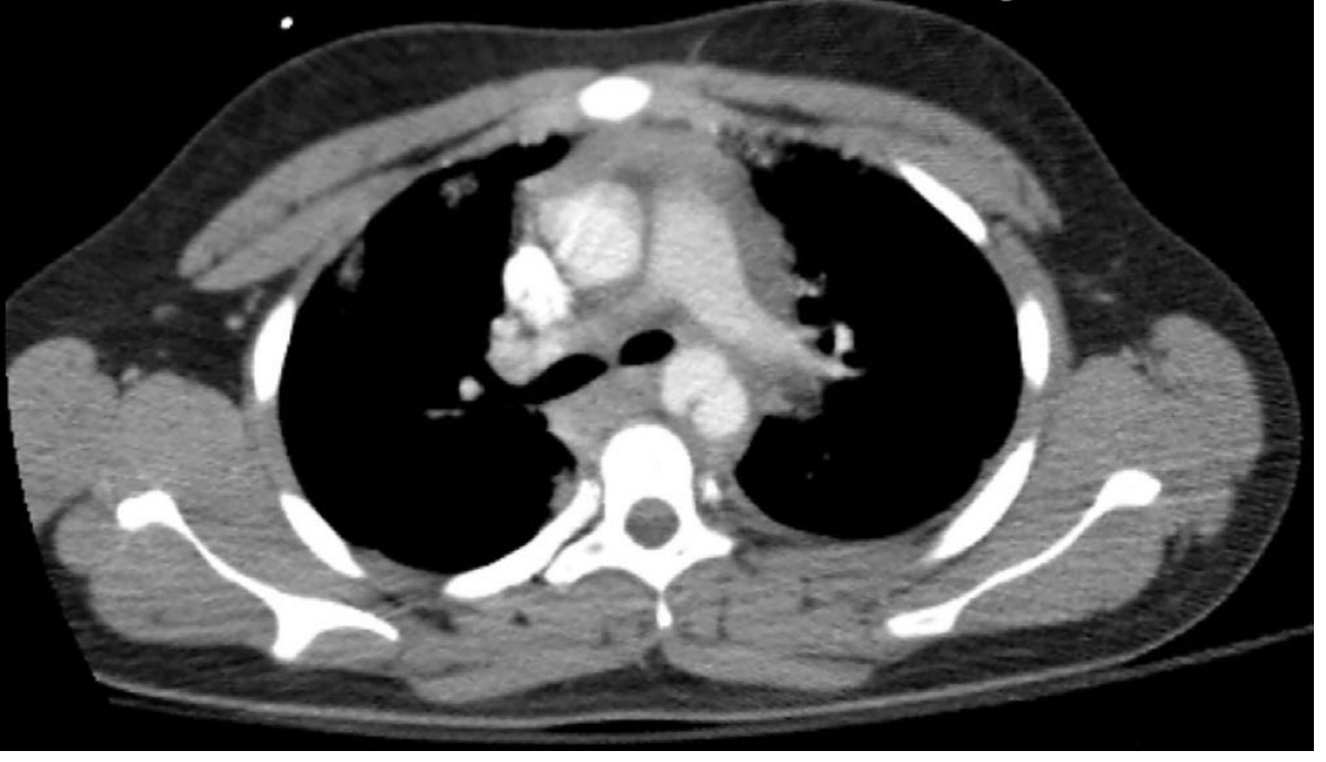
Figure: Axial CTA showing large traumatic dissection of the thoracic aorta with extensive periaortic hematoma. (Sabiston Textbook of Surgery, Fig. 36.25)
CTA direct signs of BTAI:
- Intimal flap
- Focal calibre change (pseudocoarctation) at the isthmus
- Contrast extravasation (pseudoaneurysm / frank rupture)
- Intramural hematoma (hyperdense crescent on non-contrast)
- Aortic contour abnormality
CTA indirect signs:
- Mediastinal/periaortic hematoma
- Left hemothorax (without other explanation)
CTA pitfalls - False positives:
- Ductus diverticulum - normal anatomical variation at the isthmus (smooth, well-defined concavity on the inferior aspect of the isthmus)
- Prominent superior intercostal vein ("aortic nipple")
- Bronchial artery or intercostal artery infundibulum
- Aberrant subclavian artery
- Streak artefacts (non-elevated arms, left arm IV contrast injection, monitoring lines)
- Severe aortic atherosclerosis
Key differentiator: Traumatic injuries are associated with periaortic/mediastinal hematoma; ductus diverticulum and vessel variants are not surrounded by blood.
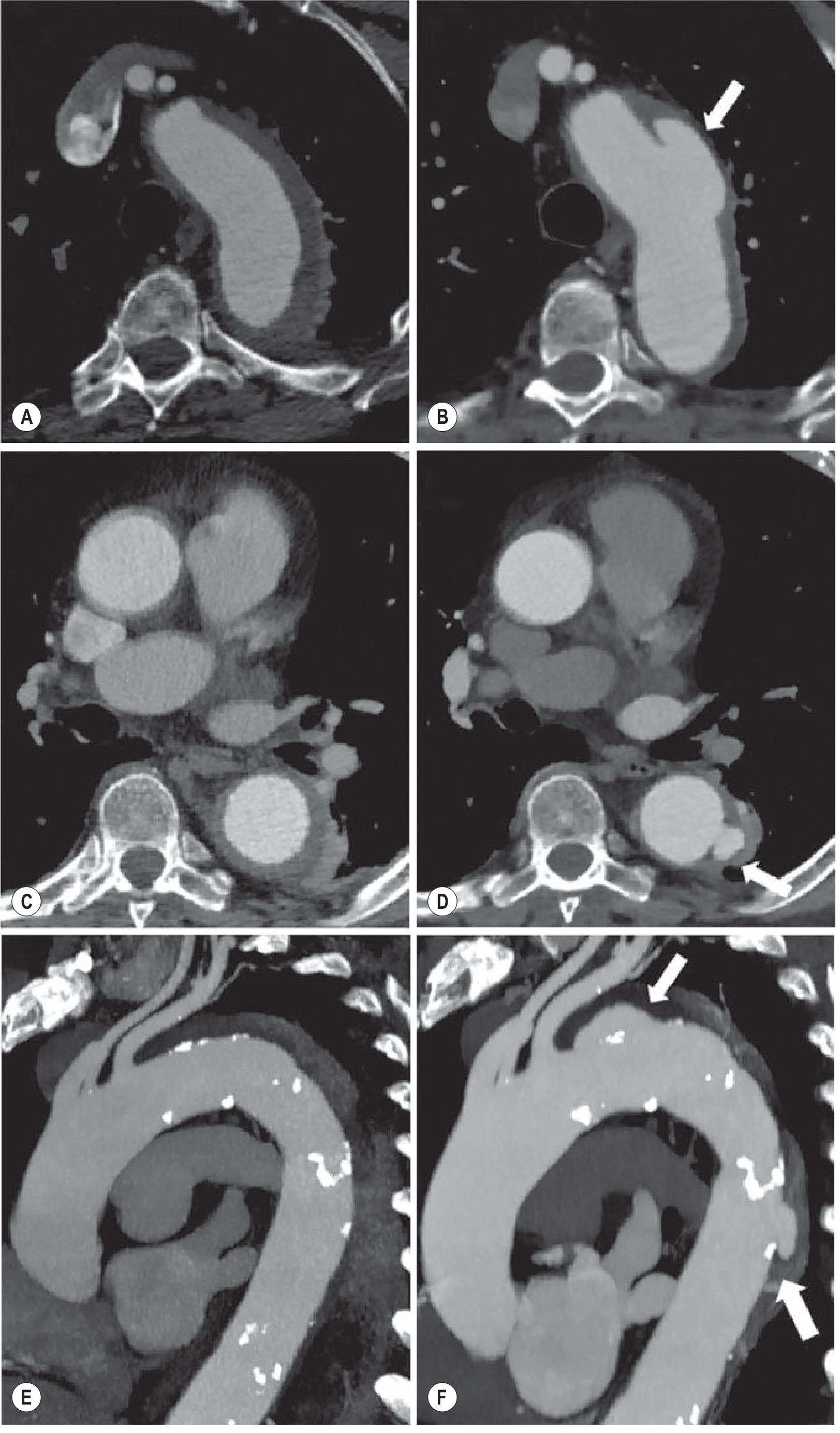
Figure: CTA axial and sagittal MPR showing evolving aortic wall injury - IMH of the descending thoracic aorta at presentation (A,C,E) and evolution to penetrating ulcer (arrows in B,D,F) at 8 days. Demonstrates the dynamic nature of aortic wall injury and need for serial imaging. (Grainger & Allison's, Fig. 17.19)
6.3 Conventional Aortography
No longer the preferred modality. Limitations:
- Lower accuracy than CTA (sensitivity 84-96%)
- False positives: ductus diverticulum, atheroma, overlapping vessel densities
- False negatives: poor aortic opacification, small intimal defects
- Does not show indirect signs (mediastinal hematoma, other injuries)
- Now reserved for: equivocal CTA findings + IVUS, or as part of endovascular intervention planning
6.4 Transesophageal Echocardiography (TEE)
- Sensitivity 91%, specificity 98% for isthmic injuries
- Performed at bedside in 15-20 minutes, even in unstable patients
- Not available in the ICU in all centers; operator-dependent
TEE findings in BTAI:
- Mural flap at the intimal disruption site
- Aortic wall deformity from contained rupture
- Gap >7 mm between probe and aortic wall at the proximal descending aorta level (with blood between aortic wall and pleura) - strongly suggests aortic disruption
- Can also detect: cardiac tamponade, left pleural effusion, myocardial contusion (wall motion abnormalities), hypovolemia (small, hyperdynamic LV)
Limitations of TEE in BTAI:
- Contraindicated in severe facial injuries, unstable cervical spine fractures, or esophageal injury
- Full circumference visualization not achieved in ~30% (aortic arch especially limited - the classic "blind spot")
- Cannot assess for other organ injuries simultaneously
Role in TEVAR: Intraoperatively, TEE provides excellent real-time guidance for precise stent-graft placement relative to arch vessel origins.
6.5 MRI
- Fastest modern sequences: oblique sagittal black-blood fast spin-echo (BFBSE) - full thoracic aorta in minutes
- Can characterize partial vs circumferential injury
- Can identify and date IMH without contrast
- Limited by: restricted urgent access, patient monitoring challenges, incompatible equipment
- Role: useful for chronic traumatic pseudoaneurysms and serial follow-up
6.6 IVUS (Intravascular Ultrasound)
- Used when CTA is equivocal (especially for Grade 1 intimal tears)
- Performed during angiography/endovascular procedure
- Confirms true lumen access, aortic diameters, branch vessel origins
- Identifies extent of injury not visible on conventional angiography
7. INITIAL RESUSCITATION AND ANTI-IMPULSE THERAPY
7.1 ATLS-Guided Resuscitation
All BTAI patients are polytrauma patients. Prioritize:
- Airway + C-spine control
- Hemorrhage control - massive transfusion protocol (MTP) if hemorrhagic shock
- FAST examination - pericardial blood, hemothorax
- CTA chest/abdomen/pelvis once sufficiently stable
7.2 Anti-Impulse Therapy
Goal: Reduce aortic wall stress (dP/dt) to prevent propagation or rupture of the contained injury. Aortic wall stress = f(heart rate × dP/dt). Reducing both is essential.
Targets:
- SBP <120 mmHg (most centers, ATF consensus)
- HR <60 bpm
- Note: In concurrent TBI (common in BTAI), aggressive BP lowering may worsen cerebral perfusion pressure - modified targets of SBP 100-140 mmHg are used based on neurosurgical guidance; this requires multidisciplinary discussion.
Drug of choice:
- Esmolol infusion (500 mcg/kg bolus → 50-200 mcg/kg/min) - short-acting, easily titratable
- Labetalol (alpha + beta blockade) - 20 mg IV bolus, then infusion
- If BP not controlled with beta-blocker alone: add sodium nitroprusside or nicardipine (avoid without beta blockade - reflex tachycardia worsens dP/dt)
Monitoring: Arterial line (right radial preferred), central venous access, Foley catheter, continuous ECG.
7.3 Damage Control Resuscitation Priorities
BTAI is rarely the immediate life-threatening injury. Prioritize:
- Active intracranial hemorrhage (neurosurgery first)
- Unstable pelvic fracture (external fixation/IR embolization)
- Solid organ injury with hemorrhagic shock
- BTAI repair - typically urgent but can be delayed 24 hours in stable patients while managing other injuries
Exception: Grade 4 BTAI (free rupture) with ongoing hemorrhagic shock requires emergent intervention.
8. MANAGEMENT BY GRADE
8.1 Grade 1 (Intimal Tear) - Expectant Management
- Anti-impulse medical therapy alone
- Serial CTA at 24-72 hours, then weekly for 4 weeks, then monthly
- Most resolve spontaneously
- SVS and ATF data support definitive non-operative management with low risk of progression
- Close monitoring for signs of injury progression: increasing mediastinal hematoma, new pseudoaneurysm, hemodynamic instability
8.2 Grade 2 (IMH) - Medical vs Interventional
- Anti-impulse medical therapy first-line
- Serial CTA monitoring
- Intervene if: progression to pseudoaneurysm, expansion of hematoma, uncontrolled pain, hemodynamic instability
- Emerging evidence (ATF registry): MAI (grades 1+2) can be safely managed non-operatively in the majority
8.3 Grade 3 (Pseudoaneurysm) - Urgent TEVAR <24 Hours
- Standard of care: TEVAR
- Timing: urgent within 24 hours; delayed repair (24-72 hours) acceptable if managing higher-priority injuries
- SVS guidelines: TEVAR preferred over OSR if anatomically suitable (regardless of age)
8.4 Grade 4 (Free Rupture) - Emergent Intervention
- Emergent TEVAR or open surgical repair
- Resuscitative Endovascular Balloon Occlusion of the Aorta (REBOA) Zone 1 may be used as a bridge to definitive repair in extremis
- In proximal aortic or arch injuries not suitable for TEVAR: emergent surgical repair via median sternotomy + CPB + DHCA
9. ENDOVASCULAR REPAIR (TEVAR) - DETAILED TECHNIQUE
TEVAR has become the treatment of choice for BTAI at most trauma centers (STS Class I recommendation, Level B evidence).
Advantages of TEVAR over open surgery in trauma:
- Avoids general anesthesia induction in unstable polytrauma patients
- No thoracotomy - avoids one-lung ventilation, positional injuries, lateral decubitus positioning
- No aortic cross-clamp (eliminates cross-clamp related paraplegia risk for short-duration cases)
- Lower blood loss, shorter operative time
- Reduced risk of spinal cord ischemia compared to clamp-and-sew
- Equivalent or better mortality compared with historical open repair
9.1 Preoperative Planning (CTA Assessment)
A dedicated high-resolution CTA of chest, abdomen and pelvis provides all necessary planning data:
- Injury location: confirm aortic isthmus; note distance from left subclavian artery (LSA) to injury
- Proximal landing zone: need ≥2 cm (15-20 mm minimum) of normal aorta proximal to injury for adequate seal
- LSA coverage in 40% of BTAI cases required because the injury is very close to the LSA origin
- Distal landing zone: ≥2 cm of normal aorta distal to injury
- Aortic diameters: at planned landing zones (oversizing 10-20% for TEVAR devices)
- Access vessels: bilateral common femoral and iliac arteries - minimum 7 mm diameter, assess for calcification/tortuosity
- Cerebrovascular anatomy: size and origin of vertebral arteries; dominant left vertebral or left vertebral terminating in PICA requires LSA revascularization before coverage
- Associated injuries: pneumothorax, hemothorax, pulmonary contusion, solid organ injury
9.2 Left Subclavian Artery (LSA) Coverage
LSA coverage is required in up to 40% of BTAI to achieve proximal seal. This is a major decision with significant implications:
Indications for LSA revascularization BEFORE coverage:
- Dominant left vertebral artery (right VA diminutive or absent)
- Left VA terminating in the PICA (posterior inferior cerebellar artery) - stroke risk
- Existing LIMA-based CABG (left internal mammary artery graft - will be devascularized)
- Functional left arm AV fistula or graft (dialysis access)
- Planned long-segment coverage (≥20 cm, especially T8-L1)
LSA coverage without prior revascularization is acceptable when:
- Urgent/emergent TEVAR required (life takes precedence)
- Normal right vertebral artery (good circle of Willis)
- No prior CABG using LIMA
Risk of adverse central neurological events after LSA overstenting without revascularization: ~10% in multiple series. Post-procedure, monitor for posterior circulation symptoms; late revascularization (carotid-subclavian bypass) can be performed if symptomatic left arm ischemia or vertebrobasilar symptoms develop.
9.3 Spinal Cord Ischemia (SCI) Risk
While SCI risk is low for BTAI TEVAR (limited coverage extent), it is elevated in:
- Prior lumbar/intercostal artery coverage (previous aortic surgery)
- Hypogastric artery occlusions
- Long-segment (≥20 cm) coverage T8-L1 (Adamkiewicz artery territory)
- Concurrent injury to intercostal supply
SCI prevention strategies in BTAI TEVAR:
- Lumbar drain (CSF pressure target <10 mmHg) - for long segment coverage
- Maintain MAP >90 mmHg post-operatively
- Selective LSA revascularization
9.4 TEVAR Operative Steps (Step-by-Step)
Setup: Hybrid operating room with fluoroscopy (ideal); can be done in IR suite or cath lab. General anesthesia standard; local anesthesia possible in selected cases.
Step 1: Access
- Patient supine, prep nipples to knees
- Percutaneous common femoral artery access via ultrasound guidance + micropuncture kit
- Serial dilation of arteriotomy (5 Fr → 8 Fr)
- "Pre-close technique": Two Perclose ProGlide devices deployed before upsizing sheath, for suture-mediated arteriotomy closure at case completion
- Place 8 Fr sheath
Step 2: Aortogram
- Advance marker pigtail catheter over soft angled glidewire to ascending aorta
- Arch aortogram at steep left anterior oblique (LAO) angulation to "open up" the arch and great vessels
- Confirm injury location; evaluate cerebrovascular anatomy (vertebral artery size, origin, and termination)
Step 3: IVUS (highly recommended)
- Confirms sizing of aorta at landing zones
- Determines adequate length of coverage
- Identifies precise locations of LSA and celiac origins
- Can identify injury extent not visible on angiography
Step 4: Anticoagulation
- Systemic heparin 100 IU/kg IV (standard weight-based dosing)
- Can be modified/withheld if multiple concurrent injuries with active hemorrhage (increased thrombosis risk without heparin)
Step 5: Stiff wire exchange
- Soft glidewire → stiff double-curved wire (e.g., Lunderquist)
- Use guide catheter or IVUS catheter as conduit to protect aorta from stiff wire trauma
- Maintain forward tension on wire to facilitate device tracking on the outer curve of the arch
Step 6: Device delivery and deployment
- Large access sheath placed over wire (or device delivered directly without sheath depending on device)
- Device advanced to target position
- Pre-deployment angiogram to confirm precise positioning relative to LSA
- Two-person technique during deployment: one fixes the device in position while the other deploys - prevents device migration
- During deployment: maintain forward tension on the stiff wire to keep device on outer curve of aortic arch
Step 7: Post-deployment assessment
- Aortic molding balloon (selective use) - only if Type Ia endoleak suspected
- Completion aortogram to confirm: adequate landing, proximal and distal sealing, no endoleak, patency of arch vessels
Step 8: Access closure
- Retrieve devices
- Suture-mediated closure via pre-placed ProGlide devices
- Assess for hemostasis; open cut-down if percutaneous closure fails
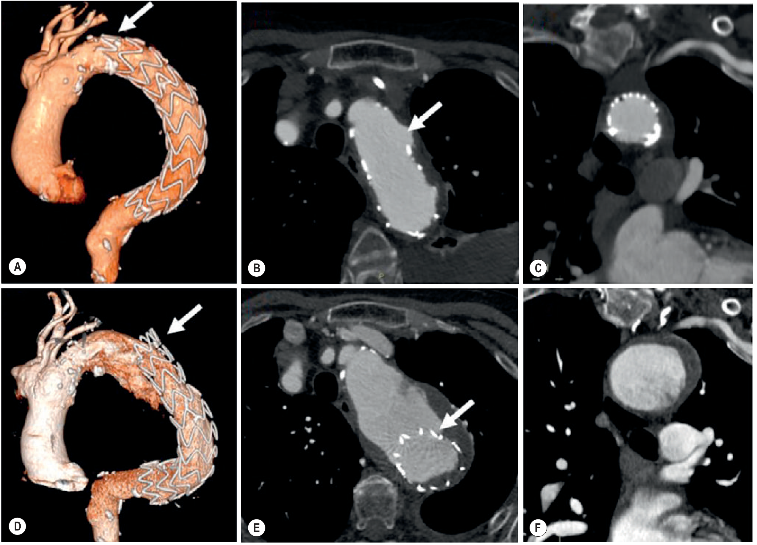
Figure: CT volume rendering and MPR imaging after TEVAR for TAI. Top row (A-C): pre-intervention showing injury (arrow in B = pseudoaneurysm/hematoma). Bottom row (D-F): post-TEVAR showing stent-graft in situ. Note the stent-graft has covered the proximal descending thoracic aorta. This sequence illustrates the planning and post-procedure assessment required. (Grainger & Allison's Diagnostic Radiology, Fig. 17.39)
9.5 Special Scenarios in TEVAR for BTAI
Ascending aortic or arch injuries (18% of BTAI):
- TEVAR is usually not anatomically feasible
- Requires open surgical repair via median sternotomy + CPB + DHCA
- Zone 0 or Zone 1 injuries need open repair; hybrid approaches with debranching + TEVAR occasionally possible
Tortuous or small access vessels:
- Iliac conduit via retroperitoneal approach to the common iliac artery
- Alternative: axillary artery access (rare)
"Pseudocoarctation":
- Severe narrowing of the true lumen at the injury site creating a pressure gradient
- Can cause upper limb hypertension and distal ischemia
- TEVAR restores lumen diameter and abolishes gradient
10. OPEN SURGICAL REPAIR
Open surgery is now reserved for:
- Ascending aortic or arch injury (not anatomically suitable for TEVAR)
- Access vessel anatomy unsuitable for TEVAR (calcified aortoiliac occlusive disease, small iliac arteries)
- Failed TEVAR (endoleak requiring conversion)
- Younger patients with genetic aortopathy (Marfan) in elective setting
- Concurrent cardiac injury requiring sternotomy (coronary laceration, aortic valve injury)
10.1 Descending Aortic Open Repair (Left Thoracotomy Approach)
Position: Right lateral decubitus. Left posterolateral thoracotomy through the 4th or 5th intercostal space.
Bypass strategy for spinal cord protection:
- Left heart bypass (centrifugal pump): Left inferior pulmonary vein → left femoral artery (or descending aorta distal to repair). Provides distal aortic perfusion maintaining spinal cord, renal, and visceral blood flow during cross-clamp. Does NOT require systemic heparinization (heparin-bonded circuit; important in polytrauma with coagulopathy).
- Femoral-femoral (femorofemoral) CPB: Also used; does require heparin.
- "Clamp-and-sew" technique: Aortic cross-clamp only, no bypass. Acceptable if cross-clamp time <30 minutes and adequate collateral supply. Associated with higher paraplegia risk (3.8-40% depending on duration). Rarely used in modern practice.
Operative steps:
- Left thoracotomy; enter pleural space; evacuate hemothorax
- Identify proximal (zone between LSCA and injury) and distal aorta for clamping
- Initiate left heart bypass
- Apply proximal clamp (ideally between LSCA and injury; sometimes distal arch clamp required)
- Apply distal clamp to descending aorta distal to injury
- Transect aorta at injury; inspect ends - confirm healthy aortic wall for anastomosis
- Interposition Dacron graft with end-to-end anastomoses (running polypropylene suture)
- Remove clamps in sequence; de-air; check for hemostasis
- Wean from left heart bypass; close thoracotomy with drains
Morbidity and mortality of open repair in BTAI:
- Historical mortality: >15-20% (emergency context + polytrauma)
- Paraplegia: 3-8% with distal perfusion techniques; up to 40% with clamp-and-sew
- Modern TEVAR has largely supplanted open repair due to significantly lower morbidity
10.2 Ascending Aorta / Arch Open Repair
Approach: Median sternotomy + CPB
Indications: Zone 0 (ascending) or Zone 1 (proximal arch) injuries
CPB strategy:
- Femoral or axillary artery cannulation (avoid ascending aorta due to injury)
- Venous return via right atrium
- Cooling to DHCA temperatures (18-20°C)
- Antegrade cerebral perfusion (ACP) via right axillary artery for arch work
- Open distal anastomosis technique under circulatory arrest
- Dacron interposition graft for aortic transection
- Aortic root preservation where possible (rarely involved in traumatic BTAI)
11. ANESTHETIC MANAGEMENT (MCh Level - Key Points)
- Full stomach: All trauma patients treated as having a full stomach - rapid sequence induction
- Induction agents: Ketamine (1-2 mg/kg) ± etomidate - preserve hemodynamic stability; avoid propofol bolus in hypotensive patients
- Two large-bore IV lines + arterial line before induction
- Right radial arterial line preferred (left subclavian may be compressed or covered by TEVAR)
- One-lung ventilation is required for left thoracotomy open repair - use double-lumen ETT or bronchial blocker
- TEE is mandatory intraoperatively for TEVAR guidance and for open repair to assess cardiac function, guide volume resuscitation, detect tamponade
- Temperature management: Active warming (hypothermia worsens coagulopathy)
- Coagulation management: TEG/ROTEM-guided MTP; avoid aggressive crystalloid (dilutional coagulopathy)
- Post-op: ICU with controlled ventilation; vasopressors to maintain MAP >90 mmHg (spinal cord protection)
Miller's Anesthesia 10e, p. 7776-7777
12. COMPLICATIONS AND THEIR MANAGEMENT
| Complication | Cause | Management |
|---|---|---|
| Spinal cord ischemia (paraplegia) | Intercostal artery coverage, hypotension | CSF drain, MAP >90 mmHg, LSA revascularization, steroids; may be reversible if treated early |
| Type Ia endoleak (proximal seal failure) | Inadequate landing zone, device undersizing | Balloon molding; proximal extension with second stent-graft; surgical conversion |
| Type II endoleak | Back-bleeding from LSCA or intercostals | Observe first; selective embolization if persistent |
| Stroke | Plaque/air embolism, covered supra-aortic vessel, air during deployment | Neurological monitoring; neuroradiology consultation; heparin |
| Stent-graft collapse | Device oversized for angulated aorta; excessive curve | Reballooning; snare technique; surgical extraction |
| Device migration | Undersizing; landing in diseased aorta; late | Proximal extension |
| Access vessel injury | Iliac dissection, rupture | Covered stent; retroperitoneal repair |
| LSA ischemia | Coverage without revascularization | Carotid-subclavian bypass or transposition |
| Pseudoaneurysm progression | Incomplete treatment; ongoing injury | Re-intervention (TEVAR extension or open repair) |
| Late false aneurysm | Incomplete coverage | Surveillance; reintervention if expanding |
| Coagulopathy | Massive transfusion + hypothermia + acidosis (lethal triad) | MTP; FFP:PLT:PRBC = 1:1:1; TXA; calcium; TEG/ROTEM guidance |
13. TIMING STRATEGY AND DAMAGE CONTROL PRINCIPLES
The "injury priority" principle for polytrauma with BTAI:
| Priority | Injury | Action |
|---|---|---|
| 1st | Traumatic brain injury with herniation | Neurosurgical decompression |
| 1st | Unstable pelvic ring fracture + hemorrhage | Pelvic binder / IR embolization / Ex-fix |
| 1st | Cardiac tamponade (ascending aortic injury) | Pericardiocentesis or sternotomy |
| 2nd | Solid organ injury with ongoing hemorrhage | Exploratory laparotomy / IR embolization |
| 2nd-3rd | Grade 3-4 BTAI (pseudoaneurysm / rupture) | TEVAR within 24 hours |
| Expectant | Grade 1-2 BTAI (intimal tear, IMH) | Medical management + serial CTA |
Delayed TEVAR (24-72 hours) is acceptable and may be preferable in severe polytrauma because:
- Allows stabilization of hemodynamics and coagulopathy
- Allows management of higher-priority injuries
- Reduces overall procedural risk
- Recent ATF evidence: timing of TEVAR does not significantly impact overall outcome; individualized approach is favored
14. FOLLOW-UP SURVEILLANCE
| Time Point | Imaging | Purpose |
|---|---|---|
| Pre-discharge (24-48 hrs post-TEVAR) | CTA chest | Confirm seal, detect endoleak, assess coverage |
| 1 month | CTA chest | Re-assess injury evolution, detect early complications |
| 6 months | CTA chest | Late endoleak, device migration, injury resolution |
| 12 months | CTA chest | Annual surveillance |
| Annually thereafter | CTA or MRA | Long-term device integrity, aneurysmal degeneration |
Grade 1-2 (medically managed) follow-up:
- CTA at 24-72 hours, 1 week, 1 month, 3 months - looking for: progression to pseudoaneurysm, hematoma expansion, hemodynamic changes
Long-term concerns after TEVAR:
- Endoleak (late type Ia especially as aortic anatomy changes with remodeling)
- Device fatigue / fracture
- Late aneurysmal degeneration (descending aorta distal to graft)
- Stent-graft collapse in hypoplastic aorta (young trauma patients - smaller native aortic diameter)
15. SPECIAL SITUATIONS AT MCh LEVEL
15.1 Young Patients (Pediatric / Adolescent)
- Aortic diameter often <20 mm - most commercial TEVAR devices have lower limit of ~17-18 mm
- Risk of stent-graft collapse in very angulated aortic arch (acute angle between arch and descending)
- Open repair may be preferable in children; can use homograft
- Long-term device surveillance is critical - the stent-graft will need re-intervention as the child grows
15.2 BTAI with Concurrent TBI
- The most challenging management dilemma: anti-impulse therapy (low BP) vs cerebral perfusion pressure maintenance (adequate BP)
- Multidisciplinary approach (neurosurgery + cardiac surgery/vascular surgery + critical care)
- Modified BP targets: SBP 100-140 mmHg (not <100 mmHg)
- Grade 1-2 BTAI with concurrent moderate TBI: usually managed medically (anti-impulse therapy at modified targets)
- Grade 3-4 BTAI with concurrent TBI: TEVAR preferred (shorter, less invasive; allows earlier ICP management)
15.3 BTAI with Concurrent Solid Organ Injury
- If splenectomy or hepatorrhaphy required: perform laparotomy first (usually faster to control bleeding)
- TEVAR second (semi-urgent within 24 hours)
- Avoid full-dose heparin during TEVAR if recent hepatic or splenic repair - consider heparin-free or reduced-dose TEVAR
15.4 Ascending Aortic BTAI
- Rare (18%) but requires sternotomy + CPB + DHCA
- Associated with massive pericardial hematoma, tamponade, acute AR, and coronary ostial avulsion
- Higher mortality than isthmic injury
15.5 Chronic Traumatic Pseudoaneurysm
- Occasionally diagnosed incidentally weeks to months after trauma
- False aneurysm contains the injury; adventitia and fibrotic reaction maintain integrity
- Still requires treatment (risk of late rupture is significant)
- TEVAR is preferred; open repair has higher risk than acute injury (fibrotic adhesions)
16. KEY NUMBERS FOR MCh EXAMINATIONS
| Parameter | Value |
|---|---|
| Prehospital mortality | ~80% |
| BTAI as cause of US MVA deaths | ~20% |
| Most common site | Aortic isthmus (50-90%) |
| Grade 3 BTAI frequency (ATF registry) | ~50% of all BTAI presentations |
| SVS: TEVAR recommended for grades | 2-4 (urgent <24 hours) |
| Proximal landing zone required | ≥2 cm (15-20 mm) |
| LSA coverage required | ~40% of BTAI TEVAR cases |
| Risk of CNS events post-LSA coverage (no revascularization) | ~10% |
| Paraplegia - clamp-and-sew technique | 3.8-40% |
| Paraplegia - left heart bypass | <5% |
| Anti-impulse SBP target | <120 mmHg |
| Anti-impulse HR target | <60 bpm |
| CTA sensitivity/specificity | ~100% (modern MDCT) |
| TEE sensitivity for isthmic injury | 91% |
| TEE specificity | 98% |
| Mediastinal width threshold on CXR | >8 cm (or M/C ratio >25%) |
17. CURRENT GUIDELINES SUMMARY
2022 ACC/AHA Guideline for Aortic Disease (PMID: 36322642, Circulation, 2022):
- TEVAR is the preferred approach for BTAI of the descending thoracic aorta when anatomically suitable (Class I)
- Anti-impulse therapy (HR <60, SBP <120 mmHg) is mandated for all BTAI during the perioperative period
- Grade 1-2 injuries can be managed with anti-impulse therapy and serial imaging
- Open repair reserved for ascending/arch injuries or when TEVAR not feasible
SVS Systematic Review 2026 (PMID: 40482894):
- Supports TEVAR over open surgical repair for descending BTAI
- Emerging evidence for definitive medical management of MAI (grades 1-2)
- Timing of TEVAR (immediate vs delayed) does not significantly impact overall outcomes
MANAGEMENT ALGORITHM
BTAI confirmed on CTA
│
├── Grade 4 (Free Rupture) → EMERGENT TEVAR or Open Surgery
│ └── Ascending/Arch → Sternotomy + CPB + DHCA
│
├── Grade 3 (Pseudoaneurysm) → URGENT TEVAR within 24 hrs
│ ├── Anti-impulse therapy NOW
│ ├── Manage higher-priority injuries first
│ ├── Assess: landing zones, access vessels, LSA, cerebrovascular anatomy
│ └── TEVAR (preferred) vs Open (if unsuitable anatomy)
│
├── Grade 2 (IMH) → Anti-impulse therapy + Serial CTA
│ ├── Consider TEVAR if: hematoma expansion, pain, hemodynamic instability
│ └── Most can be managed medically
│
└── Grade 1 (Intimal tear) → Anti-impulse therapy + Serial CTA
├── Expectant management
└── Intervene only if: progression on imaging, instability
All patients:
→ ICU admission with invasive monitoring
→ Beta-blocker infusion (esmolol preferred)
→ SBP target <120 mmHg, HR target <60 bpm
→ Polytrauma co-management (TBI, ortho, abdo)
→ CTA at 24-72 hrs, 1 week, 1 month, 3 months, 6 months, then annually
Sources for this note:
- Fischer's Mastery of Surgery, 8e, Chapter 261
- Grainger & Allison's Diagnostic Radiology, Chapters 17 & 79
- Miller's Anesthesia, 10e, Chapter 52
- Sabiston Textbook of Surgery, Chapter 36
- Schwartz's Principles of Surgery, 11e, Chapter 22
- Textbook of Clinical Echocardiography, Chapter 16
- 2022 ACC/AHA Guideline for Aortic Disease [PMID: 36322642]
- SVS Systematic Review for BTAI guidelines 2026 [PMID: 40482894]That is a complete MCh Cardiac Surgery-level note on Blunt Traumatic Aortic Injury. Here is what was covered across 17 sections:
What Was Covered:
- Epidemiology - 80% prehospital mortality; 20% of all MVA deaths; only adventitially contained injuries survive to hospital
- Mechanisms (3 biomechanical forces with diagram) - Deceleration/shear at isthmus (50-90%), osseous pinch (ascending), torsional/hydraulic. Site frequency by mechanism.
- Spectrum of pathology - Subintimal tear → IMH → pseudoaneurysm → frank transection, with cross-sectional diagram
- Classification - SVS/Azizzadeh Grades 1-4 (ATF registry frequencies) + Presley Trauma Center CT system (full table)
- Clinical presentation - High index of suspicion; physical signs; ATLS primary survey relevance; "normal BP does not rule out BTAI"
- Diagnostic imaging with real images:
- CXR - all signs with widened mediastinum photo
- CTA (gold standard) - direct + indirect signs, false positive pitfalls, pseudocoarctation
- Conventional aortography - why abandoned
- TEE - bedside value, contraindications, 7 mm probe gap sign
- MRI and IVUS roles
- Initial resuscitation - ATLS priorities, anti-impulse therapy targets, drug choices, TBI conflict resolution
- Management by grade - Grade 1 (expectant) → Grade 4 (emergent)
- TEVAR - detailed step-by-step technique - access, pre-close technique, arch aortogram, IVUS, anticoagulation, stiff wire exchange, two-person deployment, completion angiogram
- LSA coverage - 40% incidence; when to revascularize first (LIMA CABG, dominant left VA, PICA termination); when safe to cover without revascularization
- Spinal cord ischemia - risk factors, prevention strategies, Adamkiewicz territory
- Open surgical repair - left thoracotomy, left heart bypass vs clamp-and-sew, paraplegia rates; ascending injury via sternotomy/CPB/DHCA
- Anesthetic management - RSI, ketamine, one-lung ventilation, TEE, coagulopathy management
- Complications - Type Ia endoleak, SCI, stroke, stent collapse, access injury, device migration
- Timing strategy - damage control principles, injury priority hierarchy, evidence for delayed TEVAR
- Follow-up surveillance - detailed CTA schedule post-TEVAR and for medically managed cases
- Special situations - young patients, concurrent TBI (the hardest management dilemma), solid organ injury, ascending BTAI, chronic pseudoaneurysm
This is a shared conversation. Sign in to Orris to start your own chat.