
Venous drainage of heart
Reading File
The venous drainage of the heart is primarily via the coronary sinus and its tributaries, which drain into the right atrium. Now let me pull the full details from the medical library.
Searching sources...
Finding Sources
Finding Sources
Finding Sources
Finding Sources
Good - I found relevant sections. Let me read the anatomy content from the Thieme Atlas and the Barash anesthesia text on coronary anatomy, and also the Guyton physiology section.
Reading File
Reading File
Finding Sources
The Barash text has the coronary venous drainage content. Let me read more of it, and also search for dedicated anatomy content.
Reading File
Finding Sources
Excellent - I now have very rich source material. Let me read the most detailed anatomical sections from Fuster's Heart, Fischer's Surgery, Gray's Anatomy, and the Color Atlas.
Reading File
Reading File
Reading File
I now have excellent material from multiple authoritative sources. Let me also get the Gray's Anatomy content on the great cardiac vein and read the Fischer Surgery content, plus fetch the key anatomical diagram.
Reading File
Reading File
Loading Image
I also have a great cardiac veins diagram. Let me also fetch the Gray's Anatomy cardiac veins diagram for completeness.
Loading Image
I now have all the information needed for a comprehensive answer. Let me compile it.
Venous Drainage of the Heart
The venous return from the myocardium is channeled through two main systems: the greater coronary venous system (drains ~85% of venous flow) and smaller direct-drainage veins (Thebesian veins and anterior cardiac veins). The primary collector of the greater system is the coronary sinus, which drains into the right atrium.
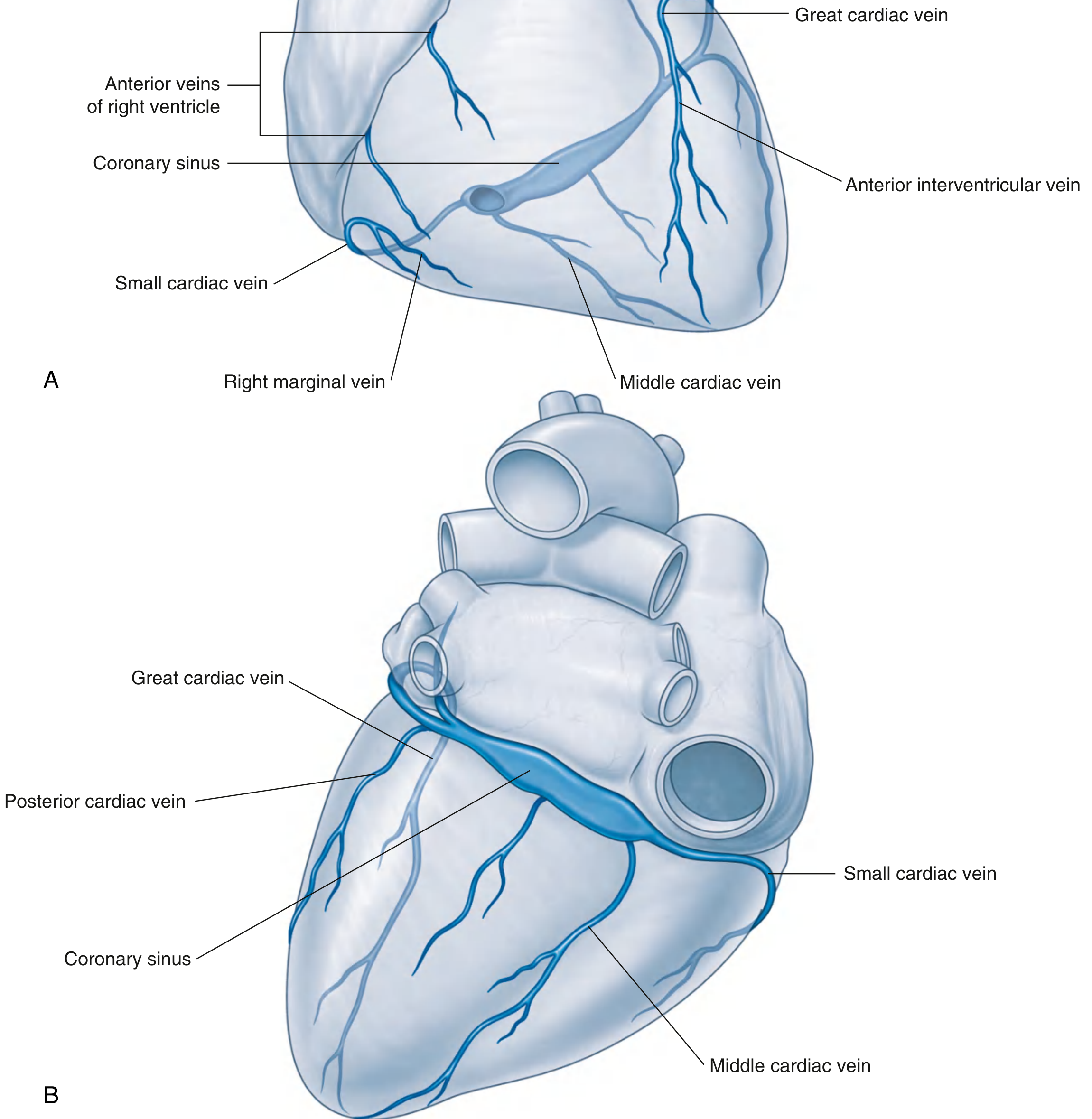
1. The Coronary Sinus
- The coronary sinus is the main venous channel of the heart, lying in the posterior part of the coronary (atrioventricular) sulcus.
- It is a wide, short (~2-3 cm) venous channel, roughly tubular, with an ostial diameter of 5-15 mm.
- It opens into the posterior wall of the right atrium, immediately above the tricuspid valve, between the inferior vena cava orifice and the atrioventricular septum (guarded by the valve of Thebesius).
- It receives most of the tributaries draining the left ventricle and left atrium.
- Venous blood flow into the coronary sinus peaks during late systole, because the contracting LV compresses major venous drainage channels.
- Approximately 85% of total coronary blood flow returning from the LV empties into the coronary sinus.
2. Tributaries of the Coronary Sinus
Great Cardiac Vein (largest tributary)
- Begins as the anterior interventricular vein, ascending alongside the left anterior descending (LAD) artery in the anterior interventricular sulcus.
- Turns at the apex to course in the left atrioventricular groove (alongside the left circumflex artery) and continues into the coronary sinus.
- Its transition to the great cardiac vein proper is marked by the valve of Vieussens (present in 80-90% of hearts) and the junction with the oblique vein of Marshall.
- It collects tributaries from the anterior LV, lateral (obtuse marginal) veins, and inferior LV veins - these lateral/posterolateral branches are important targets for LV pacing leads in cardiac resynchronization therapy.
Middle Cardiac Vein
- Starts at the apex of the heart.
- Courses in the posterior interventricular sulcus alongside the posterior descending artery (PDA).
- Drains into the right end of the coronary sinus (or occasionally directly into the right atrium).
- At the cardiac crux it passes immediately superficial to the right coronary artery.
Small Cardiac Vein
- Begins as the right marginal vein, accompanying the right marginal branch of the RCA.
- Turns posteriorly into the right atrioventricular (coronary) sulcus.
- Drains into the right end of the coronary sinus or occasionally into the middle cardiac vein.
- Receives tributaries from the right atrium and inferior wall of the right ventricle.
Posterior Vein of the Left Ventricle
- Courses on the diaphragmatic surface of the left ventricle, just to the left of the middle cardiac vein.
- Enters either the coronary sinus directly or joins the great cardiac vein.
Oblique Vein of the Left Atrium (Vein of Marshall)
- Courses on the posterior wall of the left atrium.
- Merges with the great cardiac vein to form (or enter) the coronary sinus.
- It is an embryonic remnant of the left superior vena cava (left common cardinal vein).
- Rarely exceeds 3 cm before tapering to a blind end; when wide, it can be used for left atrial ablation.
3. Veins That Bypass the Coronary Sinus
Anterior Cardiac Veins (Anterior Veins of the Right Ventricle)
- 3-4 small veins arising on the anterior surface of the right ventricle.
- Cross the coronary sulcus and enter directly into the anterior wall of the right atrium.
- Drain the anterior portion of the RV.
- The right marginal vein may be part of this group if it does not enter the small cardiac vein.
Smallest Cardiac Veins (Venae Cordis Minimae / Thebesian Veins)
- Tiny vessels draining capillary beds of the myocardium directly into the cardiac chambers.
- Most numerous in the right atrium and right ventricle.
- Occasionally associated with the left atrium; rarely found in the left ventricle.
- The remaining ~15% of LV coronary blood flow (not entering the coronary sinus) drains through these veins, predominantly into the RA > LA and LV.
Summary Table
| Vein | Course | Drains Into |
|---|---|---|
| Great cardiac vein | Anterior interventricular sulcus → left AV groove | Coronary sinus (left end) |
| Middle cardiac vein | Posterior interventricular sulcus | Coronary sinus (right end) |
| Small cardiac vein | Right AV groove (from right marginal vein) | Coronary sinus (right end) |
| Posterior vein of LV | Diaphragmatic surface of LV | Coronary sinus / great cardiac vein |
| Oblique vein of Marshall | Posterior left atrial wall | Great cardiac vein / coronary sinus |
| Coronary sinus | Posterior AV groove | Right atrium |
| Anterior cardiac veins (3-4) | Anterior RV surface | Directly into right atrium |
| Thebesian veins | Myocardial walls | Directly into cardiac chambers |
Clinical Relevance
- The coronary sinus is used as access for retrograde cardioplegia in cardiac surgery, and for placing LV pacing leads in cardiac resynchronization therapy (CRT) via the lateral/posterolateral tributaries.
- The left phrenic nerve runs close to the lateral/posterolateral LV veins, and pacing from these veins can cause phrenic capture.
- The oblique vein of Marshall can be used for alcohol ablation of the left atrial wall in certain arrhythmias.
- The muscular sleeve around the proximal coronary sinus may participate in atrial arrhythmias.
- The thebesian veins create a small physiological right-to-left shunt, contributing to normal arterial desaturation.
Sources: Fuster & Hurst's The Heart 15th Ed., Barash Clinical Anesthesia 9th Ed., Fischer's Mastery of Surgery 8th Ed., Gray's Anatomy for Students, Color Atlas of Human Anatomy Vol. 2 (Thieme)
This is a shared conversation. Sign in to Orris to start your own chat.